• 검색 결과가 없습니다.

Diagnosis and Treatment of Lower Extremity Deep Vein Thrombosis: Koeran Practice Guidelines

N/A
N/A
Protected

Academic year: 2021

Share "Diagnosis and Treatment of Lower Extremity Deep Vein Thrombosis: Koeran Practice Guidelines"

Copied!
28
0
0

로드 중.... (전체 텍스트 보기)

전체 글

(1)

Vascular Specialist International

Vol. 32, No. 3, September2016 pISSN 2288-7970 • eISSN 2288-7989

INTRODUCTION

1) Background and purpose

Deep vein thrombosis (DVT) refers to the presence of thrombus within a deep vein of the body, most frequently

in the lower extremities. Many episodes are asymptomatic and the symptoms of acute DVT, including edema, pain, and erythema, are non-specific. At least three-quarters of patients having lower extremity symptoms consistent with DVT have a non-thrombotic cause of their symptoms.

Therefore, confirmatory testing is almost always required,

Diagnosis and Treatment of Lower Extremity Deep Vein Thrombosis: Korean Practice Guidelines

Seung-Kee Min

1

, Young Hwan Kim

2

, Jin Hyun Joh

3

, Jin Mo Kang

4

, Ui Jun Park

5

, Hyung-Kee Kim

6

, Jeong-Hwan Chang

7

, Sang Jun Park

8

, Jang Yong Kim

9

, Jae Ik Bae

10

, Sun Young Choi

11

, Chang Won Kim

12

, Sung Il Park

13

, Nam Yeol Yim

14

, Yong Sun Jeon

15

, Hyun-Ki Yoon

16

, and Ki Hyuk Park

17

1Department of Surgery, Seoul National University College of Medicine, Seoul, 2Department of Radiology, Keimyung University College of Medicine, Daegu, 3Department of Surgery, Kyung Hee University School of Medicine, Seoul, 4Department of Surgery, Gachon University College of Medicine, Incheon, 5Department of Surgery, Keimyung University College of Medicine, Daegu, 6Department of Surgery, Kyungpook National University School of Medicine, Daegu, 7Department of Surgery, Chosun University College of Medicine, Gwangju, 8Department of Surgery, University of Ulsan College of Medicine, Seoul, 9Department of Surgery, Catholic University College of Medicine, 10Mint Intervention Clinic, Seongnam, 11Department of Radiology, Ewha Womans University College of Medicine, Seoul,

12Department of Radiology, Pusan National University School of Medicine, Yangsan, 13Department of Radiology, Yonsei University College of Medicine, Seoul, 14Department of Radiology, Chonnam National University College of Medicine, Gwangju, 15Department of Radiology, Inha University College of Medicine, Incheon, 16Department of Radiology, University of Ulsan College of Medicine, Seoul,

17Department of Surgery, Daegu Catholic University College of Medicine, Daegu, Korea

Received August 9, 2016 Accepted August 9, 2016

Corresponding author: Ki Hyuk Park Department of Surgery, Daegu Catholic University College of Medicine, 33 Duryugongwon-ro 17-gil, Nam-gu, Daegu 42472, Korea

Tel: 82-53-650-4053 Fax: 82-53-624-7185 E-mail: khpark@cu.ac.kr Conflict of interest: None.

This Guideline has been published jointly by invitation and consent in both the Vascular Specialist International and the Journal of the Korean Society of Radiology.

Lower extremity deep vein thrombosis is a serious medical condition that can result in death or major disability due to pulmonary embolism or post-thrombotic syndrome. Appropriate diagnosis and treatment are required to improve symptoms and salvage the affected limb. Early thrombus clearance rapidly resolves symptoms related to venous obstruction, restores valve function and reduces the incidence of post-thrombotic syndrome. Recently, endovascular treatment has been established as a standard method for early thrombus removal. However, there are a variety of views regarding the indications and procedures among medical institutions and operators. Therefore, we intend to provide evidence-based guidelines for diagnosis and treatment of lower extremity deep vein thrombosis by multidisciplinary consensus. These guidelines are the result of a close collaboration between interventional radiologists and vascular surgeons. The goals of these guidelines are to improve treatment, to serve as a guide to the clinician, and consequently to contribute to public health care.

Key Words: Guideline, Venous thrombosis, Diagnosis and treatment

Copyright © 2016, The Korean Society for Vascular Surgery

This is an Open Access article distributed under the terms of the Creative Commons Attribution Non-Commercial License (http://creativecommons.org/licenses/by-nc/4.0) which permits unrestricted non-commercial use, distribution, and reproduction in any medium, provided the original work is properly cited.

Vasc Spec Int 2016;32(3):77-104 • http://dx.doi.org/10.5758/vsi.2016.32.3.77

Review

(2)

2) Guideline development

Considering that the development of de novo domestic clinical guideline is a difficult undertaking, these guidelines were developed by adapting the pre-existing guidelines of other countries. If no existing guidelines were available, we evaluated existing good-quality articles using the systemic literature review methodology.

The steering committee was composed of the executives of The Korean Society of Interventional Radiology and The Korean Society for Vascular Surgery. The steering committee fixed the subject and the goal, assigned the members of the guideline development committee and approved the guideline development budget. The guideline development committee comprised 22 members.

Guideline development committee members discussed the purpose of the guidelines, the range of development including writing topics, the subjects of the application and user groups, development method, determined the level of evidence (LOE), classified recommendations, selected the consensus development method, internal and external review processes, revision processes and formed the committee of detail associated with guideline development during the first conference. The committee of detail was composed of the guideline evaluation committee, the writing committee, and the editing committee. The guideline evaluation committee comprised four members and evaluated the pre-existing guidelines based on Appraisal of Guidelines for Research & Evaluation II (AGREE II). The writing committee had 17 members and were responsible for drawing up the draft guidelines and the proposal of recommendations. The editing committee was composed of four members and was responsible for reviewing the recommendation levels, the LOE, and the draft guidelines by performing peer review.

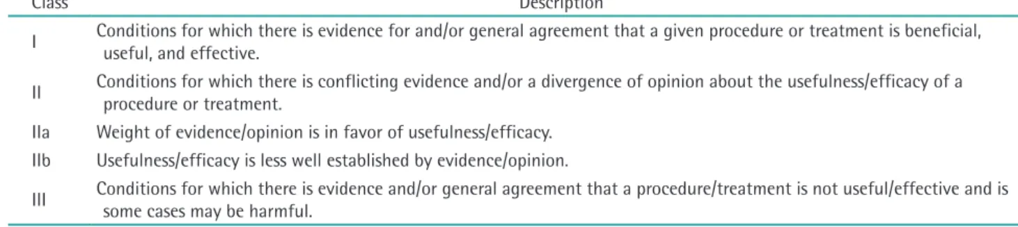
The LOE and the classification of recommendations (COR) followed the criteria used in the American College of Cardiology/American Heart Association (ACC/

AHA) 2005 guidelines [1]. COR was divided into three categories: Recommendation I (strong recommendation), Recommendation II (weak recommendation), and Recommendation III (contraindication). Recommendation I was defined as conditions for which there was solid evidence for and/or general agreement that a given proce- dure or treatment is effective, useful, and beneficial and will not be changed by further research. Recommendation II was defined as conditions with conflicting evidence and/

or a divergence of opinion about the usefulness/efficacy of a procedure or treatment. IIa was defined as cases for which the weight of evidence/opinion was in favor of usefulness/efficacy. IIb was defined as cases for which the not only to ensure appropriate treatment of those with

confirmed DVT but also to prevent the complications of inappropriate anticoagulation in those with other disorders. The aim of the treatment of DVT is to prevent its complications—pulmonary embolism (PE), recurrent DVT, post-thrombotic syndrome (PTS), and death.

The standard treatment for acute DVT is systemic anti- coagulation to decrease the propagation of the thrombus and prevent PE. However, anticoagulation alone has no significant thrombolytic activity and does not prevent PTS. Early thrombus clearance rapidly resolves symptoms related to venous obstruction, and can restore valve function so that it reduces the incidence of PTS. Recently, catheter-directed thrombolysis with or without mechanical thrombectomy has been established as a standard method for early thrombus removal. The indications for these interventional procedures are gradually increasing with the rapid development of equipment and procedures, and there are a variety of views regarding the indications and procedures among medical institutions and operators.

In the United States, the Society of Interventional Radiology, American College of Chest Physicians, Society of Vascular Surgeon, and American Venous Forum pre- sented the recommended guidelines for the interven tional procedures of acute DVT in 2006 and 2012. In Europe, the Scottish Intercollegiate Guidelines Network and National Clinical Guideline Center working group issued guidelines for management of venous thromboembolism (VTE) in 2010 and 2012. Through these efforts, by esta blishing standard procedures and raising awareness of physicians on the front lines of medicine to the importance of the procedures, a cautious approach is encouraging. Moreover, these guidelines can be used as basic standards for health insurance payment, review and assessment for reducing medical costs.

However, although a number of similar studies have

been conducted, clinical guidelines for Korea have not

yet been established. Thus, experts from academies (The

Korean Society of Interventional Radiology and The Korean

Society for Vascular Surgery) related to interventional

procedures in Korea came together and agreed to develop

clinical guidelines. We aim to propose recommendations

by presenting evidence-based treatment recommendations

through a multidisciplinary approach, which will guide

interventional procedures by providing up-to-date and

accurate information to healthcare providers working at

primary, secondary and tertiary hospitals. Furthermore, we

aim to help patients themselves choose medical services by

providing accurate information and so contribute to public

health promotion.

(3)

usefulness/efficacy was less well established by evidence/

opinion. Recommendation III was defined as conditions for which there was evidence and/or general agreement that a procedure/treatment is not useful/effective and may be harmful in some cases (Table 1). LOE was classified into three steps: A, B, and C. Evidence A was defined as data derived from multiple randomized clinical trials or meta- analysis. Evidence B was defined as data derived from a single randomized clinical trial or non-randomized studies.

Evidence C was defined as only a consensus opinion of experts, case studies, or standard of care (Table 2).

To select high-quality guidelines for use as a reference in the adaptation process, we searched for existing guidelines.

We recovered 115 documents by mixing search index words, such as VTE, diagnosis, anticoagulation, thrombolysis, thrombectomy, and guidelines, using the PUBMED, SCOPUS, and COCHRANE search engines.

The guideline evaluation committee determined the inclusion and exclusion criteria for quote-worthy documents among the obtained documents. The inclusion criteria were set as evidence-based guidelines, international guidelines written in English, and guidelines written after 2005. Guidelines that did not represent the organization, those written by one person and translations of single guidelines were excluded.

Five guidelines were chosen based on these inclusion criteria. Four members of the guideline evaluation committee evaluated these guidelines based on AGREE II, which is the most commonly used tool internationally for the quality assessment of guidelines. AGREE II comprises 23 sub-items within six assessment categories, and each item is assigned a score on a 7-point Likert scale. Based on the evaluated results after opinion coordination, three

guidelines that had standardized scores of more than 50%

in all categories were finally selected [2-4].

The guideline development committee selected the final key question after reviewing whether population, intervention, comparison, and outcome (PICO), the essential structural component of the key question, was well equipped and appropriate as a clinical question. The key question was agreed upon using the nominal group technique. Consensus was defined as more than 75% of the panel selecting 1 or 2 on the 5-point Likert scale (1, agree entirely; 2, agree generally; 3, agree partially; 4, do not agree generally; 5, do not agree entirely). If the consensus was less than 75%, a second round of voting was carried out after discussion and a modification of phrases. If agreement was not reached in the second round of voting, the key question was dismissed. Eleven members of the development committee participated as a panel, and 22 of a total of 42 key questions that were drawn up were agreed on by the committee members and were selected as the final clinical questions.

Writing committee members, who were assigned accor- ding to sub-themes, drew up the proposal of recommen- dations for clinical questions. A single draft of the proposal of recommendation was deduced by collecting common information and deleting unnecessary infor- mation after analyzing recommendations extracted from selected guidelines. If there were no data for reference in existing guidelines of clinical questions, a new proposal of recommendation was developed on the basis of the results after evaluating the quality of articles through a literature search and review. Thus, a total of 43 proposals of recommendation were drawn up. Ten of these recom- mendations were deleted because no final agreement

Table 1. Classification of recommendations

Class Description

I Conditions for which there is evidence for and/or general agreement that a given procedure or treatment is beneficial, useful, and effective.

II Conditions for which there is conflicting evidence and/or a divergence of opinion about the usefulness/efficacy of a procedure or treatment.

IIa Weight of evidence/opinion is in favor of usefulness/efficacy.

IIb Usefulness/efficacy is less well established by evidence/opinion.

III Conditions for which there is evidence and/or general agreement that a procedure/treatment is not useful/effective and is some cases may be harmful.

Table 2. Levels of evidence

Level Description

A Data derived from multiple randomized clinical trials or meta-analysis.

B Data derived from a single randomized clinical trial or nonrandomized studies.

C Only consensus opinion of experts, case studies, or standard of care.

(4)

was reached due to opinions that the information was ambiguous and difficult to understand despite modification of phrases or sentences and was impractical in Korea, etc.

The remaining 33 recommendations were chosen by means of a Delphi consensus survey. We created a recommendation data extraction form, which was used by the panel as a reference during the Delphi consensus process.

For formal mutual agreement of the final adoption of recommendations, a modified Delphi technique was applied. The panel was composed of 26 members (The Korean Society of Interventional Radiology, 14; The Korean Society for Vascular Surgery, 12). To assist the panel, the recommendation data extraction form and related references were provided by e-mail. The vote was performed anonymously. After sending the disclosure sheet on conflict of interest to all panel members, the members signed the document confirming that they did not receive any support from interest groups related to the recommendation. The degree of consensus was quantitatively analyzed using a 9-point Likert scale (1, strongly disagree; 9, entirely agree). In the response scale, scores of 7-9 points were considered to indicate agreement with the recommendations. When more than 50% of the panel agreed, the proposal of recommendations was considered to have achieved consensus. During the first round of voting, all recommendations met the agreement condition. Based on the adopted recommendations, the writing committee members assigned to specific sub- themes wrote draft guidelines. The editing committee evaluated the draft guidelines based on the selected recommendations. The guidelines were internally evaluated through an expert advisory conference to discuss the expertise of relevant academies associated with the guidelines, including problems in the recommendations, background and description of the evidence, etc.

Following this internal evaluation, the final guidelines were established by means of a public hearing involving experts in relevant fields and stakeholders.

CONTENTS

1) Symptoms

① Clinical presentation of acute lower extremity DVT The clinical presentation of acute lower extremity DVT varies with the anatomic distribution, extent, and degree of occlusion of the thrombus. Symptoms may range from absence to massive swelling and cyanosis with impending venous gangrene. Three patterns of thrombosis are usually recognized: isolated calf vein (distal), femoropopliteal, and iliofemoral thrombosis, and symptoms tend to be more

severe as thrombosis extends more proximally. However, up to 50% of patients with acute DVT may lack specific signs or symptoms [5,6]. Postoperative patients are, in particular, more likely to have small, asymptomatic, distal, non-occlusive thrombi. When present, signs and symptoms of acute lower extremity DVT may include pain, edema, erythema, tenderness, fever, prominent superficial veins, pain with passive dorsiflexion of the foot (Homan’s sign), and peripheral cyanosis. Phlegmasia cerulea dolens, characterized by the triad of massive swelling, cyanosis, and pain, is the most severe form of acute lower extremity DVT and results from complete thrombosis of an extremity’s venous outflow [7]. In advanced cases, it is marked by severe venous hypertension with collateral and microvascular thrombosis, leading to venous gangrene.

Venous gangrene is particularly associated with warfarin- mediated protein C depletion in patients with cancer or heparin-induced thrombocytopenia [8,9].

Unfortunately, the diagnosis of DVT based on clinical signs and symptoms is notoriously inaccurate. The signs and symptoms of DVT are non-specific and may be associated with other lower extremity disorders, including lymphedema, PTS, superficial venous thrombosis, cellulitis, musculoskeletal trauma, and Baker’s cysts. Among patients referred to the vascular laboratory for exclusion of DVT, only 12%-31% will have a positive ultrasound (US) study [10-12].The most common presenting symptoms have a wide range of reported sensitivities and specificities: calf pain, sensitivity 75%-91% and specificity 3%-87%; and calf swelling, sensitivity 35%-97% and specificity 8%- 88% [13-18]. None of the signs or symptoms is sufficiently sensitive or specific, either alone or in combination, to accurately diagnose or exclude thrombosis [19].

② Complications of acute lower extremity DVT

(1) Pulmonary embolism: The potentially life-threatening consequences of PE make it the most important short-term complication of acute lower extremity DVT. Symptomatic PE accompanies approximately 10% of DVT [20]. However, respiratory symptoms correlate poorly with the presence or absence of objectively documented PE, and as many as 75% of pulmonary emboli may be asymptomatic [21,22].

(2) Post-thrombotic syndrome: PTS, the symptoms of which include pain, edema, skin changes, and ulceration, is the most important late complication of acute lower extremity DVT. Older studies, many with methodological flaws, reported post-thrombotic manifestations in up to two-thirds of patients with acute lower extremity DVT.

More recent studies suggest that the PTS develops in

29.6% of those with proximal thrombosis and 30% of

those with isolated calf vein thrombosis [23]. In addition to

(5)

the substantial economic costs, the physical limitations of patients with PTS are similar to those of patients with other serious chronic medical conditions [20].

(3) Mortality after acute lower extremity DVT: Mortality after an episode of acute lower extremity DVT exceeds that expected in an age-matched population. Although the in-hospital fatality rate for DVT is only 5%, 1-, 3-, and 5-year mortality rates of 22%, 30%, and 39%, respectively, have been noted [20]. Early mortality is most frequently secondary to cancer, PE and cardiac disease. Among patients older than 45 years, cancer is the most important predictor of early death (28-day mortality rate, 25.4%) compared with 12.6% in patients without cancer [24].

While deaths among cancer patients and idiopathic DVT remain high for at least 3 years beyond the index event, that for those with secondary VTE unrelated to cancer return to those of general population after 6 months [24].

Clinically, there appears to be an association between idiopathic VTE and cardiovascular events [25]. For instance, the 10-year cumulative risk of symptomatic vascular events among patients with idiopathic DVT is 25.4%, compared with 12.9% in those with secondary VTE [26]. Patients with idiopathic DVT also have a higher prevalence of atherosclerotic risk factors (diabetes, hypertension, and hypercholesterolemia) and coronary artery calcium than control group without VTE [27].

③ Natural history of acute lower extremity DVT

(1) Venous thrombogenesis: As Virchow proposed, three factors are important in the development of venous thrombosis—abnormalities of blood flow, abnormalities of blood coagulation, and vessel wall injury. The role of structural injury to the vein wall is disputable. Overt endothelial injury appears to be neither a necessary nor a sufficient condition for thrombosis [28]. In contrast, evidence is accumulating that biologic injury to the endothelium may play a more important role in venous thrombogenesis. Although most venous thrombi originate in areas of low blood flow, stasis alone is also an inadequate stimulus in the absence of low levels of activated coagula- tion factors [29,30]. Stasis may be a permissive factor for the other events required for thrombosis.

Imbalanced activation of the coagulation system appears to be the most important factor underlying many episodes of acute lower extremity DVT. Some components of imbalanced coagulation appear to be associated with thrombotic risk factors, including age, malignancy, surgery, trauma, primary hypercoagulable states, pregnancy, and oral contraceptive use.

Lower extremity thrombi originate in areas of localized imbalanced coagulation due to stasis, such as soleal sinuses,

behind venous valve pockets, and venous confluences.

Propagation of thrombi beyond areas of stasis likely depends largely on the relative balance between activated coagulation and thrombolysis. In contrast to arterial thrombi, venous thrombi are composed largely of red cells and fibrin, with relatively few platelets.

(2) Recanalization: Once formed, the competing pro- cesses of recanalization and recurrent thrombosis charac- terize the natural history of acute lower extremity DVT. The development of chronic sequelae is closely related to the balance between recanalization and recurrence. Monocytes appear to play a particularly important role in thrombus organization and recanalization. Recanalization appears to be a complex process involving intrinsic and extrinsic fibrinolysis, peripheral fragmentation, neovascularization, and retraction. Thrombus organization begins in the attachment zone with the migration of surfacing cells, presumably derived from the endothelium, over the thrombus [31]. Most recanalization occurs within the first 6 weeks [32]. Although thrombus resolution proceeds at a similar rate in the proximal venous segment, some have found more rapid clearance from the tibial segments, perhaps reflecting the increased efficiency of thrombolysis in small veins [33].

The degree of recanalization is related to both the degree of activated coagulation and fibrinolytic inhibition. From a clinical perspective, more complete recanalization has been reported in older patients, those with asymptomatic postoperative thrombosis, and patients with involvement of only one venous segment [34]. Cancer is associated with less complete recanalization. The presence of permanent risk factors has been associated with an 11-fold higher risk of delayed recanalization [35].

(3) Recurrent venous thrombosis: Standard anticoagula- tion is effective in preventing recurrent VTE while patients are being treated. Among patients with proximal DVT, recurrent thromboembolic events occurred in 5.2% of patients treated with standard anticoagulation for 3 months [36], compared with 47% of patients inadequately treated with a 3-month course of low-dose subcutaneous heparin [37].

Not surprisingly, most symptomatic events occur after anticoagulation has been stopped. Sarasin and Bouna- meaux [38] calculated a theoretical recurrence rate of 0.9%

per month after discontinuing anticoagulant therapy for

proximal DVT, similar to annual recurrence rates of 7.0%-

12.9% [39,40]. The risk of recurrent VTE is highest over the

first 6-12 months after the index event, although cumula-

tive rates can reach 24% at 5 years and 30% at 8 years

after initial presentation [40-43]. The risk of recurrence

is at least as great in the contralateral as in the ipsilateral

extremity [42].

(6)

The risk of recurrence is highly related to the underlying thrombotic risk factors. Data from the Duration of Anti- coagulation (DURAC) trial suggest a 2-year recurrence rate of 12% in patients with idiopathic DVT or irreversible risk factors and 4.8% in patients with reversible risk factors if treated with 6 months of anticoagulants [43]. Others have similarly noted that patients with idiopathic DVT or throm- bophilia are at three-fold greater risk for recurrent VTE than those with secondary thrombosis [40,41]. Other risk factors for symptomatic recurrent DVT include advanced age, male gender, increased body mass index, lower extremity paresis and active malignancy [44].

Recurrent VTE in the setting of thrombosis isolated to the calf veins requires special consideration. Limited data suggest that isolated calf vein thrombosis is associated with less extensive activation of coagulation than proximal venous thrombosis [45]. At least two types of calf vein thrombosis may be differentiated—those with involvement of the paired posterior tibial and peroneal vein (axial calf vein thrombosis) and those isolated to the veins draining the gastrocnemial and soleal muscles (muscular calf vein thrombosis); the natural history of these types may be different. In patients with thrombosis isolated to the axial calf veins, proximal propagation occurred in 23% of untreated patients and 10% of patients treated with only intravenous heparin [46]. As US technology has improved, muscular calf vein thrombi are identified more frequently and now account for approximately 40% of isolated calf vein thrombi. More information is needed regarding the natural history and management of these thrombi.

Recognized thrombophilic states, particularly the factor V Leiden mutation, lupus anticoagulant, and homocy- steinemia, have been associated with recurrent throm- boembolic events [47-49]. Others have reported a 2.2-fold to >5-fold increased risk of recurrent thrombosis among those with incomplete recanalization [34,40]. A D-dimer level of >500 ng/mL measured 1 month after discontinuing anticoagulants was associated with a 3.3-fold increased risk of recurrence [50].

2) Diagnosis of lower extremity DVT

① Clinical probability scores (clinical scores)

Patients suspected with DVT usually present with swelling, pain, redness, and warmth in the lower extremity.

Currently, the diagnosis of DVT relies on imaging modalities such as compression and color Doppler US, and computed tomography (CT). However, only a minority of patients evaluated for suspected DVT with symptoms actually have the disease and the symptoms of many patients have other causes [51]. In addition, considering the cost and

invasiveness of diagnostic methods, the initial approach for patients with a possible DVT should be focused on the assessment of their individual pre-test probability (i.e., the likelihood that they have a DVT), and diagnostic tests should be selected according to the results of pre-test probability.

Clinical probability scores estimate the probability of DVT by incorporating signs, symptoms, and risk factors.

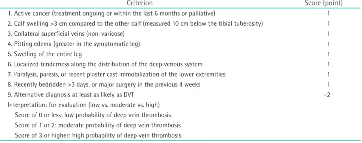
This score stratifies patients into groups according to the probability, which influences the subsequent diagnostic strategy. Currently, several structured scoring systems have been developed and introduced; the most widely used and well-studied is the Wells score [52]. The original Wells scoring system published in 1997 consisted of a nine- component clinical prediction rule for DVT and stratified patients into three categories: ‘high’ (3 or more points)

‘intermediate’ (1-2 points), and ‘low’ (less than 1 point) (Table 3). The prevalence of DVT was estimated as 5.0% (95%

confidence interval [CI], 4%-8%) in the low risk category, 17% (95% CI, 13%-23%) in the intermediate risk category, and 53% (95% CI, 44%-61%) in the high risk category [51].

In 2003, the original Wells score was modified. A further component, ‘previously documented DVT’, was added to the original Wells score, and instead of considering surgery within 4 weeks as a risk factor, the duration was extended to 12 weeks. In place of the three risk categories in the original version, this version has only two risk categories:

‘likely’ (≥2 points) or unlikely (<2 points) (Table 4) [53]. The prevalence of DVT according to this ‘two-level’ Wells was estimated as 28% (95% CI, 24%-32%) in the ‘likely’ group and 6% (95% CI, 4%-8%) in the ‘unlikely’ group [53].

According to the NICE guidelines, analysis involving

13,086 patients showed that the sensitivity and specificity

for DVT of the Wells score ranged from 77% to 98% and

37% to 58%, respectively [3]. For the purpose of ruling out

DVT, this means that 2 to 23 out of 100 patients with DVT

will be missed using the Wells score; therefore, this test

can be considered for ruling out DVT in conjunction with

another test. The specificity suggests that 42 to 63 out of

100 of people without DVT will be identified as having

the condition, suggesting that this score is not suitable for

confirming the presence of DVT without further diagnostic

testing. Thus, a clinical probability score, such as the Wells

score, cannot be used as the sole diagnostic modality

to confirm or rule out DVT. However, this score enables

stratification of subjects into different risk categories, so

that the most appropriate diagnostic or treatment pathway

can be followed. The diagnosis of DVT should be made in

conjunction with further diagnostic modalities.

(7)

② D-dimer

After thrombus formation, a fibrinolytic response is immediately activated. The resultant generation of plasmin causes the release of fibrin degradation products (predo- minantly D-dimer) into the circulation. Therefore, the level of D-dimer, a degradation product of cross-linked fibrin, is typically elevated in patients with acute DVT. A negative D-dimer assay implies that thrombosis is not occurring and thus has a role in excluding a diagnosis of DVT. However, a positive D-dimer assay should be interpreted cautiously because D-dimer levels may also be increased in a variety of nonthrombotic disorders (e.g., liver disease, inflammatory conditions, malignancy, pregnancy, and following surgery

or trauma).

A wide variety of D-dimer assays are available. D-dimer can be tested by enzyme-linked immunofluorescence assays, microplate enzyme-linked immunosorbent assays (ELISAs), quantitative latex immunoturbidimetric assays, latex semiquantitative assays and the whole-blood D-dimer assay. Among these tests, ELISAs and enzyme-linked immunofluorescence assays, along with latex immuno- turbidimetric assays, are generally termed highly sensitive due to their high sensitivity, whereas the whole blood D-dimer assay is considered moderately sensitive [51,54].

According to a meta-analysis published in 2006, the pooled sensitivity and specificity of all D-dimer tests was Table 3. Wells score criteria for assessment of suspected DVT

Criterion Score (point)

1. Active cancer (treatment ongoing or within the last 6 months or palliative) 1 2. Calf swelling >3 cm compared to the other calf (measured 10 cm below the tibial tuberosity) 1

3. Collateral superficial veins (non-varicose) 1

4. Pitting edema (greater in the symptomatic leg) 1

5. Swelling of the entire leg 1

6. Localized tenderness along the distribution of the deep venous system 1 7. Paralysis, paresis, or recent plaster cast immobilization of the lower extremities 1 8. Recently bedridden >3 days, or major surgery in the previous 4 weeks 1

9. Alternative diagnosis at least as likely as DVT –2

Interpretation: for evaluation (low vs. moderate vs. high) Score of 0 or less: low probability of deep vein thrombosis Score of 1 or 2: moderate probability of deep vein thrombosis Score of 3 or higher: high probability of deep vein thrombosis DVT, deep vein thrombosis.

Table 4. Revised Wells score criteria for assessment of suspected DVT

Criterion Score (point)

1. Active cancer (treatment ongoing or within the last 6 months or palliative) 1 2. Calf swelling >3 cm compared to asymptomatic calf (measured 10 cm below the tibial tuberosity) 1

3. Collateral superficial veins (non-varicose) 1

4. Pitting edema (greater in the symptomatic leg) 1

5. Swelling of the entire leg 1

6. Localized tenderness along the distribution of the deep venous system 1 7. Paralysis, paresis, or recent plaster cast immobilization of the lower extremities 1 8. Recently bedridden for ≥3 days, or major surgery requiring a regional or general anesthetic in the

previous 12 weeks 1

9. Previously documented deep vein thrombosis 1

10. Alternative diagnosis at least as likely as DVT –2

Interpretation: for dichotomized evaluation (likely vs. unlikely) Score of 2 or higher: deep vein thrombosis is ‘likely’

Score of less than 2: deep vein thrombosis is ‘unlikely’

DVT, deep vein thrombosis.

(8)

90.5% and 54.7%, respectively [55]. In the NICE guidelines, the sensitivity and specificity for D-dimer tests ranged from 75% to 100% and 26% to 83%, respectively [3].

These results suggest that the D-dimer test generally has high sensitivity but lower specificity. In addition, this implies that this test is not suitable for confirming the presence of DVT but can assist ruling out of DVT due to its high sensitivity. In conclusion, the D-dimer test cannot be used as a sole diagnostic modality to confirm DVT, similar to a clinical probability score. However, in conjunction with further diagnostic tests, such as a clinical probability score or ultrasonography, D-dimer tests can be helpful to rule out DVT.

③ Diagnostic strategies for suspected lower extremity DVT

Sensitivity is the most important factor in diagnostic methods for suspected DVT because tests with high sensitivity can minimize the risk of missed diagnosis that might result in no treatment for patients with DVT. For this purpose, the clinical probability score, D-dimer, and Doppler ultrasonography have been investigated either alone or in combination with other tests. In contrast to western countries, the clinical probability score and D-dimer test are not widely used to rule out DVT in Korea.

As described earlier, the clinical probability score alone cannot be used to confirm or rule out DVT. However, according to a multicenter randomized control trial of 1,723 patients published in 2013, the incidence of symptomatic VTE at 3 months was not significantly different between selective testing and uniform testing groups (0.5% in both). Selective testing (n=860) is defined as D-dimer testing for outpatients with low or moderate clinical pretest probability for DVT and ultrasonography without D-dimer testing for outpatients with high clinical pretest probability. Uniform testing (n=862) is defined as D-dimer testing for all participants. Selective testing reduced the proportion of patients who required D-dimer testing by 21.8%. In addition, it reduced the proportion of patients who required ultrasonography by 7.6% overall and by 21.0% in outpatients with a low clinical pretest probability [56]. Therefore, the sensitivity for ruling out DVT of clinical probability scores, such as the Wells score, can be enhanced by use in conjunction with other tests, and the number of diagnostic tests can be reduced.

In patients with a low or unlikely clinical probability score, further diagnostic tests, such as the D-dimer test and duplex ultrasonography, can be performed to rule out DVT. As described above, positive D-dimer test cannot be used as a sole diagnostic modality to confirm DVT, but a negative D-dimer test can be helpful to rule out DVT.

Therefore, the combination of a clinical probability score and D-dimer test has been investigated. According to a systemic review of the diagnostic properties of D-dimer in patients with suspected DVT published in 2006, the post- test probability of DVT was 0.7% in patients with a low clinical probability score and negative D-dimer test, which would result in 0.08 cases of fatal PE and 0.36 cases of nonfatal PE per 1,000 patients [54]. Moreover, the addition of a D-dimer test to a clinical probability score has been reported to decrease the rate of fatal and non-fatal PE by 90% compared to a clinical probability score alone [54]. In another cohort study, the prevalence of DVT in patients with a low clinical probability score and negative D-dimer test was 0.45% during 3 months of follow-up [57]. These results are comparable to those of other radiologic tests to rule out DVT in patients suspected to have the condition.

The prevalence of DVT during 3 months of follow-up was 1.9% in patients with negative venography to rule out DVT [58], and 0.5-0.9% in patients with negative duplex ultrasonography to rule out DVT [59-61].

In a recent meta-analysis based on these results, the authors concluded that DVT could be excluded in patients with an ‘unlikely’ clinical probability score combined with a negative D-dimer test result [62]. However, further radiologic testing is necessary in patients with a high clinical proba- bility score or positive D-dimer test considering the low specificity of these methods.

Recommendations

• A clinical probability score is useful for the diagnosis of DVT in patients with suspected lower extremity DVT.

(Class I, Level B)

• A low clinical probability score and negative D-dimer test can be used as exclusive criteria for the diagnosis of DVT in patients with suspected lower extremity DVT. (Class I, Level B)

• If the clinical probability score is high or D-dimer test is positive, a further radiologic test is necessary for confirmative diagnosis of DVT in patients with suspected lower extremity DVT. (Class I, Level B)

④ Thrombophilia testing

Thrombophilia is an acquired or inherited predisposition to venous thrombosis. One of the important acquired thrombophilia is the anti-phospholipid antibody syndrome, which can be detected as a lupus anticoagulant or anti- cardiolipin antibodies. Inherited thrombophilia includes deficiencies in one of the three natural anticoagulants—

antithrombin III, protein C and protein S, which have

been linked with familial venous thrombosis. In addition,

inherited thrombophilia can be caused by factor V Leiden

(9)

mutation or prothrombin G20210 mutation that can predispose patients to dysfunctional coagulation factors.

The intensity and duration of anticoagulation can be adjusted in patients with VTE and thrombophilia to decrease complications and mortality caused by recurrent VTE, although clinical evidence for such adjustment is currently insufficient. According to the international consensus statement published in 2005, screening for thrombophilia should be performed in 1) all patients with a first episode of spontaneous VTE; 2) patients with VTE under the age of 50 years even with a transient predisposing factor; 3) patients with VTE whose only risk factor is oral contraceptive therapy, estrogen replacement therapy, or pregnancy; and 4) patients with recurrent VTE irrespective of the presence of risk factors [63].

However, whether spontaneous VTE means the presence of thrombophilia is controversial. In a prospective cohort study by Baglin et al. [64], the prevalence of thrombophilia in 157 patients with idiopathic DVT was 32%; however, the prevalence of thrombophilia in 330 patients with clinical risk factors was also 26%. In addition, the risk of recurrence in patients with thrombophilia is similar (relative risk: factor V Leiden mutation, 1.4; prothrombin G20210A mutation, 1.7; protein S deficiency, 1.0; protein C deficiency, 1.8;

and antithrombin III deficiency, 2.6), and the annual risk of recurrent VTE is higher in patients with idiopathic VTE compared to those with VTE associated with thrombophilia (3.3% vs. 2.5%) [64-66]. On the basis of these results, genetic thrombophilia testing is not routinely recommended in all patients with DVT for reasons of cost effectiveness.

The mutation spectrum of genetic thrombophilia in Korean patients is different to that in western countries.

Activated protein C resistance caused by factor V Leiden mutation and increased plasma prothrombin caused by prothrombin G20210A mutation are the most common causes of VTE in Caucasians. However, natural anti- coagulant deficiencies—such as protein C, protein S, and antithrombin III deficiencies—are the most common causes of genetic thrombophilia in Asian patients. Several studies of Chinese and Japanese VTE patients reported a paucity of factor V Leiden mutation [67-69]; indeed, one study of 418 Koreans reported no factor V Leiden mutation [70].

The prothrombin G20210A mutation is extremely rare in Japanese and Chinese patients [71,72]. Moreover, there has been no report of the prothrombin G20210A mutation in Korean patients. A study of natural anticoagulant deficiency in 127 Korean VTE patients reported that the most common deficiency was of protein C (50.7%), followed by antithrombin III (29.6%) and protein S (19.7%) [73].

Recommendations

• Genetic thrombophilia testing is not routinely recom- mended in all patients with DVT. (Class IIb, Level B)

• Thrombophilia tests for factor V Leiden or prothrombin G20210A mutation are not recommended for Korean patients with DVT. (Class III, Level C)

3) Imaging diagnosis of lower extremity DVT

① Imaging diagnosis of acute lower extremity DVT We generally recommend against invasive diagnostic stra tegies when a comparably accurate non-invasive alter- native is available for diagnosis of lower extremity DVT.

This is because invasive tests are generally associated with greater patient discomfort, side effects (e.g., reactions to contrast) and radiation exposure than noninvasive tests. However, we recommend invasive testing over noninvasive testing if the benefits of a more accurate diagnosis outweigh these disadvantages. Individual patient preferences relating to test discomfort and tolerance for diagnostic uncertainty influence this decision.

(1) Ultrasound: US is widely used and preferred as a first- line imaging modality for the diagnosis of proximal DVT [2-4,74-76]. It is non-invasive, can easily be performed at the patient’s bedside, and sufficiently reliable for serial evaluation. The inability to fully collapse a venous segment under gentle US probe pressure is considered diagnostic of DVT. US evaluation for DVT is often combined with real- time Doppler imaging, such as duplex, continuous-wave, and color-flow Doppler imaging. Duplex US imaging can assist in the characterization of a clot as obstructive or partially obstructive.

US examination has high sensitivity and specificity for the diagnosis of symptomatic proximal lower extremity DVT when compared to conventional venography [4].

However, the diagnostic performance of US is consistent in the femoral and popliteal vein, but less consistent in the iliocaval region and below the knee [74,75]. A recent meta-analysis found US to have very high sensitivity (range, 93.2%-95.0%; pooled sensitivity, 94.2%) and high specificity (range, 93.1%-94.4%; pooled specificity, 93.8%) for diagnosing proximal DVT, but much lower sensitivity (range, 59.8%-67.0%; pooled sensitivity, 63.5%) for diagnosis of distal DVT [77].

There are two methods in US examination of lower

extremity DVT. Proximal compressive US examination

assesses the compressibility of the femoral and popliteal

veins. Whole leg US examination assesses the deep veins of

both the proximal leg and calf. Distal DVT may be present in

patients with a normal proximal compressive US; however,

it is seldom associated with important clinical sequelae such

(10)

as PE or PTS [2]. Thus, whole leg US as a stand-alone test to exclude DVT has a risk of over-treatment. Nevertheless, as distal DVT may propagate proximally and lead to PE, additional investigations, such as whole leg US or a second (serial or repeat US) proximal US may be needed to exclude distal DVT or to detect early extension into the proximal veins [60,78,79].

The pretest clinical probability score and D-dimer result have a significant effect on the usefulness of US [2,75,80]. In patients with a low pretest probability of first lower extremity DVT, if the D-dimer is negative, further testing using US or venography is not recommended. If the D-dimer is positive, US is recommended over no test or venography. Initial testing with US is preferred over D-dimer if the patient has a comorbid condition associated with elevated D-dimer levels [2,74]. Whole leg US may be preferred over proximal US in patients unable to return for serial testing and those with severe symptoms consistent with calf DVT [2]. In patients with a high pretest probability of first lower extremity DVT, US is recommended over a stand-alone D-dimer test. In patients with extensive unexplained leg swelling in a high probability group, if there is no DVT on US and D-dimer testing has not been performed or is positive, the iliac veins should be imaged to exclude isolated iliac DVT [2,4].

Recurrent leg pain is common in patients after an episode of DVT and can be caused by recurrent disease, acute exacerbation of PTS, or non-thrombotic problems.

Accurate diagnosis of recurrence is important because the consequences of misdiagnosis are considerable. US is preferable in patients suspected of having recurrent lower extremity DVT. However, persistent abnormalities of the deep veins following a first episode of thrombosis complicate evaluation using compressive US. Prospective follow-up studies have reported residual US abnormalities (non-compressibility) in approximately 80% of patients at 3 months and 50% of patients at 1 year after the diagnosis of proximal lower extremity DVT. Thus, the presence of a non-compressible venous segment on compressive US is not diagnostic of recurrent thrombosis [81]. Although the finding of a new non-compressibility of the common femoral or popliteal vein when compared with a previous US is considered diagnostic of recurrence, this finding occurs in only 10% to 20% of patients with recurrent DVT [2,81,82]. The presence of new non-compressibility of a previously normal popliteal or common femoral vein and/

or a 2 mm increase in the residual venous diameter of one of these two veins, when measured in the transverse plane during maximal compression, has a sensitivity of 91% and specificity of 97% for diagnosis of recurrent DVT [81-83].

A 4 mm increase in venous diameter during compression

compared with a previous result on venous US has a sensitivity of 71% and specificity of 100% for diagnosis of recurrent DVT [84,85].

Recommendation

• US examination is one of the primary imaging modali- ties for diagnosis of acute lower extremity DVT. (Grade I, Level B)

(2) Venography: Conventional contrast venography is the gold standard for the diagnosis of lower extremity DVT. In this technique, proximal compression tourniquets are applied, and a series of overlapping radiographs are obtained after injection of contrast medium into a dorsal vein in the foot to outline the entire deep venous system of the lower extremity. DVT is diagnosed by the presence of a constant intra-luminal filling defect that is present in more than one view; non-filling of a venous segment despite repeated injection is suspicious, but not diagnostic, of DVT [86]. However, venography is not uniformly available, uncomfortable for patients, and contraindicated in patients with renal insufficiency and severe allergic reactions to contrast medium. Further, inadequate imaging is common in venography; visualization of a venous segment is inadequate in 20% of cases. The dorsal foot vein cannot be cannulated in 5%. Also it can be difficult to interpret, and the designation of ‘DVT present’ or ‘DVT absent’ is subject to a considerable degree of intra- and inter-observer varia- tion [2]. Thus, venography is now rarely used in clinical practice and many hospitals are unable to perform the procedure. However, venography is still the reference standard test for DVT, and can be used when other tests are unable to definitely exclude the diagnosis of DVT or for planning endovascular treatment.

(3) CT venography: Although ultrasonography is widely

used for the diagnosis of DVT, CT venography has recently

been documented as a rapid and available alternative to

ultrasonography for lower extremity DVT, with sensitivity

and specificity of 89%-100% and 94%-100%, respectively

[87-92]. CT venography typically involves injection of

contrast media into an arm vein, followed by multi-detector

CT imaging timed to coincide with opacification of the

deep veins of the legs to allow assessment of these veins for

thrombus. Intravenous contrast media, ranging from 100-150

mL, is used with an injection rate of 3-4 mL/s. However, the

specifics of the protocol may vary among institutions. CT

venography can also be incorporated into a comprehensive

examination that includes pulmonary CT angiography for

evaluation for both PE and proximal DVT [93]. In patients

with suspected PE, a recent meta-analysis found CT

venography for the diagnosis of proximal DVT to have high

(11)

sensitivity (range, 71%-100%; pooled sensitivity, 95.9%) and high specificity (range, 93%-100%; pooled specificity, 95.2%), comparable to those of US [94]. CT venography is a noninvasive technique and has an inherent advantage of cross-sectional imaging to identify the extravascular sources of extrinsic compression, presumably an underlying cause of DVT. However, it shares the disadvantage with conventional contrast venography of requiring exposure to ionizing radiation and use of iodinated contrast media.

(4) Magnetic resonance venography: Magnetic resonance (MR) venography is a noninvasive imaging modality that shares many of the clinical advantages of US, such as preventing exposure to ionizing radiation or iodinated contrast media [95-98]. MR venography has the advantage of cross-sectional imaging for delineation of extravascular anatomy and identification of potential sources of extrinsic venous compression that may be an underlying cause of lower extremity DVT or suggest alternative conditions that mimic DVT. Therefore, MR venography may be the test of choice for patients in whom ultrasound is not feasible.

MR venography can be applied using a variety of pulse sequences or techniques [95-98]. Some techniques such as time-of-flight or phase-contrast venography visualize blood flow without the need for contrast media. However, the imaging of vascular structures is often improved by the use of contrast media [98]. Despite the wide variety of techniques, a recent meta-analysis found MR venography to have both high sensitivity (range, 87.5%-94.5%; pooled sensitivity, 92%) and specificity (range, 92.6%-96.5%;

pooled specificity, 95%) [98]. However, it should be noted that MR venography has fewer evaluations in studies, has contraindications, and is not recommended in certain patients, such as those with MRI unsafe devices.

Recommendation

• CT and MR venography are useful diagnostic modali- ties in patients with highly suspected iliac vein thrombosis with non-diagnostic results of US. (Class I, Level C)

② Imaging diagnosis of chronic lower extremity DVT US examination has improved diagnostic accuracy and reproducibility and is the preferred first-line diagnostic test for patients with suspected chronic DVT [99,100]. Duplex US can evaluate both venous obstruction and reflux, and includes the following components: direct visualization of deep, superficial, and perforator venous anatomic segments;

compressibility of the femoral and popliteal veins; phasic venous flow with and without augmentation maneuvers;

and documentation of venous reflux with measurement of valve closure time [101].

Duplex US of the femoral vein may provide indirect evidence of outflow iliac vein obstruction with monophasic waveforms, loss of respiratory variation in the femoral tracing, or poor augmentation of the signal with distal limb compression [102]. The diagnostic accuracy of duplex US may be improved by performing direct duplex US examination of the iliocaval veins; however, reliable and reproducible imaging is limited by body habitus, intestinal gas, and operator variability [103]. For improved diagnostic accuracy, if additional information on the iliac vein is required, patients with suspected chronic thrombotic venous obstruction should undergo additional imaging studies by conventional venography, CT venography or MR venography [104-106]. Ascending venography can accurately identify the post-thrombotic changes in the deep venous system, the collateral patterns and status of iliocaval veins, and thus is useful for determining whether endovascular or surgical intervention is needed and which procedure is feasible [107]. Descending venography can determine the extent of the reflux, and may be useful for determining whether deep venous reconstructive surgery is needed and what type of surgery is feasible [63]. Imaging with intravascular US with cross-sectional views of the vein and adjacent structures has high diagnostic accuracy for iliocaval thrombus burden or iliocaval compression due to May-Thurner syndrome or other adjacent structures, which may influence the therapeutic options [108,109].

Recommendations

• US examination is one of the primary imaging modali- ties for diagnosis of chronic lower extremity DVT. (Class I, Level B)

• CT or MR venography can be performed in patients with chronic lower extremity DVT if additional infor- mation on the state of the iliac vein is required or for planning before surgical or endovascular interventions.

(Class IIa, Level C) 4) Anticoagulation therapy

① Preliminary assessment

Before embarking upon anticoagulant therapy, consideration should be given to 1) investigating disorders underlying the development of DVT or PE, 2) ensuring it is safe to anticoagulate the patient, 3) ensuring that monitoring of anticoagulation can be carried out safely and accurately, and 4) clinical assessment of the risks of anticoagulation [4].

Good clinical history taking, physical examination and

some laboratory tests are essential in the assessment of

factors contributing to the development of DVT and the

(12)

fitness of the patient for anticoagulation. The presence of inherited thrombophilia does not influence the choice of initial anticoagulant therapy, the intensity of treatment or the duration of anticoagulation. Therefore, screening of thrombophilia is not always necessary [110]. Due to their pharmacology, the anticoagulants most often used in the management of DVT require assessment of baseline coagulation and renal function prior to embarking on therapy. Use of a vitamin K antagonist (VKA) or low- molecular-weight heparin (LMWH) in patients with impaired renal function could increase the bleeding risk. A baseline assessment of the prothrombin time (PT) and the activated partial thromboplastin time (aPTT) is required to identify prolongation of clotting times, which might contraindicate anticoagulation or complicate monitoring [111-113].

Treatment with all forms of heparin is associated with a risk of developing heparin-induced thrombocytopenia. All patients embarking on anticoagulation with heparin or LMWH should have a baseline platelet count performed before starting. This involves a simple clinical assessment combined with a full blood count and assessment of serum creatinine [114]. Patient age, a history of gastrointestinal bleeding, a history of stroke and a history of concomitant medical illness are important in the assessment of bleeding risk. There is a well-documented association between cancer and DVT. A full clinical history and physical examination for symptoms and signs of underlying malignancy should be performed in patients presenting with apparently unprovoked DVT management. Laboratory tests for tumor markers or CT scan for detection of hidden malignancy is useful; however, the cost effectiveness and effect on the mortality and morbidity due to malignancy is unknown [115].

Recommendations

• All patients presenting with lower extremity DVT should have a full clinical history and examination undertaken with the aim of detecting underlying conditions contributing to the development of throm- bosis and assessing suitability for antithrombotic therapy. (Class I, Level C)

• Patients commencing treatment with anticoagulant should have a baseline assessment of renal function, PT and aPTT. (Class IIa, Level C)

• Patients commencing treatment with VKA, heparin, or LMWH should undergo a full blood count. (Class I, Level C)

② Indications for anticoagulation

All patients diagnosed with DVT or PE should undergo anticoagulation. The effect of immediate anticoagulation

therapy was demonstrated in prospective studies [116,117].

This is also true when attempting invasive treatment.

Despite differences in the duration and intensity of anti- coagulation as well as the choice of anticoagulants according to patients, anticoagulation is the essential in DVT treatment. Certain drugs may be prohibited or used in reduced dose for specific patients. Patients with high risk of bleeding or who are currently bleeding should be excluded from anticoagulation.

Recommendation

• All patients diagnosed with lower extremity DVT require anticoagulation therapy to prevent recurrence and death unless they are bleeding or at risk of bleeding.

(Class I, Level A)

③ Choice of anticoagulation therapy

Because anticoagulation therapy requires long-term treatment (>3 months), VKA oral anticoagulant is the standard treatment [118]. Unless the patient is pregnant, which is a contraindication for VKA, there are few reasons to choose other anticoagulants [119,120]. Adjusted-dose subcutaneous unfractionated heparin (UFH) is an effective alternative to VKA, but LMWH is more popular because UFH requires initial laboratory monitoring and twice-daily injection, and is associated with osteoporosis [121,122].

Synthetic pentasaccharides, such as fondaparinux and idraparinux, are not used widely because of concern over bleeding and the lack of an antidote [123]. Recently, new oral anticoagulants (NOACs), such as rivaroxaban, dabigatran, apixaban, and edoxaban, have been evaluated for treatment of DVT and are now available in many countries. These are attractive preferred alternatives to warfarin for long- term management of patients with VTE. However, whether the NOACs have a more favorable risk-benefit profile than warfarin during long-term treatment is unclear.

Most studies regarding the choice of anticoagulant have compared them with VKA. LMWH is at least as effective as VKA for treatment of recurrent DVT, extension of thrombi, mortality and major bleeding, and was more effective than VKA in patients with cancer [124]. However, considerations favoring use of LMWH over VKA are limited because the evidence of the benefit of LMWH is of low quality, the estimated absolute advantage compared with VKA is small, and the high cost of LMWH. Recommendation to use LMWH over VKA in cancer patients with DVT is usually limited to those with metastatic disease, impaired liver function, poor nutritional status, or unstable nutritional status, and those who wish to avoid laboratory monitoring.

A single study has directly compared rivaroxaban with

paren teral anticoagulation/VKA in patients with acute DVT.

(13)

Their results have suggested that rivaroxaban is not inferior to VKA in terms of recurrent DVT, major bleeding, or death [125].

Recommendations

• VKA is the standard treatment of lower extremity DVT.

(Class I, Level A)

• UFH, LMWH, and NOACs can be considered as alternatives to VKA therapy. (Class IIa, Level B)

④ Duration of anticoagulation

Anticoagulant therapy for DVT should be discontinued if the reduction of recurrent DVT no longer clearly out- weighs the increase in bleeding or if patients prefer to stop treatment due to the financial burden, even if the reduction in DVT outweighs the increase in bleeding.

The primary goal of trials regarding the optimal duration of anticoagulation is to identify the shortest duration of therapy that results in a post-treatment risk of recurrence is as low as can be achieved [2].

Four systematic reviews have addressed the duration of anticoagulation with VKA, principally warfarin, after an episode of proximal lower extremity DVT [126-129]. A meta-analysis of 15 studies revealed that shorter-term VKA treatment (median, 1.75 months) results in more recurrences than longer-term treatment (median, 6 months) [126]. In two studies comparing 6 and 3 months of treatment with VKA in patients with VTE which was unprovoked or provoked by a reversible risk factor, there was no difference in the risk of recurrence [130,131].

An elevated plasma concentration of D-dimer measured shortly after the discontinuation of a course of VKA treat- ment for VTE identifies patients at higher risk of recurrence [4].

In the PROLONG study, patients with an abnormal D-dimer level 1 month after discontinuation of anticoagulant for treatment of unprovoked VTE were randomized to resume warfarin or not. When the primary end-points of recurrent VTE and major bleeding were assessed, there were three events among the 103 patients who resumed warfarin therapy and 18 among the 120 who did not (adjusted hazard ratio [HR], 4.26; 95% CI, 1.23-14.6; P=0.02) [132].

Recommendation

• VKA should be continued for at least 3months for treatment of lower extremity DVT. (Class I, Level A)

⑤ Monitoring during anticoagulation

VKA has a narrow therapeutic window and there is considerable inter-individual as well as temporal intra- individual variability, which necessitates regular monitoring.

The PT, with the result expressed as international normalized ratio (INR), is the best measure of intensity

of VKA therapy. Cohort studies have established that high-quality INR control is associated with better clinical outcomes. During anticoagulant therapy, particularly the first 90 days, periods of INR >4.5 are associated with increased bleeding episodes (risk ratio [RR], 5.96; 95%

CI, 3.68-9.67; P<0.0001) while periods of INR <2 are associated with increased thrombotic events (RR, 1.88; 95%

CI, 1.16-3.07; P<0.05) [133,134]. Poor-quality INR control, as assessed by the percentage of time with INR <1.5, is associated with a long-term higher risk of recurrent VTE after eventual anticoagulant cessation (RR, 2.7; 95% CI, 1.39-5.25; P=0.003) [135]. NOACs, such as dabigatran and rivaroxaban, directly inhibit thrombin or factor Xa, res- pectively. They have reproducible pharmacokinetics, which allows fixed dosing with no requirement for coagulation monitoring [4].

Recommendation

• The VKA dose should be adjusted to maintain a target INR of 2.0 to 3.0 irrespective of treatment duration in patients with lower extremity DVT. (Class I, Level B) 5) Compression therapy

PTS is a burdensome complication that develops in 25%-50% of patients after acute DVT [23,136]. Its clinical features range from minor limb swelling and discomfort to severe leg pain, edema, skin changes and even ulceration.

Because no effective treatment options are available, prevention of PTS is crucial [137]. Compression stockings can prevent PTS by reducing venous hypertension and reflux, and can be used to both mitigate leg complaints and reduce the risk of PTS after acute DVT [138].

Two previous randomized trials suggested that wearing compression stockings for 2 years after proximal lower extremity DVT reduced the risk of developing PTS by 50%

[139,140]. Guidelines for the treatment of DVT published in

2010 recommended the use of below-knee graduated elastic

compression stockings, which provide 40 mmHg at the

ankle, on the affected leg for 2 years after lower extremity

DVT to reduce the incidence of PTS [4]. Both trials were

small, performed in a single center, and were not placebo-

controlled. In contrast, Kahn and colleagues reported the

results of a multicenter randomized placebo-controlled

trial of active treatment (knee-length stockings with 30-

40 mmHg compression) versus placebo compression

stocking (knee-length sham stockings without therapeutic

compression) for 2 years in 806 patients to prevent PTS

after a first proximal lower extremity DVT. The cumulative

incidence of PTS was 14.2% for active compression

stockings versus 12.7% for placebo compression stockings

(14)

(adjusted HR, 1.13; 95% CI, 0.73-1.76; P=0.58). These findings do not support routine wearing of compression stockings after DVT [141]. The efficacy of compression stockings in terms of reducing the incidence of PTS in patients with lower extremity DVT remains controversial.

Because there is no evidence of the benefit of continuing compression stockings use in patients beyond 2 years, this is a decision for the individual patient and their physician.

However, compression stockings may be required by patients to control their PTS symptoms beyond 2 years. This is more as a treatment for PTS rather than prevention of PTS.

Regarding the length of compression stockings, there is little evidence from randomized controlled trials about the effectiveness of thigh-length stockings. Prandoni et al.’s trial [142] compared thigh-length compression stockings with below-knee compression stockings. This study failed to show any advantage of the thigh-length over the below- knee compression stockings. PTS developed in 32.6% of patients randomized to the thigh-length and in 35.6% to the below-knee compression stockings, for an adjusted HR of 0.93 (95% CI, 0.62-1.41). Furthermore, thigh-length stockings can be more difficult to fit and often roll down, creating a tourniquet effect. Below-knee compression stockings are thus preferred for prevention of PTS in patients with proximal lower extremity DVT. They have similar effectiveness as thigh-length compression stockings but better compliance and lower cost. However, physician judgment, patient preference and adherence are important issues when deciding on stocking length.

Recommendation

• Compression stockings reduce the risk of PTS in patients with lower extremity DVT. (Class IIa, Level A) 6) Endovascular treatment of acute lower extremity DVT

① Indications for endovascular treatment

DVT thrombolysis has the potential to reduce the risk of PE and the incidence of PTS. Endovascular therapy for DVT consists of catheter-directed thrombolytic therapy (CDT) and percutaneous mechanical thrombectomy (PMT). PMT is usually combined with thrombolysis and additionally involves mechanical agitation or disruption of the large thrombus. Similarly, the thrombus can be removed using suction catheters in combination with thrombolytic agents.

Carefully selected patients with low bleeding risk (often younger patients) with extensive proximal iliofemoral DVT may benefit from thrombolysis, particularly CDT, in which bleeding rates are lower than the systemic thrombolytic therapy [143-146]. CDT should be considered for patients with symptomatic iliofemoral DVT who have symptoms of

less than 14-day duration, good functional status, a life expectancy of 1 year or more and a low risk of bleeding.

Especially, when phlegmasia cerulea dolens or venous gangrene is present, aggressive endovascular thrombus removal should be performed on an emergency basis for limb salvage [147-151].

The threshold for thrombus removal strategies in acute femoropopliteal DVT should be higher than that for iliofemoral DVT. Compared with iliofemoral DVT, femoropopliteal DVT is associated with less deranged hemodynamics and a lower risk of recurrent VTE, and many femoropopliteal DVT patients experience symptom resolution or no PTS when treated with anticoagulation and compression stockings alone [139,140]. Multicenter registries have suggested a less favorable outcome for femoropopliteal than iliofemoral DVT treated with throm- bolytic therapy [152]. Therefore, endovascular therapy can be performed in selected patients with progression of acute femoropopliteal DVT despite of using anticoagulant therapy or those with severe symptoms [146].

Recommendations

• Endovascular treatment is not routinely recommended in patients with lower extremity DVT. (Class IIb, Level C)

• Endovascular treatment can be considered in patients with acute symptomatic iliofemoral DVT with a low risk of bleeding. (Class IIa, Level B)

• Endovascular treatment for femoropopliteal DVT can be considered in patients with progression of DVT despite anticoagulant therapy or those with severe symptoms. (Class IIb, Level B)

• Endovascular treatment should be considered in patients manifesting venous gangrene or phlegmasia cerulea dolens with a low risk of bleeding. (Class I, Level B)

② Catheter-directed thrombolysis

CDT inserts a catheter into the blood clot through which the thrombolytic agent is infused. Compared with systemic anticoagulation and systemic thrombolysis, CDT achieves early lysis of the blood clot, resulting in rapid relief of the symptoms, improved venous patency rates, and decreased occurrence of PE and PTS. PTS commonly occurs in acute iliofemoral DVT and subsequently, active removal of the thrombi in these circumstances is recommended. In a prospective multicenter registry, CDT using urokinase resulted in 88% successful thrombolysis in patients with acute iliofemoral DVT [152].

The results of randomized trials and meta-analyses suggest that CDT may reduce PTS and improve quality of life without an unacceptable increase in bleeding [153-156].

In a multicenter registry, the major bleeding complication

수치

Table 1. Classification of recommendations
Table 4. Revised Wells score criteria for assessment of suspected DVT

참조

관련 문서

3) A comparison of the stoichiometric equation with the experimental kinetic expression can suggest whether or not we are dealing with an elementary reaction. 4) If one

It considers the energy use of the different components that are involved in the distribution and viewing of video content: data centres and content delivery networks

2) Serum Cortisol, ACTH 3) Serum TSH and Free T4 4) Serum Prolactin and GH 5) Serum 25OHD and PTH.. What is the most common laboratory. abnormality expected in this

After first field tests, we expect electric passenger drones or eVTOL aircraft (short for electric vertical take-off and landing) to start providing commercial mobility

1 John Owen, Justification by Faith Alone, in The Works of John Owen, ed. John Bolt, trans. Scott Clark, &#34;Do This and Live: Christ's Active Obedience as the

- 우선, 외부 전원이 없는 회로, 즉 초기조건에 의해서 전류, 전압이 결정되 는 회로의 응답 : natural response.. - 다음, 직류 전압이나 전류가

Walker, D. A naturalistic model for curriculum development. Guidelines for better staff development.. A Study on the Development of Early Childhood Parental

First, analyzing the difference of enjoyment factor based on demographic attributes of elderly sport for all program’s participant and by its gender, the