A R T I C L E O p e n A c c e s s
Mitochondrial NADP
+-dependent isocitrate dehydrogenase de ficiency increases
cisplatin-induced oxidative damage in the kidney tubule cells
Min Jung Kong1, Sang Jun Han1, Jee In Kim2, Jeen-Woo Park3and Kwon Moo Park1
Abstract
Mitochondrial NADP+-dependent isocitrate dehydrogenase (IDH2) plays an important role in the formation of NADPH, which is critical for the maintenance of mitochondrial redox balance. Cis-diamminedichloroplatinum II (cisplatin), an effective anticancer drug, induces oxidative stress-related nephrotoxicity, limiting its use. Therefore, we investigated whether IDH2, which is a critical enzyme in the NADPH-associated mitochondrial antioxidant system, is involved in cisplatin nephrotoxicity. Idh2 gene-deleted (Idh2−/−) mice and wild-type (Idh2+/+) littermates were treated with cisplatin, with or without 2-(2,2,6,6-tetramethylpiperidin-1-oxyl-4-ylamino)-2-oxoethyl) triphenylphosphonium chloride (Mito-T), a mitochondria-specific antioxidant. Cisplatin-induced renal functional and morphological impairments were greater in Idh2−/−mice than in Idh2+/+mice. Mito-T mitigated those impairments in both Idh2−/−and Idh2+/+mice and this mitigation was greater in Idh2−/−than in Idh2+/+mice. Cisplatin impaired IDH2 function in the mitochondria, decreasing mitochondrial NADPH and GSH levels and increasing H2O2generation; protein, lipid, and DNA oxidation;
mitochondrial damage; and apoptosis. These cisplatin-induced changes were much more severe in Idh2−/−mice than in Idh2+/+mice. Mito-T treatment attenuated cisplatin-induced alterations in both Idh2−/−and Idh2+/+mice and this mitigation was greater in Idh2−/−than in Idh2+/+mice. Altogether, these data demonstrate that cisplatin induces the impairment of the mitochondrial IDH2-NADPH-GSH antioxidant system and IDH2 deficiency aggravates cisplatin- induced mitochondrial oxidative damage, inducing more severe nephrotoxicity. This suggests that the mitochondrial IDH2-NADPH-GSH antioxidant system is a target for the prevention of cisplatin-induced kidney cell death.
Introduction
Cisplatin (cis-diamminedichloroplatinum II) is widely used as an effective chemotherapeutic reagent for malig- nant tumors. However, its nephrotoxicity limits its use1,2. This cisplatin nephrotoxicity, which causes acute kidney injury (AKI), is associated with oxidative stress of kidney
tubular cells. Recent studies have demonstrated that cis- platin toxicity is highly associated with mitochondrial oxidative stress and subsequent mitochondrial dysfunc- tion and cell death3–5. Cisplatin accumulates in the mitochondria of renal epithelial cells during excretion of cisplatin metabolites through renal tubules and further forms a cisplatin–glutathione (GSH) complex, which is easily released to the extracellular matrix through organic cation transporter 1 (OCT1) and copper transporter (Ctr1), consequently reducing mitochondrial GSH levels6. This causes the impairment of the GSH-associated mitochondrial antioxidant system, consequently increas- ing mitochondrial susceptibility to oxidative stress3,6–8.
© The Author(s) 2018
Open Access This article is licensed under a Creative Commons Attribution 4.0 International License, which permits use, sharing, adaptation, distribution and reproduction in any medium or format, as long as you give appropriate credit to the original author(s) and the source, provide a link to the Creative Commons license, and indicate if changes were made. The images or other third party material in this article are included in the article’s Creative Commons license, unless indicated otherwise in a credit line to the material. If material is not included in the article’s Creative Commons license and your intended use is not permitted by statutory regulation or exceeds the permitted use, you will need to obtain permission directly from the copyright holder. To view a copy of this license, visithttp://creativecommons.org/licenses/by/4.0/.
Correspondence: Kwon Moo. Park (kmpark@knu.ac.kr)
1Department of Anatomy, Cardiovascular Research Institute and BK21 Plus, School of Medicine, Kyungpook National University, 680 Gukchaebosang-ro, Junggu, Daegu 41944, Republic of Korea
2Department of Molecular Medicine and MRC, Keimyung University School of Medicine, 1095 Dalgubeol-daero, Dalseogu, Daegu 42601, Republic of Korea Full list of author information is available at the end of the article Edited by E Baehrecke
1234567890():,; 1234567890():,;
Mitochondrial respiration produces reactive oxygen species (ROS), which can induce oxidative stress to cellular com- ponents, consequently leading to cell dysfunction and death.
Therefore, mitochondria are well equipped with various antioxidant systems to cope with oxidative stress. However, pathological conditions, such as cisplatin nephrotoxicity, cause functional loss of mitochondrial antioxidant systems and overproduction of ROS, overwhelming their antioxidant capacity9,10. Mitochondrial oxidative stress eventually leads to mitochondrial dysfunction and damage, which can induce cell death11–13. In normal conditions, the superoxide anion produced in the mitochondria during mitochondrial respiration is primarily converted to toxic H2O2by manga- nese superoxide dismutase (MnSOD). H2O2is then further reduced to H2O by catalase, glutathione peroxidase (GSH- Px), and thiol-containing enzymes, such as thioredoxins (Trx), thioredoxin reductases (TrR), peroxiredoxins (Prx), and glutaredoxins12,14,15. GSH-Px is a family of tetrameric enzymes that contain the unique amino acid, selenocysteine, within their active sites and use low-molecular weight thiols, such as GSH, to reduce H2O214
. NADPH is commonly required for these antioxidants to provide a reducing equivalent, it maintains catalase in the active form, and it is
used as a cofactor by TRX and glutathione reductase (GR), which convert oxidized GSH (GSSG) to GSH, a co-substrate for GSH-Px enzymes14. Therefore, NADPH is critical for the GSH-associated mitochondrial antioxidant system.
Intracellular NADPH is mainly generated by the reduction of NADP+ by glucose-6-phosphate dehy- drogenase (G6PD) in the cytosol and NADP+-dependent isocitrate dehydrogenase 2 (IDH2) in the mitochondria16. Isocitrate dehydrogenases (IDHs) catalyze the oxidative decarboxylation of isocitrate to α-ketoglutarate, accom- panied by the reduction of NAD(P)+to NAD(P)H. Three IDHs, IDH1, IDH2, and IDH3, are present in mammals17. IDH1 and IDH2 are NADP+-dependent and they are localized in the cytosol and mitochondria, respectively17. IDH3 is NAD+-dependent and is localized in the mito- chondria17. Recent evidence has demonstrated that NADPH levels generated by IDH1 and IDH2 are critical for the maintenance of redox balance via the GSH and thioredoxin systems of peroxide detoxification18–21.
Therefore, we hypothesized that IDH2 may be asso- ciated with cisplatin-induced AKI. In this study, we investigated the involvement of IDH2 in the mitochon- drial NADPH-GSH antioxidant system in cisplatin
0.0 0.5 1.0 1.5 2.0 2.5 3.0
0 50 100 150 200 250 300
0.0 0.2 0.4 0.6 0.8 1.0 1.2 1.4 1.6
Idh2+/+Idh2–/–
a
b c d
erocs egamad ralubuT BUN (mg/dL) PCr(mg/dL)
Idh2+/+ Idh2–/– Idh2+/+ Idh2–/– Idh2+/+ Idh2–/–
*
#
#
*
# # *
#
#
V M C C+M V M C C+M V M C C+M V M C C+M V M C C+M V M C C+M
*† *† *†
Vehicle Mito-T Cisplatin Cisplatin+Mito-T
*
*
*
* *
*
*
*
*
*
* *
* *
*
*
*
* *
**
*
*
* *
*
*
* *
*
*
*
*
*
*
*
*
*
*
*
*
*
*
*
*
*
Fig. 1 IDH2 deficiency aggravates renal histological and functional impairments after cisplatin administration. Idh2‒/‒mice and wild-type (Idh2+/+) littermates were intraperitoneally injected with either cisplatin (C, 20 mg/kg B.W.) or 0.9% saline (vehicle, V) once. Some mice were treated with Mito-T (M, 0.7 mg/kg B.W.) daily, beginning 7 days before cisplatin injection and continuing until experiments were completed. Three days after cisplatin injection, renal functional and histological impairment were determined. a Kidney sections were stained with periodic acid Schiff reagent.
Asterisks indicate damaged tubules. b Tubular damage score was obtained as described in the“Materials and methods” section. c, d Concentrations of blood urea nitrogen (BUN) and plasma creatinine (PCr) were determined 3 days after cisplatin injection. Results are expressed as means ± SE (n= 5). Scale bars: a 100 µm. *p < 0.05 vs. respective V;#p < 0.05 vs. respective C;§p < 0.05 vs. V in Idh2+/+;†p < 0.05 vs. C in Idh2+/+
nephrotoxicity using Idh2 gene-deleted mice. Here, we report that cisplatin impairs the IDH2-NADPH-GSH- associated antioxidant system in the mitochondria, lead- ing to mitochondrial oxidative stress and eventually, kidney tubular cell death and kidney dysfunction.
Results
IDH2 deficiency aggravates renal morphological and functional impairments after cisplatin administration
To investigate whether IDH2 deletion affects cisplatin- induced AKI, we evaluated kidney morphology and
0.00 0.05 0.10 0.15 0.20 0.25
H2O2(O.D. 560 nm)
a
Idh2+/+Idh2–/–
8-OHdG+- )%(aera
0.0 0.5 1.0 1.5 2.0 2.5 3.0 3.5 4.0
b
*
*
*†
*†
#
#
#
#
Vehicle Mito-T Cisplatin Cisplatin+Mito-T
V M C C+M V M C C+M
0.0 1.5 3.0 4.5
V M CC+M V M CC+M
*
COX IV Prx-SO3
0.0 0.5 1.0 1.5 2.0 2.5 3.0
*
Idh2+/+ Idh2–/–
Idh2+/+ Idh2–/–
§
#
#
#
#
*†
*†
25
15 kDa
CuZnSOD MnSOD
d
V M CC+M V M CC+M Idh2+/+ Idh2–/–
e
h
f
Histone H1
Mito Cyto
0 1 2 3 4 5
25 40
g
4-HNE/GAPDH (vs. veh-Idh2+/+)
* #
*†
j
35 Mitochondria kDa
COX IV 25 40
15 kDa
V M C V M C
Idh2+/+ Idh2–/–
Cytosol
GAPDH
i
4-HNE 4-HNE
V M CC+M V M CC+M Idh2+/+ Idh2–/–
4-HNE/COX IV (vs. veh-Idh2+/+)
C+M C+M
V M C V M C
Idh2+/+ Idh2–/–
C+M C+M
V MCC+M V M CC+M Idh2+/+ Idh2–/–
V MCC+M V M CC+M Idh2+/+ Idh2–/–
c
Prx-SO3/COX IV (vs. veh-Idh2+/+) 25
25 kDa 25
Nu
Fig. 2 IDH2 deficiency exacerbates hydrogen peroxide formation and oxidation of DNA in the kidney after cisplatin administration. Idh2‒/‒
mice and wild-type (Idh2+/+) littermates were intraperitoneally injected with either cisplatin (C, 20 mg/kg B.W.) or 0.9% saline (vehicle, V) once. Some mice were treated with Mito-T (M, 0.7 mg/kg B.W.) daily, beginning 7 days before cisplatin injection and continuing until experiments were completed. Kidneys were harvested 3 days after cisplatin injection. a H2O2was measured in whole kidney tissue (n= 4-5). b Kidney sections were immunostained with an anti-8-hydroxy-2′-deoxyguanosine (8-OHdG) antibody. c 8-OHdG-positive area (%) was measured. More than 10 fields per kidney section were analyzed (n= 3). d Fractions were confirmed by western blot analysis using anti-MnSOD for the mitochondria, -CuZnSOD for the cytosol, and -histone H1 for the nucleus. e, h Expression levels of Prx-SO3and 4-hydroxynonenal (4-HNE) were determined in the mitochondrial fraction of kidneys by western blot analysis. f, h Band densities were normalized to COX IV band densities using the ImageJ program. COX IV bands in
“e” and “g” are same band produced by same blot. i Expression of 4-HNE was detected in cytosolic fraction of kidneys by western blot analysis.
GADPH was used as a loading control. j Band density was normalized to GAPDH band density using the ImageJ program. Results are expressed as means ± SE (n= 3–5 per group). Scale bars: b 50 µm. *p < 0.05 vs. respective V;#p < 0.05 vs. respective C;§p < 0.05 vs. V in Idh2+/+;†p < 0.05 vs. C in Idh2+/+. Nu nucleus, Mito mitochondria, Cyto cytosol
function. Cisplatin induced the loss of the brush border in tubular epithelial cells and dilation and congestion of tubules in both Idh2+/+ and Idh2‒/‒ mice (Fig. 1a, b).
Tubular cell damage was greatest in the proximal tubular cells compared with other tubular cells (Fig. 1a). Con- sistent with tubular damage, blood urea nitrogen (BUN) and plasma creatinine (PCr) levels were markedly increased in the cisplatin-treated mice (Fig.1c, d). These morphological and functional effects in the kidney after cisplatin injection were higher in Idh2‒/‒ mice than in wild-type littermates (Fig. 1). Mito-T, a mitochondria- targeting antioxidant molecule, ameliorated this cisplatin- induced histological and functional damage in both Idh2+/+and Idh2‒/‒mice and this amelioration was more profound in Idh2‒/‒mice than in Idh2+/+ mice (Fig. 1);
Mito-T treatment reduced tubular damage score by ~45%
and 27% in cisplatin-injected Idh2‒/‒and Idh2+/+ mice, respectively (Fig.1b). The decreases in BUN after Mito-T treatment were ~68% and 30% in Idh2‒/‒ and Idh2+/+
mice, respectively (Fig.1c). Reductions of PCr after Mito- T treatment were ~51% and 28% in Idh2‒/‒and Idh2+/+
mice, respectively (Fig. 1d). These results indicate that IDH2 deficiency elevates cisplatin nephrotoxicity.
IDH2 deficiency augments mitochondrial oxidative stress after cisplatin administration
To investigate whether high susceptibility to cisplatin in Idh2-gene-deficient mice is associated with oxidative stress, wefirst determined ROS formation in the kidney.
Cisplatin injection increased H2O2 production and 8- OHdG, an index of oxidized DNA, signals in both Idh2+/+
and Idh2‒/‒ mouse kidneys and these cisplatin-induced increases were higher in Idh2‒/‒ mice than in Idh2+/+
mice (Fig. 2a–c). The 8-OHdG antibody binds to DNA damaged by oxidation in mitochondria and nuclei22. Therefore, these increased 8-OHdG signals indicate increased nuclear and mitochondrial DNA oxidation.
Next, we measured the mitochondrial oxidative stress by western blot analysis using anti-Prx-SO3, an oxidized form of Prx, and -4-hydroxynoneal (4-HNE), an oxidized lipid, antibody in the mitochondria. Mitochondrial and cytosolic fraction was confirmed through western blot analysis using anti-manganese superoxide dismutase (MnSOD) for the mitochondria, -copper-zinc superoxide dismutase (CuZnSOD) for the cytosol, and -histone H1 for the nucleus antibodies, respectively (Fig. 2d). Mito- chondrial Prx-SO3 and 4-HNE expression also increased in cisplatin-injected mice and these increases were greater in Idh2‒/‒ mice than in Idh2+/+ mice (Fig. 2e–h). In addition, we determined 4-HNE expression in cytosol, since mitochondrial oxidative stress can extend into the cytosol, and vice versa. 4-HNE expression in the cytosol also increased in cisplatin-injected mice and this increase was greater in Idh2‒/‒mice than in Idh2+/+mice (Fig.2i, j).
These cisplatin-induced increases in H2O2, 8-OHdG, Prx-SO3, and 4-HNE were significantly attenuated by Mito-T treatment in both Idh2‒/‒ and Idh2+/+ mouse kidneys (Fig. 2). Attenuation by Mito-T was greater in Idh2‒/‒mice than in Idh2+/+mice (Fig.2). Taken together, these results indicate that cisplatin induces mitochondrial oxidative stress and IDH2 deficiency augments cisplatin- induced mitochondrial oxidative injury. Therefore, increased susceptibility caused by Idh2 gene deletion may be associated with the increased mitochondrial damage.
Cisplatin impairs the IDH2-NADPH-GSH-associated mitochondrial antioxidant system and Idh2 gene deletion exacerbates this cisplatin-induced impairment
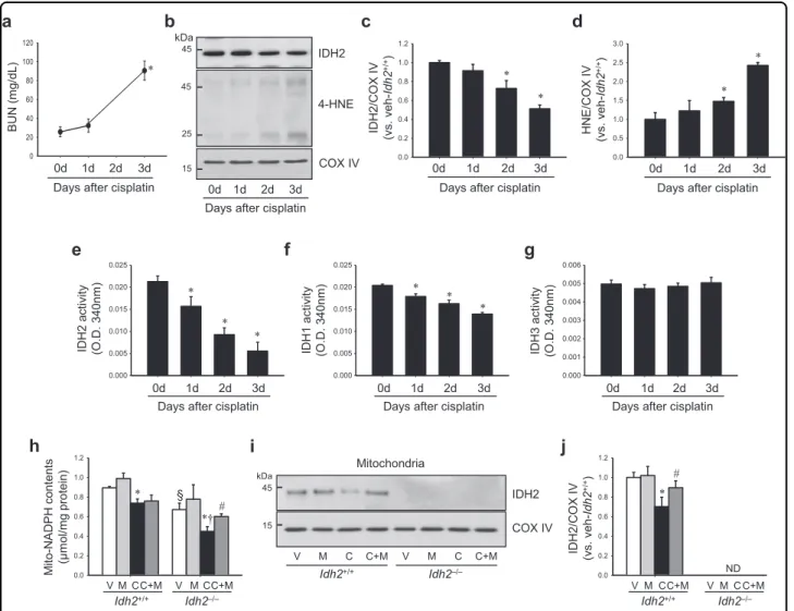
First, we determined whether cisplatin affects IDH2 function and expression. Cisplatin injection greatly decreased the activity of IDH2 in the mitochondria from 1 day after injection with mild or no increase in the expression level (Fig. 3b, c, e), even when increases of BUN and 4-HNE expression were not observed, or very mild (Fig. 3a, b, d). The decrease of IDH2 activity was exacerbated overtime in a time-dependent manner (Fig. 3e). IDH2 expression also decreased after cisplatin injection in the same pattern as activity (Fig.3b, c). Cis- platin also decreased IDH1 activity in cytosol. This decrease in IDH1 activity was less when compared with the decrease of IDH2 activity (Fig. 3f). However, IDH3 activity in the mitochondria was not significantly changed after cisplatin injection (Fig. 3g). Although these results cannot provide an answer regarding causality, the above data suggest that the decrease of IDH2 function through cisplatin could be a cause at least in part.
Next, we determined whether cisplatin decreased NADPH levels in the mitochondria in both Idh2+/+mice and Idh2−/−mice (Fig. 3h). Compared to Idh2+/+ mice, Idh2−/−mice showed a greater decrease in NADPH level after cisplatin injection (Fig. 3h). NADPH levels in the mitochondrial fraction of vehicle-treated Idh2‒/‒ mouse kidneys were lower than that in vehicle-treated Idh2+/+
mice (Fig. 3h). In addition, cisplatin reduced IDH2 expression and this reduction was prevented by Mito-T treatment (Fig. 3i, j). These results indicate that IDH2 regulates NADPH level in the mitochondria and cisplatin impairs the NADPH-producing system of mitochondria.
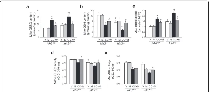
Further, we determined the ratio of oxidized glutathione (GSSG) to total glutathione (tGSH) in the mitochondrial fraction of kidneys, because NADPH plays a critical role in the reduction of GSSG to GSH. After cisplatin treat- ment, mitochondrial GSSG levels increased in both Idh2+/+and Idh2‒/‒ mouse kidneys, whereas GSH levels decreased (Fig.4a, b). These changes increased the GSSG/
tGSH ratio in both Idh2+/+and Idh2‒/‒mice, showing a higher increase in Idh2‒/‒mouse kidneys than in Idh2+/+
mouse kidneys (Fig. 4c). Mito-T significantly attenuated the cisplatin-induced changes in GSSG and GSH in both mice. However, these mitigations after Mito-T treatment were more prominent in Idh2‒/‒ mice compared with their wild-type littermates (Fig.4a–e).
Finally, we determined the activity of GSH-Px, which uses GSH as a substrate, and GR, which reduces GSSG to GSH using NADPH12. Mitochondrial GSH-Px and GR activities decreased in both Idh2+/+and Idh2‒/‒mice after cisplatin injection (Fig. 4d, e). These reduced activities were greater in Idh2‒/‒ mice than in Idh2+/+ mice (Fig. 4d, e). The reduction in GSH-Px and GR activities
was significantly attenuated by Mito-T and this attenua- tion was higher in Idh2‒/‒than in Idh2+/+mice (Fig. 4d, e). These data indicate that cisplatin impairs the mito- chondrial NADPH-GSH antioxidant system.
IDH2 deficiency accelerates mitochondrial damage following cisplatin administration
Oxidative stress in mitochondria induces mitochondrial dysfunction and a shift of the mitochondrial dynamics towardfission, leading to mitochondrial fragmentation and activation of the apoptotic signal pathway23,24. Therefore, we investigated whether increased susceptibility to cisplatin in
0.0 0.2 0.4 0.6 0.8 1.0 1.2
0.0 0.5 1.0 1.5 2.0 2.5 3.0
0.000 0.005 0.010 0.015 0.020 0.025
0.000 0.005 0.010 0.015 0.020 0.025
0.000 0.001 0.002 0.003 0.004 0.005 0.006
1d 2d 3d
0d 0d 1d 2d 3d 0d 1d 2d 3d
Days after cisplatin Days after cisplatin Days after cisplatin
*
*
*
* *
* IDH2
4-HNE
45
25
1d 2d 3d 0d
Days after cisplatin
45
c d
IDH2/COX IV (vs. veh-Idh2+/+) HNE/COX IV (vs. veh-Idh2+/+)
1d 2d 3d
0d
Days after cisplatin
1d 2d 3d
0d
Days after cisplatin
*
* *
*
COX IV
0 20 40 60 80 100 120
1d 2d 3d 0d
Days after cisplatin
a
IDH2 activity (O.D. 340nm) IDH1 activity (O.D. 340nm) IDH3 activity (O.D. 340nm)
kDa
/gm( NUBdL)
b
0.0 0.2 0.4 0.6 0.8 1.0 1.2
0.0 0.2 0.4 0.6 0.8 1.0 1.2
IDH2/COX IV (vs. veh-Idh2+/+)
V MCC+M V MCC+M
V M C C+M
ND
V M C C+M
Mito-stnetnoc HPDAN (μmol/mg protein)
IDH2
COX ൘
V MCC+M V MCC+M
Idh2+/+ Idh2–/–
Idh2+/+ Idh2–/–
Idh2+/+ Idh2–/–
*
#
§
*†
*
#
45
15 kDa
i j
h
Mitochondria
e f g
15
*
Fig. 3 Cisplatin administration decreases IDH2 expression and NADPH levels. Idh2‒/‒mice (h–j) and wild-type (Idh2+/+) littermates (a–j) were intraperitoneally injected with either cisplatin (C, 20 mg/kg B.W.) or 0.9% saline (vehicle, V) once. Some mice were treated with Mito-T (M, 0.7 mg/kg B.
W.) daily, beginning 7 days before cisplatin injection and continuing until experiments were completed. Kidneys were harvested at the indicated time after cisplatin injection. a BUN concentration was determined at the indicated time (n= 4). b IDH2 and 4-HNE expression in mitochondrial fraction was determined by western blot analysis. c, d Band densities were measured by the ImageJ program. COX IV was used as loading control (n= 4). e–g Activities of IDH2 (e), IDH1 (f), and IDH3 (g) were measured in mitochondria fraction (e, g) and cytosol fraction (f), respectively (n= 4). h NADPH concentration in the kidney mitochondrial fraction was measured as described in the“Materials and methods” section. i Expression of IDH2 in mitochondrial fraction was detected by western blot analysis. j Band density was normalized to COX IV band using the ImageJ program. Results are expressed as means ± SE (n= 3–4 per group). *p < 0.05 vs. respective V;#p < 0.05 vs. respective C;§p < 0.05 vs. V in Idh2+/+;†p < 0.05 vs. C in Idh2+/+. ND non-detectable
Idh2‒/‒ mice is associated with mitochondrial damage. Cis- platin induced damage of mitochondria in the proximal tubular cells of both Idh2+/+and Idh2‒/‒mouse kidneys (Fig.5a). This mitochondrial damage was more severe in Idh2‒/‒mice than in Idh2+/+ mice together with higher mitochondrial aspect ratio [(major axis/minor axis)] in the Idh2‒/‒ mice than Idh2+/+ mice (Fig. 5a, b). Because mitochondrial fragmentation is associated with regulatory proteins of mitochondrial fusion and fission9, we deter- mined the expression of mitochondrial fusion andfission regulatory proteins. Cisplatin decreased the expression of Opa1, a regulator of mitochondrial fusion, in both Idh2+/+
and Idh2‒/‒mouse kidneys (Fig.5c, d). Conversely, Drp1, a mitochondria fission protein, was increased by cisplatin administration (Fig. 5c, e). These cisplatin-induced alterations in Opa1 and Drp1 expression were greater in Idh2‒/‒ than in Idh2+/+ mice (Fig. 5c–e). Mito-T treat- ment preserved mitochondrial morphology (Fig. 5a) and significantly mitigated the changes in Opa1 and Drp1 expression in both Idh2+/+and Idh2‒/‒mice (Fig. 5c–e).
These Mito-T effects were more dramatic in Idh2‒/‒mice than in their wild-type littermates (Fig. 5c–e). These results indicate that Idh2 gene deletion exacerbates cisplatin-induced mitochondrial damage.
IDH2 deficiency augments apoptosis after cisplatin administration
Because mitochondrial oxidative stress activates apop- tosis signaling pathways13,25, we examined whether Idh2
gene deletion affects apoptosis after cisplatin injection.
First, we investigated the responses of the apoptosis reg- ulatory signal pathway after cisplatin injection in both Idh2+/+ and Idh2‒/‒ mouse kidneys. Cisplatin increased Bax expression, whereas it decreased Bcl-2 expression in both Idh2+/+ and Idh2‒/‒ mouse kidneys. Changes in expression were greater in Idh2‒/‒than in Idh2+/+ mice (Fig. 6a–c). Mito-T treatment significantly inhibited the cisplatin-induced increase in Bax, but not Bcl-2 expres- sion (Fig. 6a–c). Cisplatin induced the release of cyto- chrome c from mitochondria into the cytosol in both Idh2+/+and Idh2‒/‒mice, and this release was greater in Idh2‒/‒ mice than in Idh2+/+ mice (Fig. 6d–f). Cleaved caspase-3 expression levels were also elevated by cisplatin injection in both Idh2+/+and Idh2‒/‒mice, and this ele- vation was greater in Idh2‒/‒ mice than in Idh2+/+ mice (Fig. 6g, h). Mito-T reduced cytochrome c release and cleavage of caspase-3 in both Idh2‒/‒and Idh2+/+ mice, showing a greater reduction in Idh2‒/‒ mice than in Idh2+/+mice (Fig.6d–h).
Finally, we determined the apoptosis of kidney tubular cells by terminal deoxynucleotidyl transferase dUTP nick end labeling (TUNEL) analysis. Cisplatin injection increased the number of TUNEL-positive tubular epi- thelial cells (Fig. 7a). This increase in TUNEL-positive cells was greater in Idh2‒/‒ mice than in their wild-type littermates (Fig. 7a, b). Mito-T reduced the cisplatin- induced increase in TUNEL-positive cells in both Idh2‒/‒
and Idh2+/+mice (Fig.7a, b). This effect of Mito-T was
0 5 10 15 20
0 3 6 9 12 15
0.0 0.5 1.0 1.5 2.0 2.5
0.00 0.02 0.04 0.06
0.08 e
0.000 0.005 0.010 0.015 0.020
Mito-GSH-Pxactivity (O.D. 340nm) Mito-GR activity (O.D. 340nm)
#
V MCC+M V MCC+M V MCC+M V MCC+M
*
#
Mito-GSSG/tGSH (vs. veh-Idh2+/+)
d
c
V MCC+M V MCC+M
*
Idh2+/+ Idh2–/– Idh2+/+ Idh2–/–
Idh2+/+ Idh2–/–
Mito-GSSG content (pmole/μg protein) Mito-GSH content (pmole/μg protein)
V MCC+M V MCC+M V MCC+M V MCC+M
Idh2+/+ Idh2–/– Idh2+/+ Idh2–/–
*
# # #
a b
*†
*†
#
§
§ §
*†
*
*†
*
§
#
*†
#
#
Fig. 4 Cisplatin administration impairs the mitochondrial GSH-mediated antioxidant system in the kidney. Idh2‒/‒mice and wild-type (Idh2+/
+) littermates were intraperitoneally injected with either cisplatin (C, 20 mg/kg B.W.) or 0.9% saline (vehicle, V) once. Some mice were treated with Mito-T (M, 0.7 mg/kg B.W.) daily, beginning 7 days before cisplatin injection and continuing until experiments were completed. Kidneys were harvested 3 days after cisplatin injection. a–c Oxidized GSH (GSSG) levels (a), reduced GSH levels (b), and the GSSG/tGSH ratio (c) were determined in mitochondrial fractions. d, e GSH-Px (d) and GR (e) activities were measured in mitochondrial fractions. Results are expressed as means ± SE (n= 4 per group). *p < 0.05 vs. respective V;#p < 0.05 vs. respective C;§p < 0.05 vs. V in Idh2+/+;†p < 0.05 vs. C in Idh2+/+
greater in Idh2‒/‒mice than in Idh2+/+mice (Fig.7a, b).
These results indicate that IDH2 deficiency exacerbates cisplatin-induced apoptosis, which is associated with mitochondrial oxidative stress.
Discussion
In the present study, we have reported, for thefirst time, that cisplatin impairs the mitochondrial IDH2-NADPH- GSH antioxidant system, leading to mitochondrial oxi- dative stress and consequently, renal cell death and dys- function. In addition, Idh2 gene deletion reduces mitochondrial NADPH levels and exacerbates cisplatin- induced mitochondrial oxidative stress and kidney injury.
Furthermore, the mitochondria-targeting antioxidant, Mito-T, mitigates cisplatin nephrotoxicity. Thesefindings suggest that IDH2 is associated with cisplatin-induced nephrotoxicity and the IDH2-NADPH-GSH axis con- sidered as a target pathway to develop preventive treat- ments for cisplatin nephrotoxicity and AKI.
When cisplatin is excreted through urination, cisplatin accumulates in kidney cells by uptake through OCT1 and Ctr1, which are highly expressed on kidney tubular epi- thelial cells6,7. Cisplatin metabolites have a positive charge by hydration and therefore, easily penetrate the
mitochondrial membrane, because the mitochondria has a negative membrane potential (~180 mV). The metabolites then accumulate in the mitochondria and combine with negatively charged mitochondrial components, such as DNA, RNA, and proteins, leading to loss of function, including their antioxidant functions26,27. Therefore, mitochondria-rich cells, such as proximal tubular cells, are very susceptible to cisplatin25. In the present study, we found that proximal tubular cell damage is most severe among kidney tubular cells, suggesting that cisplatin nephrotoxicity is associated with mitochondrial density and damage. Because IDH2 is abundant in the proximal tubular cells16, Idh2-gene-deleted mice may be more susceptible to cisplatin toxicity in these cells.
Cisplatin induces ROS formation by inhibiting com- plexes I–IV of the respiratory chain and inhibiting GR function, which leads to decreased GSH levels, conse- quently resulting in increased mitochondrial ROS and oxidative stress, which can expand to entire cells28. Many studies have demonstrated that the reduction of mito- chondrial ROS attenuates renal injury during cisplatin administration11,29,30. On the contrary, weakening of antioxidant capacity, such as by GSH deletion, exacer- bates cisplatin nephrotoxicity30. Furthermore, cisplatin
0.0 0.5 1.0 1.5 2.0 2.5 3.0
0.0 0.5 1.0 1.5 2.0 2.5 3.0 3.5 4.0
Idh2+/+Idh2–/–
0.0 0.2 0.4 0.6 0.8 1.0 1.2
Opa1/β-actin (vs. veh-Idh2+/+) Opa1
Drp1
β-actin
Drp1/β-actin (vs. veh-Idh2+/+)
c d e
V MCC+M V MCC+M
a
V M C C+M V M C C+M V MCC+M V MCC+M
Idh2+/+ Idh2–/–
Idh2+/+ Idh2–/– Idh2+/+ Idh2–/–
*†
*
#
*†
*
#
# Vehicle Mito-T Cisplatin Cisplatin+Mito-T
100
70
40 kDa
b
Aspect ratio (Mito. Branch length)
V MCC+M V MCC+M Idh2+/+ Idh2–/–
*†
§ #
Fig. 5 IDH2 deficiency augments mitochondrial damage after cisplatin administration. Idh2‒/‒mice and wild-type (Idh2+/+) littermates were intraperitoneally injected with either cisplatin (C, 20 mg/kg B.W.) or 0.9% saline (vehicle, V) once. Some mice were treated with Mito-T (M, 0.7 mg/kg B.
W.) daily, beginning 7 days before cisplatin injection and continuing until experiments were completed. a Two days after cisplatin administration, mitochondrial structures were examined by transmission electron microscopy (TEM). Higher magnification is shown by the dash-lined rectangles.
Scale bar indicates 2μm. b The mitochondrial aspect ratio [(major axis)/(minor axis)] was computed using 30 mitochondria per cell. c Expressions of OPa1 and Drp1 were determined by western blot analysis.β-actin was used as a loading control. d, e OPa1 (d) and Drp1 (e) band densities were measured using the ImageJ program. Results are expressed as means ± SE (n= 3–4 per group). Scale bars: a 2 µm. *p < 0.05 vs. respective V;#p < 0.05 vs. respective C;§p < 0.05 vs. V in Idh2+/+;†p < 0.05 vs. C in Idh2+/+
decreased mitochondrial GSH levels and GSH-Px and GR activity, leading to increases in mitochondrial H2O2levels;
oxidation of DNA, proteins, and lipids; mitochondrial swelling; cristae loss; and a shift to fission in proximal tubular cells. These cisplatin-induced changes were enhanced by Idh2 gene deletion, whereas a mitochondria- targeting antioxidant inhibited these cisplatin-induced changes in both Idh2‒/‒ and Idh2+/+ mice, but with a greater effect in Idh2‒/‒mice. In addition, in this present study we found that the activity of IDH2 declined from 1 day after cisplatin injection, when renal functional impairment and oxidative stress are very mild or absent.
This result indicates that cisplatin impairs IDH2 function,
suggesting that the decline of IDH2 activity is not only a secondary response to oxidative stress, but also a primary response in cisplatin-nephrotoxicity. However, to define how cisplatin impairs IDH2 function, further studies are required.
NADPH is essential for the GSH-associated mitochon- drial antioxidant system by providing a reducing equiva- lent, maintaining catalase in the active form, and acting as a cofactor for Trx and GR, which convert GSSG to GSH, a substrate for the GSH-Px enzymes14. NADPH does not shuttle between the mitochondria and the cytosol14,31. We found that IDH1 activity declined after cisplatin injection, although the decline was relatively milder than the decline
0 2 4 6 8 10
0.0 0.2 0.4 0.6 0.8 1.0 1.2
0.0 1.5 3.0 4.5 6.0 7.5
Bcl-2
Cleaved casp-3 GAPDH
Bax (5B7)
a b c
g
Bcl-2/GAPDH (vs. veh-Idh2+/+)
Bax/GAPDH (vs. veh-Idh2+/+) Cleaved casp-3/GAPDH (vs. veh-Idh2+/+)
V M C C+M V M CC+M V M CC+M V M CC+M
V M C VC+M M C C+M
0 1 2 3 4 5 6 7 8
0.0 0.2 0.4 0.6 0.8 1.0 1.2
cyto c GAPDH cyto c
COX IV
Mito-cytoc/COX IV (vs. veh-Idh2+/+)
d e
Cytosol-cytoc/GAPDH (vs. veh-Idh2+/+)
MitolosotyC
f
V M C VC+M M C C+M V M CC+M V M CC+M V M CC+M V M CC+M
h
V M CC+M V M CC+M
GAPDH
V M C VC+M M C C+M
Idh2+/+ Idh2–/– Idh2+/+ Idh2–/– Idh2 +/+ Idh2–/–
Idh2+/+ Idh2–/–
Idh2+/+ Idh2–/–
Idh2+/+ Idh2–/–
Idh2+/+ Idh2–/–
Idh2+/+ Idh2–/–
* #
*†
*
#
§
* *†
# §
*†
*
#
§
#
*†
#
* #
*†
25
25
40
25
15
25
35
15
35 kDa
kDa
kDa
Fig. 6 IDH2 deficiency exacerbates apoptosis after cisplatin administration. Idh2‒/‒mice and wild-type (Idh2+/+) littermates were
intraperitoneally injected with either cisplatin (C, 20 mg/kg B.W.) or 0.9% saline (vehicle, V) once. Some mice were treated with Mito-T (M, 0.7 mg/kg B.
W.) daily, beginning 7 days before cisplatin injection and continuing until experiments were completed. Kidneys were harvested 3 days after cisplatin injection. a–c Bax and Bcl-2 expression in whole kidney lysates were determined by western blot analysis. GAPDH was used as a loading control. b, c Bax (b) and Bcl-2 (c) band densities were measured using the ImageJ program. d–f Mitochondria and cytosol were fractioned as described in the
“Materials and methods” section. Cytochrome c expression was determined in mitochondrial and cytosolic fractions by western blot analysis. e, f Band densities were measured using the ImageJ program and normalized to COX IV and GAPDH. g Cleaved caspase-3 was detected in whole kidney lysates by western blot analysis. h Band density was measured using the ImageJ program and normalized to GAPDH. Results are expressed as means
± SE (n= 3–5 per group). *p < 0.05 vs. respective V;#p < 0.05 vs. respective C;§p < 0.05 vs. V in Idh2+/+;†p < 0.05 vs. C in Idh2+/+