https://doi.org/10.12750/JET.2017.32.3.111
Effects of Roscovitine on In Vitro Development of Porcine Oocyte Using Brilliant Cresyl Blue
Pantu Kumar Roy, Xun Fang, Bahia MS Hassan, Sang Tae Shin†, Jong Ki Cho† College of Veterinary Medicine, Chungnam National University, Daejeon 34134, Republic of Korea
ABSTRACT
The objective of this experiment was to explore the effects of Roscovitine (Rosco) prior to in vitro maturation (IVM) of immature pig oocyte. Brilliant cresyl blue test has been used to select the good quality of oocyte.
Specifically, the effects of Rosco exposure on nuclear and cytoplasmic maturation, diameter, intracellular glutathione (GSH) and reactive oxygen species (ROS), and embryonic development after parthenogenetic activation (PA) and somatic cell nuclear transfer (SCNT), and gene expression levels in SCNT embryos have been measured. Cumulus oocyte complexes (COCs) have been exposed in 75 µM of Rosco for 22 and 44 h. The COCs that were matured in the IVM for 44 h without Rosco used as control group. Diameter of matured porcine oocytes 44 h culture with Rosco was significantly lower than 22 h culture with Rosco and control groups. GSH was higher in control group than 22 h and 44 h with Rosco but reduction of ROS in 22 h than 44 h with Rosco. In PA, exposure with Rosco 44 h oocytes group has been significantly lower than 22 h and control group in rates of maturation, cleavage and blastocyst formation. Similarly, in SCNT embryos rates of maturation, cleavage and formation of blastocyst have been also significantly lower in 44 h Rosco treated group than other two groups. SCNT embryos treated with Rosco 22 h showed greater expression levels of POU5F1, DPPA2 and NDP52Il mRNA compared with other two groups. Our results demonstrate that Rosco treatment with 22 h prior to IVM improves the development competence of porcine oocyte.
(Key words: Brilliant cresyl blue, Cloning, embryo, Parthenogenesis, Roscovitine)
†Correspondence: Sang Tae Shin & Jongki Cho Phone: +82-42-821-6788, Fax.: +82-42-821-8903 E-mail: [email protected] & [email protected]
INTRODUCTION
Oocytes maturation is a well-organized process that results in the ovulation of one or few oocytes from a unit of developing follicles (Hennet and Combelles, 2012; tradis et al., 2006). In vitro embryo production is an essential technique which is used for increasing the genetic gain on the basis of adult animals performance (Wang et al., 2016b). This procedure is followed to improve the fertility of immature oocytes from small follicles of animals (Chian et al., 2004;
Jurema and Nogueira, 2006; Shu et al., 2008). In vivo development competence of matured oocytes is higher than that of in vitro matured oocytes (Gilchrist, 2011). However, in vitro production (Catala et al., 2011) in the field is a major challenge to produce more competent oocytes units (Gilchrist, 2011Mehlmann et al., 2002).
Appropriate assessment of oocytes quality is a crucial for in
vitro development rate to the effective foundation of pregnancy after transfer to recipients and development of terms of assisted reproductive techniques (ARTs). To identification of probable predictors of oocytes quality is crucial for the efficient embryo in vitro production. However, proper assessment of oocytes in vitro is still an obstacle. The inefficiency of in vitro embryo production were attributed to the lack of oocytes quality at the onset of maturation (Manjunatha et al., 2007). That’s why brilliant cresyl blue (BCB) test has been used for the selection of grown oocytes to identify more competent oocytes for production of embryo.
Oocyte maturation is a discerning and main tool for further development of embryo. Over the last decade many researchers are trying to gain more knowledge about maturation and development of embryos but the efficiency of in vitro matured oocytes with proper development of embryos still limited (Abeydeera, 2002). Nuclear and cytoplasmic
maturation of the oocytes must be synchronized to reach full development of potency. Nuclear maturation changes during meiosis have been found in porcine maturing oocytes in vivo or in vitro (Motlik and Fulka, 1976) but cytoplasmic maturation need more time in vitro than in vivo maturing oocytes. Cumulus cells also play a crucial role in oocytes maturation (Tanghe et al., 2002) and contribute to resumption of meiosis (Isobe and Terada, 2001) and development of oocytes cytoplasmic maturation (Sun et al., 2001). During the maturation process of oocyte, synchronization between nuclear and cytoplasmic maturation of in vitro matured oocyte cumulus cells will result into long-term attachment (Schoevers et al., 2005). For this synchronization activation of M-phase promoting factor (MPF) is the crucial step for meiotic resumption of oocytes (Eppig et al., 1996). To synchronize the nuclear maturation, MPF activation in oocyte was prohibited by the high level of intracellular cAMP and dbcAMP (Miyano et al., 1995) through the nonspecific prohibition phosphorylation of protein with 6-dimethylaminopurine (Avery et al., 1998). A newly found purine derivative, roscovitine, that is an ATP binding pocket of CDK1 is blocked by interposes with MPF kinase activity (De Azevedo et al., 1997), chromosome condensation prevention (Krischek and Meinecke, 2001), and division of cell (Meijer and Kim, 1997).
Roscovitine reversely prohibited porcine oocyte meiotic resumption and expansion of cumulus cells (Le Beux et al., 2003). After removal of these meiotic inhibitors, combination of roscovitine and butyrolactone I showed better bovine embryo development in short time duration cultured oocytes exposure during IVM (Ponderato et al., 2001). The periodic prohibition resumption of meiosis results into synchronized development of nucleus and cytoplasm of the oocytes might be obtained.
The reprogramming-related genes POU5F1, NDP52I1, DPPA2 were compared in somatic cell cloned embryos (You et al., 2010b). In cloned embryos gene expression show aberrant gene expression when compared with cloned embryos due to incomplete reprogramming of donor nuclei in pigs (Zhang et al., 2009). POU5F1 and other reprogramming- related genes, such as NDP52I1, DPPA2 were identified as some markers in nuclear reprogramming in cloned pigs and mouse embryos (Boiani et al., 2002; Loh et al., 2006).
Expression levels of POU5F1 and other related reprogramming genes enhanced the embryonic development imposed to the
exact reprogramming of donor nuclei after reactivation of genes important for embryonic development (You et al., 2010b).
The objectives of this experiment were to investigate development competence of porcine oocytes are improved by cytoplasmic maturation time extension and synchronous exposure of roscovitine by the prohibited meiotic resumption for improved preimplantation development in porcine parthenogenetic and cloned embryos. For these objectives, GSH and ROS level of matured oocytes and their diameter after selection for IVM by BCB staining 22 h and 44 h prior to IVM was compared. Furthermore, preimplantation development of parthenogenetic and cloned embryos and genes expression related reprogramming in cloned embryos were investigated.
MATREIALS AND METHODS
1. Chemicals and reagents
All chemicals and reagents were purchased from Sigma- Aldrich (St. Louis, MO, USA) unless otherwise indicated.
2. In vitro maturation
1) Oocytes collection and BCB staining
Porcine ovaries were collected from a local slaughterhouse and transported to the laboratory in sterile normal saline (0.9%
NaCl) solution at 38°C within 4 h. Ovaries were washed with pre-warm saline two times, cumulus-oocyte complexes (COCs) were aspirated from the follicles (3-8mm in diameter) of ovaries using 10 mL syringes with 18-gauges’ needle. Then the aspirated fluid was put into a 15 mL conical tube and kept for 5 min to allow them to settle down. After settling down the fluid was washed with HEPES-buffered Tyrode’s (TLH) medium containing 0.05% (w/v) polyvinyl alcohol (TLH-PVA;
Sigma-Aldrich Co., St. Louis, MO, USA) (Bavister et al., 1983) then under stereomicroscope selected COCs which had three or more compact cumulus cells layers with uniform cytoplasm. After selection of oocytes were used for the experiments. When COCs collection was finished, they were washed in two-three times Dulbecco’s phosphate-buffered saline (DPBS; Gibco) which was modified by the addition of 0.4% BSA (A-3311; mDPBS). The COCs were then abandoned to 13 µm of BCB (B-5388; Sigma-Aldrich Co., St.
Louis, MO, USA) supplemented with mDPBS incubated for 90 min at 5% CO2 and 38.5°C in a humidified atmosphere (Fu et al., 2015). After exposure in BCB, COCs were washed two times in mDPBS and classified as BCB+ (blue cytoplasm) and BCB- (colorless/without blue cytoplasm). Only BCB+ oocytes were used for this experiment. Control oocytes were washed three times in IVM media and placed immediately in the in vitro maturation (IVM) medium.
2) IVM of oocytes
After COCs classification BCB+ oocytes have been washed in maturation medium. The maturation medium TCM-199 (Gibco) supplemented with 10% (v/v) of porcine follicular fluid (pFF), 0.6mM cysteine, 0.91mM sodium pyruvate, and 75 µg/mL kanamycin, 10 ng/mL epidermal growth factor (EGF), 1 µg/mL insulin, 10 IU/mL human chorionic gonadotrophin (hCG; Intervet International BV, Holland), 10 IU/mL pregnant mare serum gonadotrophin (PMSG) (Fu et al., 2015). Roscovitine (Sigma R7772) solution was prepared as 5mM stock in DMSO (Ju et al., 2003) and store at -20°C.
COCs have been cultured with 75 µM roscovitine for 22 h and 44 h into the IVM medium. Control oocytes were cultured in IVM medium without treatment of roscovitine. Oocytes have been then placed into the maturation medium 50 oocytes/well dishes containing 500 µL supplemented with hormone 22 h and without hormone 22 to 44 h at 5% CO2 and 38.5°C in a humidified atmosphere.
3) Oocyte diameter and measurement of GSH and ROS To determine the diameter, intracellular glutathione (GSH) and reactive oxygen species (ROS) levels of matured oocytes were denuded after 44 h of IVM. Diameter of matured oocytes were measured under microscope (200x magnification) by Leica Application Suite X (LAS X) (Wetzlar, Germany). In formerly described that GSH and ROS levels were determined (You et al., 2010a) and (Nasr-Esfahani et al., 1990). In shortly, 2′, 7′-dichlorodihydrofluorescein diacetate (H2DCFDA; Invitrogen Corp.) was used to determine the intracellular levels of ROS as a color of green fluorescence. 4-chloromethyl-6.8-difluoro- 7-hydroxycoumarin (CellTracker Blue; CMF2HC; Invitrogen Corp.) was used to determine the intracellular levels of GSH as a color of blue fluorescence. In the dark from each group of treatment 10 oocytes were incubated in TLH-PVA supplemented with 10 μM H2DCFDA and 10 μM CellTracker
Blue for 30 min. Oocytes were washed with Dulbecco's phosphate buffered saline (DPBS) (Invitrogen Corporation) containing 0.1% (wt/vol) polyvinyl alcohol (PVA) after following incubation, 10-μL droplets were used for oocytes, and under an epifluorescence microscope (Leica DM IRB, Wetzlar, Germany) with UV filters (460 nm for ROS and 370 nm for GSH) fluorescence was evaluated. After evaluating fluorescent images were saved as graphic files in TIFF format for next observation. ImageJ software (Version 1.41; National Institutes of Health, Bethesda, MD, USA) was used to detect the fluorescence intensities of oocytes and normalized to control oocytes (Kwak et al., 2012).
3. Production of embryos 1) Parthenogenetic embryos
After 44 h of culturing in the maturation medium, cumulus cells were removed by placing COCs into IVM medium supplemented with 0.1% (w/v) hyaluronidase by pipetting gently and repeatedly. After denuding, matured good quality oocytes were activated with 120 V/cm of direct current with 2 pulses for 60 μsec in 280 mM mannitol solution containing a concentration of 0.01 mM CaCl2 and 0.05 mM MgCl2 using a BTX 2001 Electro-cell Manipulator (BTX, San Diego, CA, USA) for parthenogenetic activation (PA). After electrically activated PA oocytes were cultured for post activation with 10 μg/mL cytochalasin (CB)+6’ dimethylaminopurine (DMAP) for 4 h with 5% CO2 humidified atmosphere at 39°C.
2) Cloned embryos by somatic cell nuclear transfer Primary cell culture prepares from Sinclair's kidney cut into small pieces and centrifuge several times and culture in the incubator until 3/4 passages. Fibroblasts were cultured in 60 mm tissue culture dish with DMEM (Dulbecco’s Modified Eagle Medium) (You et al., 2012) of Sigma-Aldrich containing 10%
(v/v) fetal bovine serum (FBS) till the formation of complete monolayer cells. G0/G1 stages of donor cells cycle synchronized for 48-72 h and a similar number of passages were used for each replicate (3-8 passages). Prior to nuclear transfer by using 0.4% (w/v) BSA with TLH prepared donor cells resuspension from trypsinization of cultured cells. COCs were transferred into IVM medium without hormone by using 0.1% (w/v) hyaluronidase with pipetting gently repeated for removing cumulus cells after 44 h cultured in the maturation medium.
After denuding oocytes were washed three times in hormone
free IVM medium and put into an incubator for 15 min with 5 µg/mL Hoechst 33342 medium of manipulation, then put into manipulation media which overlaid by mineral oil. Metaphase II and first polar body (PB) were removed with 17 µm beveled glass pipette (Humagen, Charlottesville, VA, USA) from metaphase II oocytes enucleating, after that enucleation confirmed by using epifluorescence microscope. After enucleating, inserted a single cell into the space between zona pellucida and cell membrane. Reconstructed SCNT oocytes were fused by electric cell fusion with 2 DC pulses of 160V/cm with 40 μsec, alternative current of 2V/cm, 2 sec using a BTX 2001 Electro-cell Manipulator (BTX, San Diego, CA, USA) 280 mM mannitol solution with low Ca concentration (0.001 mM). After 30 min later of fusion, good quality embryos were activated with 120 V/cm of direct current with two pulses for 60 μsec where media was used 280 mM mannitol solution having concentration of 0.01 mM CaCl2 and 0.05 mM MgCl2 for SCNT.
After electrically activated SCNT oocytes were cultured with 10 μg/mL CB+6’DMAP for 4 h with 5% CO2 humidified atmosphere at 39°C.
4. In vitro Culture of PA and SCNT embryos
PZM-5 (porcine zygote medium) was used for IVC medium that was made by 25 μL IVC droplet covered with mineral oil.
Embryos were washed three times in PZM-5 medium and put into an incubator for 6 days with 39°C, 5% CO2 humidified atmosphere, 5% O2, and 90% N2. Day of PA or SCNT were designated as day 0, whereas cleavages and formation of blastocysts evaluated on day 2 and 6, respectively. After Hoechst 33342 staining total cells number in blastocysts were counted under stereomicroscope.
5. Gene expression by qPCR
1) Total RNA extraction and cDNA synthesis
Embryos were harvested at different stages for analysis for total RNA transcript of different reprogramming genes (POU5F1, NDP52I1, DPPA2, and control gene ß-actin). For homogenization of the sample, used 10% volume of TRI REAGENT (MOLECULAR RESEARCH CENTER, Ohio, USA). Store the homogenate 2-3 min at room temperature (RT). Supplemented with 500 μL chloroform/1mL TRI REAGENT, vigorously shaked by hand for 15 sec spin at 12000 rpm, 4°C at 15 min. Transferred the 60% of colorless upper phase in a clean eppendorf tube. Adding 500 μL 0.5 mL
isopropyl alcohol and 20 µg glycogen. Mixed well by hand and stored at 4°C overnight. Centrifuged the stored sample 12000 rpm for 10 min, 4°C. Discarded the upper fluid slowly and washed with 75% of EtOH of RNA pellet. Following the manufacturer’s instructions RNA were converted to cDNA 20 µL with 10 µL of 2X RT Reaction Solution, 1 μL Enzyme mix solution, 5 μL Template RNA, 4 μL DNase/RNase free water (cDNA synthesis kit, iNtRON Bio Inc.). The cDNA synthesis completed reverse transcription at 50°C, 30 min, and RTase inactivation at 85°C, 5 min.
2) Gene expression analysis by real-time polymerase chain reaction (RT-PCR)
The transcript abundance of POU5F1, NDP5F1, DPPA2 mRNA in SCNT blastocysts were analyzed by RT-PCR. For PCR amplification mRNA was used at same concentration. The reprogramming genes (POU5F1, NDP5F1, DPPA2, and control gene ß-actin) were quantified using 40 cycles. The cDNA extended with 20 µL of PCR reaction supplemented with 2.5 U i-StarTaqTM DNA polymerase, 2.5 mM dNTPs (iNtRON Bio. Inc.) including 10 pmol/µL specific primer. Initially, denaturation at 95°C for 2 min, denatured at 95°C with 20 sec, annealing at 62°C with 10 sec, extended at 72°C with 40 sec and finally extended at 72°C with 5 min. PCR reaction for oligo primers was listed on Table 1. By using 1.5 %, agarose gel PCR reactions fractionated and stained by ethidium bromide and illumination under UV light. Pictures were taken, analyzed by Gel Doc EQ system (Bio-Rad Laboratories, Inc.).
6. Experimental designs
In experiment 1, the effects of treatment of porcine oocytes with Roscovitine or without (Control) 75 µM Roscovitine, followed by 22 and 44 h of IVM, on diameter of matured oocytes, intracellular oocyte GSH and ROS levels was determined. After denuding the oocytes from cumulus cells diameter, GSH and ROS were calculated in the previously described method. The experiment was repeated 8 times. In experiment 2, in vitro development of PA and SCNT embryos, which were derived from Roscovitine treated oocytes. In experiment 3, the effects of Roscovitine on different cell number of in vitro PA and SCNT blastocysts. In experiment 4, the effects of Roscovitine treatment during IVM on reprogramming related genes expression of POU5F1, NDP52I1, DPPA2, and control gene ß-actin were analyzed in Cloned embryos.
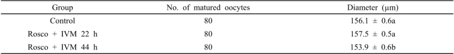
Table 2. Diameter of Roscovitine (Rosco) treated porcine matured oocytes
Group No. of matured oocytes Diameter (µm)
Control 80 156.1 ± 0.6a
Rosco + IVM 22 h 80 157.5 ± 0.5a
Rosco + IVM 44 h 80 153.9 ± 0.6b
Values in the same column with different superscript letters (a-b) are significantly different (P<0.05). The number of replicates (n=8). Results of data were expressed as mean ± S.E.M.
Table 1. Primers with a base pair (bp) used for reverse transcriptase qPCR.
Genes Sequences (5′-3′) Fragment size (bp) Annealing temperature (°C) x
cycle number ß-actin F: CCC TGG AGA AGA GCT ACG AG
R: TCC TTC CTG ATG TCC ACG TC 172 62 x 40
POU5F1 F: AGT GAG AGG CAA CCT GGA GA
R: TCG TTG CGA ATA GTC ACT GC 166 62 x 40
NDP5F1 F: TGC TGA GTT ACA TGG GTC TGG
R: ACC AAG GTC TGA TTT GCA GGT 182 62 x 40
DPPA2 F: TGA GAG AGG GGA AAA GAC CAA
R: TGG CAG AAA GGT CTC AAC AGA 151 62 x 40
7. Statistical analysis
Every experiment repeated at least eight times for embryonic development and data analyzed by Origin version 8.1 (OriginLab corporation, Northampton, USA) with a general linear model with one-way ANOVA. A probability of p-value at <0.05. All experimental data percentage presented as the mean ± SEM (standard error of the mean).
RESUTLS
1. Comparison of diameter of roscovitine (Rosco) treated matured oocytes with zona pellucida
In Table 2 shown that comparison of diameter with zona pellucida of Rosco treated oocytes after IVM of oocytes. Rosco with 22 h oocytes group (157.5 µm) was significantly greater than Rosco treated 44 h oocytes group (153.9 µm). Control oocytes group (156.1 µm) was also significantly higher than 44 h Rosco treated oocytes group but no significant differences were found among control and Rosco treated 22 h group.
2. Effect of exposure of porcine oocytes with Rosco or without (Control) 75 µM Rosco, followed by 22 and 44 h of IVM, on in vitro development rate of PA embryos
To determine the effects of Rosco to improve the development of PA embryos, COCs were cultured with Rosco 22 and 44 h. Rates of maturation, cleavage, development to blastocysts stage, and blastocysts cell numbers have been counted and the results established that 22 h of Rosco treatment enhance the development of embryos (Table 3).
When COCs have been treated with Rosco 22 and 44 h, Rosco with 44 h group has been significantly lower than 22 h and control groups in rates of maturation (76.3 vs. 87.5, 83.4%, respectively), cleavage (73.5 vs. 84.9, 82.9%, respectively), blastocysts (18.1 vs. 30.3, 28.5%, respectively). In control oocytes group was not significantly different from Rosco with 22 h group of oocytes but Rosco with 22 h group has tendency to increase than control group of oocytes.
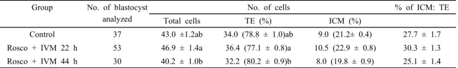
3. Comparison of different cells of blastocysts with treatment of Rosco on the in vitro development of porcine PA embryos Comparison of total number cells, percentages of trophactoderm (TE) cells, ICM cells and ICM:TE cells in PA were shown in Table 4. In total number of cells Rosco with 22 h and control oocytes group (46.2, 43.0%, respectively) was significantly higher than Rosco with 44 h oocytes groups (40.2%) whereas control and Rosco with 22 h group were not significant. Similarly, in number of TE cells Rosco with 22 h
Table 3. Effects of treatment of porcine oocytes with Roscovitine (Rosco) or without (Control) 75 µM Rosco, followed by 22 and 44 h of IVM, on in vitro development rate of PA embryos
Group No. of COCs No. of matured oocytes (%)
No. of embryos
cultured Cleaved (%) Developed to BL (%)
Control 320 267 (83.4±1.3)a 267 221 (82.9±1.8)a 76 (28.5±0.9)a
Rosco + IVM 22 h 320 280 (87.5±1.1)a 280 238 (84.9±1.5)a 85 (30.3±0.9)a
Rosco + IVM 44 h 320 244 (76.3±1.6)b 244 179 (73.5±1.4)b 44 (18.1±0.6)b
Values in the same column with different superscript letters (a-b) are significantly different (P<0.05). The number of replicates (n=8). Results of data were expressed as mean ± S.E.M.
Table 4. Effects of Roscovitine (Rosco) on different cell number of in vitro PA blastocysts Group No. of blastocyst
analyzed
No. of cells % of ICM: TE
Total cells TE (%) ICM (%)
Control 37 43.0 ±1.2ab 34.0 (78.8 ± 1.0)ab 9.0 (21.2± 0.4) 27.7 ± 1.7
Rosco + IVM 22 h 53 46.9 ± 1.4a 36.4 (77.1 ± 0.8)a 10.5 (22.9 ± 0.8) 30.3 ± 1.3 Rosco + IVM 44 h 30 40.2 ± 1.0b 32.2 (80.2 ± 0.9)b 8.0 (19.8 ± 0.9) 25.1 ± 1.4 Values in the same column with different superscript letters (a-b) are significantly different (P<0.05). The number of replicates (n=4). Results of data were expressed as mean ± S.E.M.
Table 5. Effects of treatment of porcine oocytes with Roscovitine (Rosco) or without (Control) 75 µM Roscovitine, followed by 22 and 44 h of IVM, on in vitro development rate of SCNT embryos
Group No. of
COCs
No. of matured oocytes (%)
No. of embryos
cultured Cleaved (%) Developed to BL (%)
Control 200 165 (82.5±1.7)a 136 110 (81.0±1.6)a 34 (25.0±0.8)a
Rosco + IVM 22 h 200 174 (87.0±2.1)a 144 122 (84.8±0.8)a 41 (28.4±1.0)a
Rosco + IVM 44 h 200 148 (74.0±1.3)b 118 89 (75.5±1.6)b 16 (13.6±1.3)b
Values in the same column with different superscript letters (a-b) are significantly different (P<0.05). The number of replicates (n=8). Results of data were expressed as mean ± S.E.M.
oocytes group (77.1%) was significantly higher than Rosco with 44 h oocytes group (80.2%) but was not significant difference among control and Rosco with 44 h oocytes group. There were no significant differences in percentages of ICM and ICM:TE cells among the three groups but Rosco with 22 h oocytes group has tendency to improve than other two groups.
4. Effect of treatment of porcine oocytes with Rosco or without (Control) 75 µM Rosco, followed by 22 and 44 h of IVM, on in vitro development rate of SCNT embryos
To determine the effects of Rosco to improve the development of SCNT embryos, COCs were cultured with Rosco 22 and 44 h. Rates of maturation, cleavage, development to blastocysts stage, and cell numbers in blastocysts have been counted and the results established that 22 h of Rosco treatment
enhance the development of embryos (Table 5). When COCs have been treated with Rosco 22 and 44 h, Rosco with 44 h group was significantly lower than 22 h and control groups in rates of maturation (74.0 vs. 87.0, 82.5%, respectively), cleavage (75.5 vs. 84.8, 81.0%, respectively), blastocysts (13.6 vs. 28.4, 25.0%, respectively). In control oocytes group was not significantly differ from Rosco with 22 h group of oocytes but Rosco with 22 h group has tendency to increase than control group of oocytes.
5. Comparison of different cells of blastocysts with treatment of Rosco on in vitro development of porcine clone embryos Comparison of total number cells, percentages of TE cells, ICM cells and of ICM:TE cells in SCNT embryos were shown in Table 6. In total number of cells Rosco with 22 h and
Table 6. Effects of Roscovitine (Rosco) on different cell number of in vitro SCNT blastocysts Group No. of blastocyst
analyzed
No. of cells
% of ICM: TE
Total cells TE (%) ICM (%)
Control 26 42.6±1.9ab 33.3 (78.0±1.2) 9.3 (22.0±1.2) 29.1±2.2
Rosco + IVM 22 h 33 46.5±1.6a 35.8 (76.2±1.1) 10.8 (23.8±1.1) 31.9±1.9
Rosco + IVM 44 h 12 39.8±2.8b 31.3 (78.2±1.2) 8.4 (21.8±1.2) 28.1±1.9
Values in the same column with different superscript letters (a-b) are significantly different (P<0.05). The number of replicates (n=8). Results of data were expressed as mean ± S.E.M.
Fig. 1. Epifluorescence photo micrographic images of in vitro matured porcine oocytes. (A) Oocytes were stained with (a-c) Cell Tracker Blue and (d-f) 2',7'dichlorodihydrofluorescein diacetate (H2DCFDA) to detect intracellular levels of glutathione (GSH) and reactive oxygen species (Rosner et al.), respectively whereas (a-d) control and (b-e) Roscovitine + IVM 22 h and (c-f) Roscovitine + IVM 44 h matured oocytes. (B) Effects on intracellular levels in in vitro matured porcine oocytes. Values in the same column with different superscript letters (a-b) are significantly different (P<0.05). GSH samples, N=80; ROS samples, N=80. Experiment was replicated eight times.
control oocytes group (46.5, 42.6%, respectively) was significantly higher than Rosco with 44 h oocytes groups (39.8%) whereas control and Rosco with 22 h group were not significant. There were not found significant differences in percentages of TE, ICM and ICM:TE cells among the three groups but Rosco
with 22 h oocytes group has tendency to improve than other two groups.
6. Measurement of GSH and ROS levels in porcine matured oocytes After IVM denuded the oocytes which have first polar body
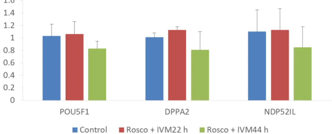
Fig. 2. Mean ± SEM expression levels of POU5F1, DPPA2, and NDP52Il mRNA levels in somatic cell nuclear transfer blastocysts that were treated with Rosco (75 µM). Values in the same column with different superscript letters (a-b) are significantly different (P<0.05). The number of replicates (n=3).
were measured the GSH and ROS levels. GSH levels were significantly higher in control oocytes group than Rosco with 22 h and 44 h oocytes group but ROS levels were significant lower in Rosco with 22 h oocytes group than control oocytes group but there was not found significant differences between Rosco with 22 and 44 h oocytes groups (Fig. 1).
7. Effect of Rosco on the expression of POU5F1, DPPA2, and NDP52IL mRNA
During IVM of Rosco with 22 h treated oocytes group was increased the expression levels of the POU5F1, DPPA2, and NDP52IL genes in SCNT blastocysts compared with Rosco with 44 h and control oocytes group (Fig. 2). The relative transcript abundance of POU5F1, DPPA2, and NDP52IL mRNA was increased by Rosco with short duration treatment compared with other two oocytes group.
DISCUSSION
In order to observe the growing stage of oocytes BCB staining was used. This test conducts the reaction of the BCB stain which is blue color and used for selection of oocytes either blue color cytoplasm or colorless cytoplasm depending on glucose-6- phosphate dehydrogenase (G6PDH) activity (Catala et al., 2011).
BCB has a beneficial effects for selection of oocytes for IVM were shown in pigs (Kempisty et al., 2011; Roca et al., 1998;
Wongsrikeao et al., 2006), cows (Alm et al., 2005; Pujol et al., 2004), goats (Rodriguez-Gonzalez et al., 2003; Rodriguez-Gonzalez
et al., 2002; Urdaneta et al., 2003a; Urdaneta et al., 2003b), bovines (Mota et al., 2010), sheep (Catala et al., 2011; Wang et al., 2016a), and dogs (Rodrigues et al., 2009). In our study, we used only BCB+ oocytes for development of embryos.
During the last decade the efficiency of in vitro embryo production and viable offspring after transfer to recipients are still low (Crocomo et al., 2016). One of the main reason of this low development of embryo potential is incomplete cytoplasmic maturation in vitro cultured of oocytes (Crocomo et al., 2016; Krisher et al., 2004). As a result, the temporary arrest of meiosis with cyclin-dependent kinase inhibitors, Rosco, was proposed as offer additional time for oocytes maturation (Gharibi et al., 2013). Therefore, we examined the diameter of matured oocytes, GSH and ROS, development of embryos and reprogramming related gene expression.
GSH and ROS affects the nuclear and cytoplasmic maturation of oocytes and embryonic development. During oocytes maturation, GSH and ROS accumulation stimulates the cytoplasmic and nuclear maturation and inhibits oxidative stress of oocytes. The GSH content level was high at 22 h of culture with Rosco than at 44 h (Yoshida et al., 1993) who have been reported during IVM with continuous increase in GSH content levels (Fig. 1). The diameter of control and Rosco with 22 h was significantly higher than Rosco with 44 h but there were no significant differences between control and Rosco with 22 h of oocytes groups (156.1 vs. 157.5 vs. 153.8 µM, respectively).
In PA, number of matured oocytes, cleavage rates and developed to blastocyst rates were significantly higher in
control and Rosco with 22 h oocytes groups than Rosco with 44 h oocytes group (83.4, 87.5 vs. 76.3 %; 82.9, 84.9 vs.
73.5%; and 28.5, 30.3 vs. 18.1%, respectively) whereas control and Rosco with 22 h oocytes group was not significant (Table 2). In the previous studies involving sheep (Crocomo et al., 2016) and cattle (Selokar et al., 2012) had also shown that treatment with 22 h roscovitine prior to IVM improved the embryonic development whereas treatment with Rosco 44 h dramatic reduced embryo production. In a case of cell number of blastocysts is very important for embryonic development and birth offspring (van Soom et al., 1997). Staining of blastocysts for PA mean total number of cells, number of TE cells were significantly greater in control and Rosco with 22 h oocytes group than Rosco with 44 h oocytes group (43.0, 46.9 vs. 40.2;
78.8, 77.1 vs. 80.2%, respectively) whereas control group was not significant with Rosco with 22 h group. There were no significant differences in percentages of ICM and ICM:TE cells among the three groups (Table 3). Similarly, Table 4 shown that in SCNT, number of matured oocytes, cleavage rates and developed to blastocyst rates were significantly higher in control and Rosco with 22 h oocytes groups than Rosco with 44 h oocytes group (82.5, 87.0 vs. 74.0 %; 81.0, 84.8 vs.
75.5%; and 25.0, 28.4 vs. 13.6%, respectively) whereas control and Rosco with 22 h oocytes group were not significant. In the mean total number of cells significant differences were found between the Rosco with 22 h and 44 h oocytes groups (46.5 vs. 39.8) but were not significant with control (42.6).
Percentages of TE, ICM and ICM:TE cells had no significant differences among the three groups (Table 5) but increase tendency in Rosco with 22 h group than other two groups. In sheep the kinetics of blastocysts development and embryo quality have not been altered with the treatment of roscovitine (Crocomo et al., 2016).
Additionally, gene expression analysis has showed that increased expression levels of different reprogramming related genes (POU5F1, DPPA2, and NDP52I1) in clone pig embryos.
It was reported that reactivation of POU5F1 and other transcription factor genes (DPPA2 and NDP52I1) are the essential steps that allows normal development of embryos in pig (You et al., 2010b), mouse (Bortvin et al., 2003;
Kawasumi et al., 2009) and human (Cauffman et al., 2005).
We identified that POU5F1 related-genes and compared their expression patterns in blastocyst stages (Lee et al., 2006) of control, Rosco with 22 h and 44 h treatment oocytes groups.
POU5F1 is a maternally expressed protein that is also present in the pre-implantation and post-implantation embryos (Scholer et al., 1990b). OCT-4 also known as POU5F1 is transcription factor which belongs to the POU (Pit, Oct, Unc) family of domain factors (Okamoto et al., 1990; Rosner et al., 1990;
Scholer et al., 1990a). It is an octamer-binding protein which binds by the POU domain of promoter and enhancer regions of various genes (Hansis et al., 2000). In mice, transcription factors are developmentally-regulated (Hansis et al., 2000).
DPPA2 is a harbor SAP DNA-binding domain (Engelen et al., 2015, Western et al., 2011). The genomic-wide binding sites have not been determined so far (Engelen et al., 2015). The inner cell mass of the early embryo is more expressed then in developing germ line, and in cells (Bortvin et al., 2003). In Fig. 2 has been shown that Rosco with 22 h oocytes group was expressed more than control and Rosco with 44 h oocytes groups in POU5F1 gene similarly in DPPA2 and NDP52Il but no significant differences were found among the three groups.
However, have to tendency more express in Rosco with 22 h oocytes group than other two groups.
In conclusion, we demonstrated that Rosco is a good model for studying the relationship between nuclear and cytoplasmic maturation in porcine oocytes and Rosco with 22 h in maturation medium can be used to improve embryo developmental competence of porcine embryos in vitro.
ACKNOWLEDGEMENTS
This research was supported by a grant from the Korea Health Technology R & D Project through the Korea Health Industry Development Institute (KHIDI), funded by the Ministry of Health & Welfare, Republic of Korea (grant number: HI13C0954).
REFERENCES
Abeydeera LR. 2002. In vitro production of embryos in swine.
Theriogenology 57:256-273.
Alm H, Torner H, Lohrke B, Viergutz T, Ghoneim IM and Kanitz W. 2005. Bovine blastocyst development rate in vitro is influenced by selection of oocytes by brillant cresyl blue staining before IVM as indicator for glucose-6-phosphate
dehydrogenase activity. Theriogenology 63:2194-2205.
Avery B, Hay-Schmidt A, Hyttel P and Greve T. 1998.
Embryo development, oocyte morphology, and kinetics of meiotic maturation in bovine oocytes exposed to 6-dimethylaminopurine prior to in vitro maturation. Mol.
Reprod. Dev. 50:334-344.
Bavister BD, Leibfried ML and Lieberman G. 1983. Development of preimplantation embryos of the golden hamster in a defined culture medium. Biol. Reprod. 28:235-247.
Boiani M, Eckardt S, Scholer HR and McLaughlin KJ. 2002.
Oct4 distribution and level in mouse clones: consequences for pluripotency. Genes Dev. 16:1209-1219.
Bortvin A, Eggan K, Skaletsky H, Akutsu H, Berry DL, Yanagimachi R, Page DC and Jaenisch R. 2003. Incomplete reactivation of Oct4-related genes in mouse embryos cloned from somatic nuclei. Development 130:1673-1680.
Catala MG, Izquierdo D, Uzbekova S, Morato R, Roura M, Romaguera R, Papillier P and Paramio MT. 2011. Brilliant Cresyl Blue stain selects largest oocytes with highest mitochondrial activity, maturation-promoting factor activity and embryo developmental competence in prepubertal sheep. Reproduction 142:517-527.
Cauffman G, Van de Velde H, Liebaers I and Van Steirteghem A. 2005. Oct-4 mRNA and protein expression during human preimplantation development. Mol. Hum. Reprod.
11:173-181.
Chian, R.C., Lim, J.H., and Tan, S.L. 2004. State of the art in in-vitro oocyte maturation. Cur. Opin. Obst. Gyne.
16:211-219.
Crocomo LF, Ariu F, Bogliolo L, Bebbere D, Ledda S and Bicudo SD. 2016. In vitro Developmental Competence of Adult Sheep Oocytes Treated with Roscovitine. Reprod.
Domest. Anim. 51:276-281.
De Azevedo WF, Leclerc S, Meijer L, Havlicek L, Strnad M and Kim SH. 1997. Inhibition of cyclin-dependent kinases by purine analogues: crystal structure of human cdk2 complexed with roscovitine. Eur. J. Biochem. 243:518-526.
Engelen E, Brandsma JH, Moen MJ, Signorile L, Dekkers DH, Demmers J, Kockx CE, Ozgur Z, van IWF, van den Berg DL and Poot RA. 2015. Proteins that bind regulatory regions identified by histone modification chromatin immunoprecipitations and mass spectrometry. Nat. Commun.
6:7155.
Eppig JJ, O'Brien M and Wigglesworth K. 1996. Mammalian
oocyte growth and development in vitro. Mol. Reprod.
Dev. 44:260-273.
Fu B, Ren L, Liu D, Ma JZ, An TZ, Yang XQ, Ma H, Zhang DJ, Guo ZH, Guo YY, Zhu M and Bai J. 2015. Subcellular Characterization of Porcine Oocytes with Different Glucose-6- phosphate Dehydrogenase Activities. Asian-Aust. J. Anim sci.
28:1703-1712.
Gharibi S, Hajian M, Ostadhosseini S, Hosseini SM, Forouzanfar M and Nasr-Esfahani MH. 2013. Effect of phosphodiesterase type 3 inhibitor on nuclear maturation and in vitro development of ovine oocytes. Theriogenology 80:302-312.
Gilchrist RB. 2011. Recent insights into oocyte-follicle cell interactions provide opportunities for the development of new approaches to in vitro maturation. Reprod. Fertil. Dev.
23:23-31.
Hansis C, Grifo JA and Krey LC. 2000. Oct-4 expression in inner cell mass and trophectoderm of human blastocysts.
Mol. Hum. Reprod. 6:999-1004.
Hennet ML and Combelles CM. 2012. The antral follicle: a microenvironment for oocyte differentiation. Int. J. Dev.
biol. 56:819-831.
Isobe N and Terada T. 2001. Effect of the Factor inhibiting germinal vesicle breakdown on the disruption of gap junctions and cumulus expansion of pig cumulus-oocyte complexes cultured in vitro. Reproduction 121:249-257.
Ju JC, Tsay C and Ruan CW. 2003. Alterations and reversibility in the chromatin, cytoskeleton and development of pig oocytes treated with roscovitine. Mol. Reprod. Dev. 64:482-491.
Jurema MW and Nogueira D. 2006. In vitro maturation of human oocytes for assisted reproduction. Fertil. Steril.
86:1277-1291.
Kawasumi M, Unno Y, Matsuoka T, Nishiwaki M, Anzai M, Amano T, Mitani T, Kato H, Saeki K, Hosoi Y, Iritani A, Kishigami S and Matsumoto K. 2009. Abnormal DNA methylation of the Oct-4 enhancer region in cloned mouse embryos. Mol. Reprod. Dev. 76:342-350.
Kempisty B, Jackowska M, Piotrowska H, Antosik P, Wozna M, Bukowska D, Brussow KP and Jaskowski JM. 2011.
Zona pellucida glycoprotein 3 (pZP3) and integrin beta2 (ITGB2) mRNA and protein expression in porcine oocytes after single and double exposure to brilliant cresyl blue test. Theriogenology 75:1525-1535.
Krischek C and Meinecke B. 2001. Roscovitine, a specific inhibitor of cyclin-dependent protein kinases, reversibly
inhibits chromatin condensation during in vitro maturation of porcine oocytes. Zygote 9:309-316.
Krisher K, Brown SD and Traczewski M. 2004. Quality control parameters for broth microdilution tests of anidulafungin. J. Clin. Microbiol. 42:490.
Kwak SS, Cheong SA, Jeon Y, Lee E, Choi KC, Jeung EB and Hyun SH. 2012. The effects of resveratrol on porcine oocyte in vitro maturation and subsequent embryonic development after parthenogenetic activation and in vitro fertilization. Theriogenology 78:86-101.
Le Beux G, Richard FJ and Sirard MA. 2003. Effect of cycloheximide, 6-DMAP, roscovitine and butyrolactone I on resumption of meiosis in porcine oocytes. Theriogenology 60:1049-1058.
Lee E, Lee SH, Kim S, Jeong YW, Kim JH, Koo OJ, Park SM, Hashem MA, Hossein MS, Son HY, Lee CK, Hwang WS, Kang SK and Lee BC. 2006. Analysis of nuclear reprogramming in cloned miniature pig embryos by expression of Oct-4 and Oct-4 related genes. Biochem.
Biophys. Res. Commun. 348:1419-1428.
Loh YH, Wu Q, Chew JL, Vega VB, Zhang W, Chen X, Bourque G, George J, Leong B, Liu J, Wong KY, Sung KW, Lee CW, Zhao XD, Chiu KP, Lipovich L, Kuznetsov VA, Robson P, Stanton LW, Wei CL, Ruan Y, Lim B and Ng HH. 2006. The Oct4 and Nanog transcription network regulates pluripotency in mouse embryonic stem cells. Nat.
Genet. 38:431-440.
Loutradis D, Kiapekou E, Zapanti E and Antsaklis A. 2006.
Oocyte maturation in assisted reproductive techniques.
Ann. Ny. Acad. Sci. 1092:235-246.
Manjunatha BM, Gupta PS, Devaraj M, Ravindra JP and Nandi S. 2007. Selection of developmentally competent buffalo oocytes by brilliant cresyl blue staining before IVM. Theriogenology 68:1299-1304.
Mehlmann LM, Jones TL and Jaffe LA. 2002. Meiotic arrest in the mouse follicle maintained by a Gs protein in the oocyte. Science 297:1343-1345.
Meijer L and Kim SH. 1997. Chemical inhibitors of cyclin-dependent kinases. Methods Enzymol. 283:113-128.
Miyano T, Ebihara M, Goto Y, Hirao Y, Nagai T and Kato S. 1995. Inhibitory action of hypoxanthine on meiotic resumption of denuded pig follicular oocytes in vitro. J.
Exp. Zool. 273:70-75.
Mota GB, Batista RI, Serapiao RV, Boite MC, Viana JH, Torres
CA and de Almeida Camargo LS. 2010. Developmental competence and expression of the MATER and ZAR1 genes in immature bovine oocytes selected by brilliant cresyl blue.
Zygote 18:209-216.
Motlik J and Fulka J. 1976. Breakdown of the germinal vesicle in pig oocytes in vivo and in vitro. J. Exp. Zool.
198:155-162.
Nasr-Esfahani MH, Aitken JR and Johnson MH. 1990. Hydrogen peroxide levels in mouse oocytes and early cleavage stage embryos developed in vitro or in vivo. Development 109:501-507.
Okamoto K, Okazawa H, Okuda A, Sakai M, Muramatsu M and Hamada H. 1990. A novel octamer binding transcription factor is differentially expressed in mouse embryonic cells.
Cell 60:461-472.
Ponderato N, Lagutina I, Crotti G, Turini P, Galli C and Lazzari G. 2001. Bovine oocytes treated prior to in vitro maturation with a combination of butyrolactone I and roscovitine at low doses maintain a normal developmental capacity. Mol. Reprod. Dev. 60:579-585.
Pujol M, Lopez-Bejar M and Paramio MT. 2004. Developmental competence of heifer oocytes selected using the brilliant cresyl blue (BCB) test. Theriogenology 61:735-744.
Roca J, Martinez E, Vazquez JM and Lucas X. 1998.
Selection of immature pig oocytes for homologous in vitro penetration assays with the brilliant cresyl blue test.
Reprod. Fertil. Dev. 10:479-485.
Rodrigues BA, Rodriguez P, Silva AE, Cavalcante LF, Feltrin C and Rodrigues JL. 2009. Preliminary study in immature canine oocytes stained with brilliant cresyl blue and obtained from bitches with low and high progesterone serum profiles.
Reprod. Domest. Anim. 44 Suppl 2:255-258.
Rodriguez-Gonzalez E, Lopez-Bejar M, Izquierdo D and Paramio MT. 2003. Developmental competence of prepubertal goat oocytes selected with brilliant cresyl blue and matured with cysteamine supplementation. Reprod. Nutr. Dev. 43:179-187.
Rodriguez-Gonzalez E. Lopez-Bejar M, Velilla E and Paramio MT. 2002. Selection of prepubertal goat oocytes using the brilliant cresyl blue test. Theriogenology 57:1397-1409.
Rosner MH, Vigano MA, Ozato K, Timmons PM, Poirier F, Rigby PW and Staudt LM. 1990. A POU-domain transcription factor in early stem cells and germ cells of the mammalian embryo. Nature 345:686-692.
Scholer HR, Dressler GR, Balling R, Rohdewohld H and Gruss
P. 1990a. Oct-4: a germline-specific transcription factor mapping to the mouse t-complex. EMBO. J. 9:2185-2195.
Scholer HR, Ruppert S, Suzuki N, Chowdhury K and Gruss P.
1990b. New type of POU domain in germ line-specific protein Oct-4. Nature 344:435-439.
Selokar NL, Saini M, Muzaffer M, Krishnakanth G, Saha AP, Chauhan MS, Manik R, Palta P, Madan P and Singla SK.
2012. Roscovitine treatment improves synchronization of donor cell cycle in G0/G1 stage and in vitro development of handmade cloned buffalo (Bubalus bubalis) embryos.
Cell Reprogram 14:146-154.
Shu YM, Zeng HT, Ren Z, Zhuang GL, Liang XY, Shen HW, Yao SZ, Ke PQ and Wang NN. 2008. Effects of cilostamide and forskolin on the meiotic resumption and embryonic development of immature human oocytes. Hum.
Reprod. 23:504-513.
Sun QY, Lai LX, Bonk A, Prather RS and Schatten H. 2001.
Cytoplasmic changes in relation to nuclear maturation and early embryo developmental potential of porcine oocytes:
Effects of gonadotropins, cumulus cells, follicular size, and protein synthesis inhibition. Mol. Reprod. Dev. 59:192-198.
Tanghe, S., Van Soom, A., Nauwynck, H., Coryn, M., de Kruif, A., 2002. Minireview: Functions of the cumulus oophorus during oocyte maturation, ovulation, and fertilization. Mol.
Reprod. Dev. 61:414-424.
Urdaneta A, Jimenez-Macedo AR, Izquierdo D and Paramio MT.
2003a. Supplementation with cysteamine during maturation and embryo culture on embryo development of prepubertal goat oocytes selected by the brilliant cresyl blue test. Zygote 11:347-354.
Urdaneta A, Jimenez AR, Izquierdo D and Paramio MT.
2003b. Effect of the addition of glutathione and glucose to the culture medium on embryo development of IVM-IVF prepubertal goat oocytes. Zygote 11:131-138.
van Soom A, Ysebaert MT and de Kruif A. 1997. Relationship between timing of development, morula morphology, and cell allocation to inner cell mass and trophectoderm in in vitro-produced bovine embryos. Mol. Reprod. Dev. 47:47-56.
Wang L, Jiang X, Wu Y, Lin J, Zhang L, Yang N and Huang J. 2016a. Effect of milrinone on the developmental competence
of growing lamb oocytes identified with brilliant cresyl blue.
Theriogenology 86:2020-2027.
Wang L.Q, Jiang XJ, Wu YS, Lin JP, Zhang L, Yang N and Huang JC. 2016b. Effect of milrinone on the developmental competence of growing lamb oocytes identified with brilliant cresyl blue. Theriogenology 86:2020-2027.
Western PS, Ralli RA, Wakeling SI, Lo C, van den Bergen JA, Miles DC and Sinclair AH. 2011. Mitotic arrest in teratoma susceptible fetal male germ cells. PloS one 6:e20736.
Wongsrikeao P, Otoi T, Yamasaki H, Agung B, Taniguchi M, Naoi H, Shimizu R and Nagai T. 2006. Effects of single and double exposure to brilliant cresyl blue on the selection of porcine oocytes for in vitro production of embryos.
Theriogenology 66:366-372.
Yoshida M, Ishigaki K, Nagai T, Chikyu M and Pursel VG.
1993. Glutathione concentration during maturation and after fertilization in pig oocytes: relevance to the ability of oocytes to form male pronucleus. Biol. Reprod. 49:89-94.
You J, Kim J, Lim J and Lee E. 2010a. Anthocyanin stimulates in vitro development of cloned pig embryos by increasing the intracellular glutathione level and inhibiting reactive oxygen species. Theriogenology 74:777-785.
You J, Lee J, Hyun SH and Lee E.,2012. L-carnitine treatment during oocyte maturation improves in vitro development of cloned pig embryos by influencing intracellular glutathione synthesis and embryonic gene expression. Theriogenology 78:235-243.
You J, Lee J, Kim J, Park J and Lee E. 2010b. Post-fusion treatment with MG132 increases transcription factor expression in somatic cell nuclear transfer embryos in pigs.
Mol. reprod. Dev. 77:149-157.
Zhang L, Wang SH, Dai YP and Li N. 2009. Aberrant gene expression in deceased transgenic cloned calves. Anim.
Reprod. Sci. 112:182-189.
Received August 18 2017, Revised August 23, 2017, Accepted August 29, 2017