Copyright ⓒ 2012, The Microbiological Society of Korea
파밤나방의 미생물적 방제를 위한 병원성 곰팡이 Nomuraea rileyi의 특성 및 병원성 검정
이원우․신태영․고승현․최재방․배성민․우수동*
충북대학교 농업생명환경대학 농생물학과
Characteristics and Virulence Assay of Entomopathogenic Fungus Nomuraea rileyi for the Microbial Control of Spodoptera exigua
(Lepidoptera: Noctuidae)
Won Woo Lee, Tae Young Shin, Seung Hyun Ko, Jae Bang Choi, Sung Min Bae, and Soo Dong Woo*
Department of Agricultural Biology, College of Agriculture, Life & Environment Sciences, Chungbuk National University, Cheongju 361-763, Republic of Korea
(Received September 21, 2012 / Accepted October 22, 2012)
To date, chemical control remains the most common way to reduce beet armyworm (Spodoptera exigua) populations. However, this insect has become more tolerant or resistant to many chemical insecticides and the insect larvae usually hide inside hollow, tube-like leaves of host plant so they were difficult to kill by spraying insecticides.
The use of viral and bacterial insecticide to solve these problems has not been successful because of their novel feeding habit. To overcome these problems, in this study, the biological characteristics and virulence of an entomopathogenic fungus isolated from the cadaver of larvae beet armyworm were investigated. Isolated entomopathogenic fungus was identified as Nomeraea rileyi (Farlow) Samson by morphological examinations and genetic identification using sequences of the ITS, β-tubulin gene and EF1-α regions. This fungus was named as N.
rileyi SDSe. Virulence tests against 3rd larvae of beet armyworm were conducted with various conidial suspensions from 1×10⁴ to 108 conidia/ml of N. rileyi SDSe in laboratory conditions. Mortality rate of beet armyworm showed from 20 to 54% and the virulence increased with increasing conidial concentrations. Although N. rileyi SDSe showed low mortality rate against beet armyworm, it is expected that N. rileyi SDSe will be used effectively in the integrated pest management programs against the beet armyworm.
Keywords: Nomuraea rileyi, Spodoptera exigua, biocontrol, entomopathogenic fungi
*For correspondence. E-mail: [email protected]; Tel.: +82-43-261- 2553; Fax: +82-43-271-4414
전 세계적으로 발생하는 파밤나방(Spodoptera exigua Hübner) 은 나비목(Lepidoptera), 밤나방과(Noctuidae)에 속하는 해충으 로 채소, 화훼, 과수 등을 포함한 거의 모든 농업 작물을 가해하 여 경제적 손실을 심각하게 일으키는 대표적인 광식성 해충이다 (Moulton et al., 1999; Smagghe et al., 2003). 또한, 내한성 기작 으로 월동이 가능하게 되고 시설 재배지의 확대와 최근의 기온 상승으로 발생세대수가 증가하면서 해마다 피해범위와 정도가 증 대되고 있는 실정이다(Kim and Kim, 1997; Zheng et al., 2011).
비교적 살충제에 감수성이 높은 어린 유충 시기에 줄기 속으로 들어가 안쪽에서 가해를 하여 약제에 노출될 기회가 적고, 3령 유충 이상이 되면 유기합성 살충제에 대한 저항성이 강해지므로
방제가 어려운 난방제 해충으로 여겨지고 있다(Park and Goh, 1992). 최근까지 이 해충의 방제를 위해서는 주로 유기인계, 카 바메이트계, 피레스로이드계 등의 화학살충제를 사용하고 있지 만, 화학살충제의 지속적인 사용은 환경오염 문제 유발, 해충의 약제 저항성 획득 그리고 사람들의 건강에 대한 우려로 친환경 적인 방제 방법에 대한 관심이 높아지고 있다(Hernández- Martínez et al., 2009). 현재까지 파밤나방 방제를 위한 친환경 적 방제 방법의 일환으로 곤충병원성 바이러스 및 세균을 이용 한 미생물 살충제가 개발되어 화학살충제의 대안으로 활용되고 있으나, 이들 역시 줄기 속으로 들어가는 가해습성으로 인하여 효과적인 방제가 이루어지지는 못하는 것으로 평가되고 있다 (Moar et al., 1995; Narayanan, 2004). 따라서 파밤나방의 가해 습성을 극복하고 효율적인 방제를 위한 곤충병원성 곰팡이의 연 구가 많은 나라에서 활발히 이루어지고 있다(Kao and Tsai,
1989; Studdert and Kaya, 1990; Choi et al., 2009). 곤충병원성 곰팡이는 대부분 불완전균류(Hyphomycetes)에 속하며 사람과 동, 식물에는 무해하면서 대상으로 하는 목적해충에는 특이적으로 병원성을 나타내는 곰팡이로서 자연상태에서 숙주의 밀도를 조절 하는 역할을 한다(Lacey et al., 2001). 곤충병원성 곰팡이의 곤충 숙주에 대한 침입 기작은 숙주의 섭식을 통해 감염되는 곤충병원 성 세균이나 바이러스와는 달리 곰팡이 포자가 숙주 표피에 부착 되는 것을 시작으로 표피를 물리적, 화학적 작용으로 뚫고 내부로 침입하여 곤충의 면역작용을 차단하고 독성물질을 분비하여 숙주 를 치사 시킨다(Bidochka and Khachatourians, 1987; Charnley, 1997). 현재까지 약 750여 종 이상의 곤충병원성 곰팡이가 알려져 있으며, 그 중 Beauveria bassiana (Bals.) Vuill, Metarhizium anisopliae (Metsch.) Sorokin, Nomuraea rileyi (Farlow) Samson, Lecanicillium lecanii (Zimmermann) Zare & W. Gams가 대표 적으로 미생물 살충제로 개발되어 여러 난방제 해충 방제에 이 용되고 있다(Hajek and St. Leger, 1994; Supakdamrongkul et al., 2010).
본 연구에서는 파밤나방 사충에서 분리한 곤충병원성 곰팡이 균주를 이용하여 파밤나방의 방제를 위한 새로운 방제제로서의 가능성을 제시하기 위해 분리 균주의 기본적인 특성과 파밤나방 에 대한 생물검정을 수행하였다.
재료 및 방법
곤충사육
본 실험에 사용된 파밤나방은 충북대학교 곤충생태 및 독성 학 실험실에서 분양 받아 온도 25℃, 광조건 16L:8D, 상대습도 70% 조건으로 사육하였고, 인공사료(120 g wheat germ, 16 g yeast powder, 16 g casein, 2.4 g methyl-4-hydroxybenzoate, 1.5 g sorbic acid, 7.2 g ascorbic acid, 6 g mineral mix, 4 g vitamin mix, 1.8 ml formaldehyde solution, 2 ml linseed oil, 20 g agar, 570 ml 증류수)를 공급하여 누대 사육하였다.
곤충병원성 곰팡이의 분리
2010년 9월 파밤나방 누대 사육 중 초록색 곰팡이로 뒤덮여 사 멸한 파밤나방 사충으로부터 포자를 수거하여, 곤충병원성 곰팡이 선택배지인 SDA-D50 [Sabouraud Dextrose Agar (SDA; DifcoTM, USA), 100 μg/ml Chloramphenicol, 50 μg/ml Streptomycin, and 50 μg/ml dodine](Shin et al., 2010)에 접종하고 25℃ 암 조 건에 7일간 배양하였다. 균주의 생장상 조사를 위한 배지로는 SDAY (Sabouraud Dextrose Agar with 1% yeast extract), PDA (Potato Dextrose Agar) 그리고 SMAY (40 g maltose, 10 g neopeptone, 10 g yeast extract, 15 g agar/L) 배지를 사용하였 다. 생물검정을 위한 곰팡이 포자의 준비는 SMAY 배지에 분리 균주를 접종하고 2주간 25℃에서 배양 후 포자를 수거하였다 (Vimala Devi and Prasad, 2001). 장기보관을 위한 냉동 보관은 얻어진 포자에 최종농도 20% glycerol 상태로 -80℃에 보관하 면서 필요 시 다시 배양하여 사용하였다.
형태학적 관찰
분리 균주의 형태학적 관찰을 위하여 slide glass culture 방법 을 이용하였다(Sigler and Gibas, 2005). SMAY 배지에서 25℃, 3–4일간 배양된 균체를 배지와 함께 약 6 mm로 자른 후 그 위에 지름 22 mm의 cover glass를 덮고 다시 25℃, 3–4일간 배양하였 다. 배양 후 포자형성이 유도된 것을 확인하고 400× 위상차 현미 경을 이용하여 균사 및 포자를 관찰하였다(Canon PowerShot A640, Japan).
분리 균주의 전자현미경 관찰은 SMAY 배지에서 25℃, 2주 간 배양된 균체를 이용하여 관찰하였다. 배양된 균체는 변형을 방지하기 위해 4%의 glutaraldehyde 용액으로 24시간 고정한 후 0.05 M phosphate buffer (pH 7.3)로 세척하고 ethyl alcohol을 이용하여 탈수시켰다. 시료는 건조시켜 시료대에 고정한 후 Ion-Sputter로 도금하여 Carl Zeiss LEO-1530으로 관찰하였다.
Genomic DNA 추출 및 PCR
곰팡이 genomic DNA 추출은 균사로부터 화학적 lysis 방법을 일부 수정하여 추출하였다(St. Leger and Wang, 2009). SMAY 에서 2주간 자란 곰팡이 균체 일부를 fungal DNA extraction buffer [0.2 M Tris-Cl; pH 7.5, 0.5 M NaCl, 10 mM EDTA; pH 8.0 and 1% (w/v) SDS]를 처리한 후 phenol-chloroform-isoamyl alcohol (25:24:1)로 원심 분리하여 DNA를 정제하였다. 정제된 DNA 용액은 냉에탄올을 이용하여 침전시키고 원심분리 후 멸 균 증류수에 녹여 실험에 이용하였다.
분리 균주의 분자생물학적 동정을 위한 PCR primers는 ITS1 (5′-TCCGTAGGTGAACCTGCGG-3′)와 ITS4 (5′
-TCCTCCGCTTATTGATATGC-3′)(White et al., 1990), tubF (5′-TGGGCYAARGGYCACTACACYGA-3′)와 tub-R (5′
-TCAGTGAACTCCATCTCRTCCAT-3′)(Tartar et al., 2002), 1577F (5′-CARGAYGTBTACAAGATYGGTGG-3′)와 2218R (5′-ATGACACCRACRGCRACRGTYTG-3′)(Rehner and Buckley, 2005)를 사용하여 각각 internal transcribed spacer (ITS1-5.8S-ITS2), β-tubulin 그리고 EF1-α 부분을 증폭시켰다.
PCR 반응은 AccuPOWERTM PCR PreMix (Bioneer Co., Korea)를 이용하여 94℃ 30초, 53℃ 30초, 72℃ 1분 35 cycles 조건으로 수행하였다. PCR 반응 후 증폭 산물은 1.0% agarose gel을 이용하여 전기영동 분석하고 Power Gel Extraction Kit (Dyne Bio Inc., Korea)를 이용하여 순수 정제하였다.
염기서열 분석 및 계통수
각 PCR 산물은 pCR2.1-TOPO (Invitrogen Co., USA)를 이 용하여 cloning 한 후 염기서열을 의뢰 결정(Solgent Co., Korea) 하고, Clustal X (Larkin et al., 2007)을 이용하여 정렬한 다음 Blast search tool을 이용하여 기 보고된 다른 곰팡이들 서열과 비교·분석하였다.
계통수는 각각의 ITS, β-tubulin 그리고 EF1-α 영역의 Blast search 결과에 따라 상동성이 높은 5개의 균주와 GenBank에 보 고된 대표적인 곤충병원성 곰팡이들을 이용하여 작성하였다. 계 통수 분석에 이용된 각각의 곰팡이 균주와 GenBank accession
(A) (B) (C)
(D) (E)
Fig. 1. The growth on SMAY media at 25°C (A–C) and the conidia and mycelia observed under phase contrast microscope (D) and scanning electron microscope (E) of isolated fungus. Fungus was inoculated on the center of SMAY media and cultured at 25℃ for 5 days (A), 10 days (B) and 15 days (C). The growth of white with flocculent mycelium was observed at 5 days after inoculation and the green sporulation was partially observed in center of mycelium at 10 days after inoculation. The green sporulation was almost observed whole of mycelium at 15 days after inoculation. Short and divergent chains of conidia observed under phase contrast microscope (400×) (D) and scanning electron microscope (1000×) (E). Scales bars (A, B, C): 5 cm and (D): 5 μm.
번호는 다음과 같다. ITS 서열 분석에는 N. rileyi 균주들로 MAFF 830007 (AB268359), Korea (FJ824809), RCEF0292 (AF368501), NBRC8560 (AB100361) 그리고 CG129 (EU553337)가 이용되 었으며, Pochonia sp. CBS 203.86 (DQ516079), M. flavoviride var. novazealandicum FI-1124 (AF139852), P. lilacinus CBS 284.36 (FR734101), B. bassiana ARSEF 1040 (HQ880758), Isaria farinosa CBS 240.32 (AY624178) 그리고 L. attenuatum CBS 402.78 (AJ292434)가 이용되었다. β-Tubulin 분석에는 N.
rileyi UFIF178-6 (DQ079608), M. anisopliae 1 (AY995134)와 P. reniformis에 속하는 ARSEF 577 (DQ079606), ARSEF429 (DQ079605) 및 Ind.Tett (DQ096478) 그리고 L. tenuipes CBS 309.85 (DQ522502), B. bassiana ARSEF 501 (DQ092752)와 I.
fumosorosea ARSEF1626 (DQ090739)가 이용되었다. EF1-α 영역 분석에는 N. rileyi CBS 806.71 (EF468787), M. lepidiotae ARSEF 7488 (EU248865) 그리고 M. guizhouense 균주들로 CBS 258.90 (EU248862), ARSEF 7502 (EU248861) 및 ARSEF 4321
(EU248860), P. lilacinus IBT27820 (GU979991), B. bassiana ARSEF 1040 (AY531881), I. farinosa OSC 111006 (EF469065) 그리고 L. attenuatum CBS 402.78 (EF468782)가 각각 이용되 었다. 각각의 염기서열들은 Clustal X (Larkin et al., 2007) 이용 하여 배열하였고, MEGA 4.1 프로그램 내의 neighbor-joining 방법을 사용하여 계통분석 및 계통도를 구축하였다(Saitou and Nei, 1987). 통계적 유의성을 가지기 위해서 bootstrap은 2000번 수행하였다.
생물검정
SMAY 배지에서 2주 동안 배양된 곰팡이 표면을 긁어 수확 된 포자를 0.02% Tween 80을 이용하여 포자현탁액을 만들고 hemocytometer를 이용하여 계수하였다. 생물검정 전 포자의 viability는 기 보고된 방법(Fernandes et al., 2008)에 준하여 SDAY 배지에 0.05% benomyl [95% active ingredient (Sigma, USA)]이 첨가된 SDAY+B 배지에서 발아 여부를 확인하여
(A)
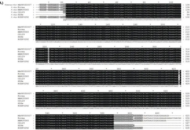
Fig. 2. Aliments of nucleotide sequences of ITS (A), β-tubulin (B), and EF1-α (C) region specific PCR products from isolated fungus (SDSe) with high similarity sequences of those from NCBI, respectively. Accession numbers of the sequences for ITS analysis were N. rileyi MAFF 83000 (AB268359), Korea (FJ824809), RCEF 0292 (AF368501), NBRC 8560 (AB10036), and CG 129 (EU553337). Accession numbers of the sequences for β-tubulin analysis were N. rileyi UFIF178-6 (DQ079608), Metarhizium anisopliae 1 (AY995134), Paecilomyces reniformis ARSEF 577 (DQ079606), P. reniformis ARSEF 429 (DQ079605) and P. reniformis Ind.Tett (DQ096478). Accession numbers of the sequences for EF1-α analysis were N. rileyi CBS 806.71 (EF468787), M. lepidiotae ARSEF 7488 (EU248865), M. guizhouense CBS 258.90 (EU248862), M. guizhouense ARSEF 7502 (EU248861) and M. guizhouense ARSEF 4321 (EU248860). Black and gray shadows indicate the identical and partial identical sequences, respectively.
90% 이상 viability를 보이는 것만을 생물검정에 이용하였다. 생 물검정은 1×10⁴–108 conidia/ml로 맞춘 포자현탁액 20 ml을 50 ml conical tube에 넣고 30마리의 파밤나방 3령 유충을 동시에 포자현탁액에 15–20초간 dipping하는 방법으로 접종하였다. 접 종 후 파밤나방 유충들은 개별적으로 insect breeding dish (SPL Co., Korea)에 옮겨 온도 25℃, 광조건 16L:8D, 습도 70% 조건 으로 유지시키고 10일 동안 매일 관찰하였다. 사충들은 표피에 서 곰팡이의 발생이 육안으로 관찰 될 경우에만 접종한 곰팡이 에 의한 치사로 인정하였으며, 대조구로는 0.02% Tween 80 용 액만을 처리하였다. 생물검정은 3반복 수행하였다.
결 과
곤충병원성 곰팡이의 분리 및 형태학적 조사
사육 중 녹색 곰팡이로 뒤덮여 이병된 파밤나방 사충으로부 터 선택배지인 SDA-D50을 이용하여 곰팡이를 분리한 결과, 오 직 한 종류의 colony만 형성됨을 확인할 수 있었다. 분리 균주를 SDAY, PDA 그리고 SMAY 배지에 옮겨 생장을 관찰한 결과, SMAY 배지에서만 흰색으로 균사생장을 하다가 시간이 지나면
서 사충에서 분리한 곰팡이의 색깔과 같은 녹색의 포자를 형성 하는 것을 관찰하였다(Fig. 1). SMAY 배지는 maltose라는 당을 함유하여 Nomuraea 속의 곰팡이 포자형성을 위한 배지로 잘 알 려져 있으며(Bell, 1975; Holdom and Van de Klashorst, 1986;
Vimala Devi et al., 2000), 또한 포자의 색이 녹색인 점과 나비 목 유충을 숙주로 한다는 것을 감안할 때 분리 균주가 1차적으로 Nomuraea 속의 곰팡이 임을 추정할 수 있었다. 현미경적 관찰 결과에서는 포자의 형태가 타원형으로 크기는 4.3±0.3 × 2.6±1.5 μm (n=20)이였으며, 3–4개의 짧고 뭉툭한 phialides를 가지고 분생포자 역시 짧고 뭉툭하여 격막 주위에서 가지를 가진 채 연쇄상으로 뻗어나가는 특징을 확인할 수 있었다(Fig. 1). 비록 3–4개의 짧고 뭉툭한 형태의 phialides를 가진다는 것이 Nomuraea 속의 특징으로 볼 수 있었으나, 연쇄상으로 포자가 뻗어 나가는 특징은 Paecilomyces 속과도 매우 유사한 특징으로 형태학적 수 준에서는 본 분리 균주의 최종 동정이 어려우며 추가적으로 분 자생물학적 동정이 필요함을 보여 주었다(Samson, 1974).
분리 균주의 분자 생물학적 동정
분자생물학적 동정을 위한 PCR primers로서 ITS1과 ITS4,
(B)
Fig. 2. continued.
tub-F와 tub-R, 1577F와 2218R을 사용하여 각각 internal transcribed spacer (ITS1-5.8S-ITS2), β-tubulin 그리고 EF1-α 부분을 증폭 한 PCR 산물을 전기영동 한 결과, ITS 영역은 약 500 bp, β -tubulin 영역은 약 900 bp, EF1-α 영역은 약 500 bp 근처에서 예 상된 크기의 밴드가 형성되었다(자료 미제시). 각각에 대해 염기 서열을 결정한 결과, ITS 영역은 580 bp, β-tubulin 영역은 938 bp, EF1-α 영역은 517 bp로 나타났으며, 분리균주의 ITS 영역은 N. rileyi MAFF 83000 (AB268359), Korea (FJ824809)와 100% 상동성을 N. rileyi RCEF 0292 (AF368501), NBRC 8560 (AB10036), CG 129 (EU553337)와 99%로 높은 상동성을 나타 내었다(Fig. 2A). 또한 β-tubulin 영역은 N. rileyi UFIF178-6 (DQ079608)와 97%로 가장 높은 상동성을 보였으며 그 뒤로 M.
anisopliae 1 (AY995134), P. reniformis ARSEF 577 (DQ079606),
ARSEF 429 (DQ079605) 및 Ind.Tett (DQ096478)가 88% 상동 성을 보였다(Fig. 2B). 마지막으로 EF1-α 영역은 N. rileyi CBS 806.71 (EF468787)와 99% 높은 상동성을 보였으며 다수의 Metarhizium 속의 곰팡이 M. lepidiotae ARSEF 7488 (EU248865), M. guizhouense CBS 258.90 (EU248862), ARSEF 7502 (EU248861), ARSEF 4321 (EU248860)와 각각 95%의 상동성 을 나타내었다(Fig. 2C). 분석된 염기서열을 이용하여 다른 곰팡 이들과의 관계를 알아보기 위하여 계통수를 그려본 결과, 분리 곰팡이는 3가지 영역의 염기서열을 토대로 한 계통수에서도 모 두 N. rileyi와 단일계통으로 묶이는 것으로 확인되었다(Fig. 3).
이로써, 형태학적 및 분자생물학적 조사를 통해 분리 균주는 N.
rileyi로 최종 동정하였으며, N. rileyi SDSe로 명명하였다.
(A)
(B)
(C)
Fig. 3. Phylogenetic tree based on the nucleotide sequences of ITS (A), β-tubulin (B), and EF1-α (C) region of isolated fungus (SDSe, closed squares) with related fungal species from NCBI. The tree was constructed by the neighbor-joining method based on genetic distances calculated by the maximum composite likelihood method.
The reliability of the tree was assessed by bootstrap analysis with 2,000 replicates. Bootstrap values ≥70% are labeled. Accession numbers of the sequences of each fungus used to generate the phylogenetic trees are shown at material and methods.
(A)
(B) (C)
Fig. 4. Accumulated mortality of N. rileyi SDSe against S. exigua larvae (A). The S. exigua 3rd instar larvae were inoculated by a 15-20 s immersion in a suspension of 1×10⁴–1×10⁸ conidia/ml. The S.
exigua larvae completely covering with white mycelium by N. rileyi SDSe at 3 days after inoculation (B) and, the green sporulation was observed at 5 days (C) after death. Scale bars: 2 cm.
파밤나방에 대한 N. rileyi SDSe의 병원성 검정
포자 현탁액(1×10⁴–108 conidia/ml)을 이용하여 파밤나방 유 충에 대하여 생물검정을 실시한 결과, 20–54%의 다양한 살충률 을 나타냈으며, 포자현탁액의 농도가 높을수록 살충률도 증가하 였다(Fig. 4A). 사멸한 파밤나방 유충은 사멸 2–3일 후에 미이라 처럼 딱딱한 형태로 굳어지면서, 체내에 침입하여 발아된 균사가 파밤나방 표피를 뚫고 나와 하얀색의 균사로 완전히 덮이는 전형 적인 곤충병원성 곰팡이의 병징을 보였다(Fig. 4B). Nomuraea rileyi를 대표하는 녹색 포자는 파밤나방 유충 사멸 5일 후부터 표피에서 형성되는 것을 관찰할 수 있었다(Fig. 4C).
고 찰
친환경적 방제 수단으로써의 곤충병원성 곰팡이는 난방제 해충 중에서도 식물을 직접 섭식하지 않는 진딧물이나 매미목 해충을
중심으로 효과적인 미생물 살충제로써 여러 나라에서 연구, 개 발되어 이용되고 있다(Srisukchayakul et al., 2005; de Faria and Wraight, 2007). 흡즙성 해충을 중심으로 주로 곤충 병원성 곰팡 이가 살충제로 고려 되는 이유는, 곰팡이 이외의 곤충 병원성 세 균 또는 바이러스의 경우에는 곤충의 섭식에 의해서만 효과를 보일 수 있으나, 흡즙성 해충은 섭식의 기회가 거의 없어 이들의 적용이 어렵기 때문에 접촉만으로도 효과를 보일 수 있는 곤충 병원성 곰팡이가 개발의 대상이 되고 있다(Shah and Pell, 2003;
de Faria and Wraight, 2007). 본 연구에서 방제 대상이 된 대표 적 난방제 해충인 파밤나방의 경우에는 비록 흡즙 하지 않고 식 물을 직접 가해하는 습성을 지니고 있으나, 식물의 줄기 속으로 파고드는 습성으로 인해 외부 섭식의 기회가 적기 때문에 줄기 속으로 들어가기 이전의 접촉만으로 병원성을 보일 수 있는 곰 팡이가 그 방제원으로써 연구 개발이 이루어지고 있다(Kao and Tsai, 1989; Studdert and Kaya, 1990; Choi et al., 2009). 본 연구 에서는 파밤나방의 이병충으로 부터 분리하여 형태학적 및 분자 생물학적 동정을 거쳐, 최종적으로 분리 균주가 N. rileyi임을 확인 할 수 있었다. 분자생물학적 동정 방법이 개발되기 이전까지는 형태학적 동정만으로 곤충 병원성 곰팡이의 동정이 이루어졌으나, 포자의 색이나 형태 만으로는 많은 동정의 오류가 지적되어 최근 에는 본 연구에서와 같이 형태학적 동정과 동시에 비교적 잘 보 존되는 지역으로 알려진 ITS, β-tubulin 그리고 EF1-α 지역의 염기 서열 비교를 통한 분자생물학적 동정을 병행하고 있다(Han et al., 2002; Rehner and Buckley, 2005). 특정 보존 지역의 서열비 교를 통한 동정에는 염기서열의 비교결과 99% 이상 상동성을 보일 경우에만 의미 있는 결과로 인정되고 있으며(Qun et al., 2011), 본 연구 결과의 분리 균주 역시 100, 97 및 99%의 상동성 확인을 통해 N. rileyi로 최종 동정할 수 있었다(Fig. 2).
Nomuraea rileyi는 30종이 넘는 나비목 유충에 대한 병원성을 보이며 밤나방과(Noctuidae)에 속하는 유충들에 감염을 잘 일으 켜 난방제 나비목 해충 방제를 위해서 많은 연구가 이루어 지고 있다(Ignoffo, 1981; Ignoffo and Boucias, 1992; Sanchez-Peña, 2000; Martins et al., 2005). 파밤나방에 대한 기보고는 확인할 수 없었으나 같은 밤나방과인 담배거세미나방(S. litura)에 대해 서는 많은 보고가 이루어져 있는데, Lin 등(2007)의 보고에 따르 면 3령 충에 대한 N. rileyi의 살충성은 LT50 값이 19.63일 그리 고 누적 살충률이 52.2%를 보임으로써 본 연구의 분리 균주인 N. rileyi SDSe 결과와 비슷한 경향을 보였으나(Fig. 4A), 2령충 을 대상으로 한 경우에는 각각 4.1일 및 95.2%의 높은 살충성을 보임으로써 유충의 영기에 따라 살충성이 매우 큰 차이를 보였 다. 따라서 N. rileyi SDSe의 경우에도 파밤나방 3령충이 아닌 2 령충을 대상으로 할 경우 그 살충성이 매우 높아질 가능성은 충 분히 있으며, 이후 그에 대한 추가 연구가 필요할 것으로 여겨진 다. 다만, 담배거세미나방이나 파밤나방을 비롯한 거의 모든 해 충들의 방제에 있어서 해충 발생 초기에 살충제를 처리하면 가 장 효과적인 것은 잘 알려진 사실이나, 실질적인 적용 면에서는 해충의 발생 시기를 정확히 예측할 수 없기 때문에 해충의 발생이 비교적 용이하게 가시화되는 3령충 이상의 시기에 실질적인 방 제가 많이 이루어진다는 점에서, 현재의 살충성을 보완 또는 증대 시킬 수 있는 방안의 모색도 필요한 것으로 여겨진다. 곤충 병원성 곰팡이는 곤충 체내 침입 후 증식과정을 통해 살충성을 보임으 로써 그 효과가 늦게 발현되는 단점을 가지고 있으므로, 그 자체 를 살충제로 개발하는 연구도 이루어지고 있으나 곰팡이 살충제 와의 병행 처리를 통해 관행적으로 사용되던 화학살충제의 사용을 최소화 할 수 있는 적용방법에 대한 연구가 많이 이루어지고 있 다(Jaramillo et al., 2005; Santos et al., 2007; Brito et al., 2008;
Paula et al., 2011; Sun et al., 2012). 또한 다른 친환경 방제제인 여러 가지 친환경 추출물 또는 천적 곤충과 곰팡이 살충제를 함 께 처리함으로써 곰팡이 살충제의 낮은 살충력을 보완 하면서 동시에 두 가지 모두 살충효과를 높일 수 있는 것에 대해 많은 연 구가 활발히 이루어지고 있다(Wekesa et al., 2007; Rashki et al., 2009; Park and Kim, 2012). 따라서 N. rileyi SDSe의 경우 에도 그 자체만으로는 충분한 살충효과를 기대하기 어렵다고 볼 때, 화학살충제나 다른 친환경 방제제와의 혼용을 통해 그 효과 를 극대화 시킬 수 있는 연구가 더욱 필요한 것으로 여겨진다.
미생물 살충제 개발의 소재로 활용되는 곤충 병원성 미생물 은 일반적으로 그 숙주 범위가 매우 좁아 목적으로 하는 해충 만을 방제할 수 있으며 안전성이 용이하게 확보되는 장점이 있으나, 그와 동시에 좁은 숙주범위는 동시 다발 하는 여러 가지 해충의 방제에는 비효율적이라는 단점을 동시에 지니고 있다(Weinzierl et al., 1998; de Faria and Wraight, 2007). 특히, 곤충 바이러스 의 경우에는 한 종류의 바이러스가 1종 내지 수 종의 곤충에만 유효하며, 미생물 살충제로 가장 많이 개발된 세균의 경우에도 그 숙주범위가 세균 별로 수 종으로 제한되어 매우 좁은 특성을 보이는 반면, 곤충 병원성 곰팡이의 경우에는 이들에 비해 상대 적으로 수 종에서 수십 종 까지 비교적 넓은 숙주범위를 가지고 있는 것으로 알려져 있다(Hajek and St. Leger, 1994; Charnley,
1997; Lacey et al., 2001; Shah and Pell, 2003). Nomuraea rileyi의 경우에도 비교적 넓은 숙주 범위를 가지고 있으며 숙주 에 따라 병원력과 특이성이 다양하다는 보고(Ignoffo and Boucias, 1992)가 있으므로, N. rileyi SDSe에 대해서도 난방제 해충을 중심으로 다양한 해충에 대해 병원성 검정이 필요하다고 여겨진다.
본 연구 결과 분리된 곤충 병원성 곰팡이 N. rileyi SDSe가 파 밤나방에 대해 높은 살충력을 보이지는 못하였으나, 파밤나방의 방제에 있어서 문제점으로 지적되는 여러 가지 한계점을 극복할 수 있는 대안으로 제시될 수 있으며, 파밤나방의 종합적 방제방 법(integrated pest management: IPM)의 일환으로 유용하게 이 용될 수 있을 것으로 기대한다.
적 요
파밤나방(Spodoptera exigua)은 유기합성 농약을 이용한 화 학 살충제 외에 뚜렷한 방제방법이 알려져 있지 않으나, 그에 대 한 저항성과 더불어 식물을 가해하는 방법에 있어 그들만의 독 특한 숙주 가해습성으로 인해 난방제 해충으로 알려져 있다. 화 학살충제에 대한 저항성의 해결을 위하여 곤충병원성 바이러스 및 세균을 이용한 살충제가 개발되어 있으나, 이들 역시 파밤나 방 유충이 줄기 속으로 들어가는 가해습성으로 인하여 효과적이 지 못한 실정이다. 그러므로 본 연구에서는 그들의 가해습성과 저항성을 극복할 수 있는 새로운 방제원으로써, 체벽과의 일시 적인 접촉을 통해 살충성을 발휘할 수 있는 파밤나방에 대한 병 원성 곰팡이를 분리하고 그 특성 및 살충성을 조사하였다. 파밤 나방 병원성 곰팡이는 파밤나방 누대사육 중 곰팡이병 증상을 보이며 이병된 사충으로부터 분리하였으며, 분리 곰팡이는 배지 에서의 증식상 및 현미경적 관찰을 통한 형태학적 동정과 더불어 ITS, β-tublin 및 EF1-α 부분의 염기서열 분석을 통해 Nomuraea rileyi로 최종 동정하고 N. rileyi SDSe로 명명하였다. 분리 균주 의 파밤나방에 대한 살충력 검정은 1×10⁴–108 conidia/ml까지의 다양한 포자현탁액에 파밤나방 3령 유충을 침지하는 조건으로 수행한 결과, 20–54%의 살충률을 나타냈으며 포자현탁액의 농 도가 증가할수록 살충력도 증가하는 양상을 나타냈다. 본 연구 결과 분리된 N. rileyi 균주가 비록 높은 살충력을 보이지 않았으 나, 파밤나방이 난방제 해충임을 감안할 때 다른 방제방법과 함 께 종합적 해충 방제 방법에 있어 효과적인 방제수단의 일환이 될 수 있을 것으로 기대된다.
감사의 말
이 논문은 2012년도 정부(교육과학기술부)의 재원으로 “한국연 구재단의 지원을 받아 수행된 기초연구사업임(No. 2012-0004572)”.
참고문헌
Bell, J.V. 1975. Production and pathogenicity of fungus, Spicaria rileyi from solid and liquid media. J. Invertebr. Pathol. 26, 129–130.
Bidochka, M.J. and Khachatourians, G.G. 1987. Purification and properties of an extracellular protease produced by the entomopathogenic fungus Beauveria bassiana. Appl. Environ.
Microbiol. 53, 1679–1684.
Brito, E.S., Paula, A.R., Vieira, L.P., Dolinski, C., and Samuels, R.I.
2008. Combining vegetable oil and sub-lethal concentrations of Imidacloprid with Beauveria bassiana and Metarhizium anisopliae against adult guava weevil Conotrachelus psidii (Coleoptera:
Curculionidae). Biocontrol. Sci. Technol. 18, 665–673.
Charnley, A.K. 1997. Entomopathogenic fungi and their role in pest control, pp. 185–201. In Wicklow, D.T. and Soderstrom, B.E. (eds.), The Mycota IV environmental and microbial relationships, Berlin, Springer.
Choi, S.U., Cheong, S.S., and Hwang, C.Y. 2009. Mycelial growth and pathogenicity of entomopathogenic fungus Nomuraea rileyi. J.
Agric. Life Sci. 40, 32–38.
de Faria, M.R. and Wraight, S.P. 2007. Mycoinsecticides and Mycoacaricides: A comprehensive list with worldwide coverage and international classification of formulation types. Biol. Control. 43, 237–256.
Fernandes, E.K., Rangel, D.E., Moraes, A.M., Bittencourt, V.R., and Roberts, D.W. 2008. Cold activity of Beauveria and Metarhizium, and thermotolerance of Beauveria. J. Invertebr. Pathol. 98, 69–78.
Hajek, A.E. and St. Leger, R.J. 1994. Interactions between fungal pathogens and insect hosts. Annu. Rev. Entomol. 39, 293–322.
Han, Q., Inglis, G.D., and Hausner, G. 2002. Phylogenetic relationships among strains of the entomopathogenic fungus, Nomuraea rileyi, as revealed by partial beta-tubulin sequences and inter-simple sequence repeat (ISSR) analysis. Lett. Appl. Microbiol. 34, 376–383.
Hernández-Martínez, P., Ferré, J., and Escriche, B. 2009. Broad-spectrum cross-resistance in Spodoptera exigua from selection with a marginally toxic Cry protein. Pest. Manag. Sci. 65, 645–650.
Holdom, D.G. and Van de Klashorst, G. 1986. Inexpensive culture media and methods for Nomuraea rileyi. J. Invertebr. Pathol. 48, 246–248.
Ignoffo, C.M. 1981. The fungus Nomuraea rileyi a microbial insecticide, pp. 513–538. In Burges, H.D. (ed.), Microbial Control of Pests and Plant Diseases 1970–1980. Academic Press, New York & London.
Ignoffo, C.M. and Boucias, D.B. 1992. Relative activity of geographical isolates of Nomuraea bioassayed against the cabbage looper and velvetbean caterpillar. J. Invertebr. Pathol. 59, 215–217.
Jaramillo, J., Borgemeister, C., Ebssa, L., Gaigl, A., Tobon, R., and Zimmermann, G. 2005. Effect of combined applications of Metarhizium anisopliae (Metsch.) Sorokin (Deuteromycotina:
Hyphomycetes) strain CIAT 224 and different dosages of imidacloprid on the subterranean burrower bug Cyrtomenus bergi Froeschner (Hemiptera: Cydnidae). Biol. Control. 34, 12–20.
Kao, C.W. and Tsai, Y.S. 1989. Control of beet armyworm with entopathogenic fungi. Chinese J. Entomol. 4, 214–225.
Kim, Y. and Kim, N. 1997. Cold hardiness of the beet armyworm, Spodoptera exigua (Noctuidae: Lepidoptera). Environ. Entomol. 26, 1117–1123.
Lacey, L.A., Frutos, R., Kaya, H.K., and Vail, P. 2001. Insect pathogens as biological control agents: Do they have a future? Biol. Control. 21, 230–248.
Larkin, M.A., Blackshields, G., Brown, N.P., Chenna, R., McGettigan, P.A., McWilliam, H., Valentin, F., Wallace, I.M., Wilm, A., Lopez, R., and et al. 2007. Clustal W and Clustal X version 2.0.
Bioinformatics 23, 2947–2948.
Lin, H.F., Yang, X.J., Gao, Y.B., and Li, S.G. 2007. Pathogenicity of
several fungal species on Spodoptera litura. Ying Yong Sheng Tai Xue Bao. 18, 937–940.
Martins, T., Oliveira, L., and Garcia, P. 2005. Larval mortality factors of Spodoptera littoralis in the Azores. Biocontrol. 50, 761–770.
Moar, W.J., Pusztai-Carey, M., Faassen, M.N., Bosch, D., Frutos, R., Rang, C., Luo, K., and Adang, M.J. 1995. Development of Bacillus thuringiensis CryIC resistance by Spodoptera exigua (Hubner) (Lepidoptera: Noctuidae). Appl. Environ. Microbiol. 61, 2086–2092.
Moulton, J.K., Pepper, D.A., Dennehy, J., Dugger, P., and Richter, D.
1999. Studies of resistance of beet armyworm (Spodoptera exigua) to spinosad in field populations from the southern USA and southeast Asia. Proceedings of the Beltwide Cotton Conferences. Orlando. FL.
USA 2, 884–887.
Narayanan, K. 2004. Insect defense: its impact on microbial control of insect pests. Curr. Sci. India 86, 800–814.
Park, J.D. and Goh, H.G. 1992. Control of beet armyworm, Spodoptera exigua (Lepidoptera: Noctuidae), using synthetic sex pheromone. I.
Control by mass trapping in Allium fistulosum Field. Kor. J. Appl.
Entomol. 31, 45–49.
Park, J.A. and Kim, Y. 2012. Phospholipase A(2) inhibitors in bacterial culture broth enhance pathogenicity of a fungus Nomuraea rileyi. J.
Microbiol. 50, 644–651.
Paula, A.R., Carolino, A.T., Paula, C.O., and Samuels, R.I. 2011. The combination of the entomopathogenic fungus Metarhizium anisopliae with the insecticide Imidacloprid increases virulence against the dengue vector Aedes aegypti (Diptera: Culicidae). Parasit
& Vectors 4, 8.
Qun, L.C., Huang, B.L., Qiao, M.J., Wei, J.G., and Ding, B. 2011.
Entomopathogenic fungi on Hemiberlesia pitysophila. PLoS ONE 6, e23649.
Rashki, M., Kharazi-pakdel, A., Allahyari, H., and van Alphen, J.J.M.
2009. Interactions among the entomopathogenic fungus, Beauveria bassiana (Ascomycota: Hypocreales), the parasitoid, Aphidius matricariae (Hymenoptera: Braconidae), and its host, Myzuspersicae (Homoptera: Aphididae). Biol. Control. 50, 324–328.
Rehner, S.A. and Buckley, E. 2005. A Beauveria phylogeny inferred from nuclear ITS and EF1-a sequences: evidence for cryptic diversification and links to Cordyceps teleomorphs. Mycol. 97, 84–98.
Saitou, N. and Nei, M. 1987. The neighbor-joining method: a new method for reconstructing phylogenetic trees. Mol. Biol. Evol. 4, 406–425.
Samson, R.A. 1974. Paecilomyces and some allied hyphomycetes. Stud.
Mycol. 6, 80–83.
Sanchez-Peña, S.R. 2000. Entomopathogens from two Chihuahuan desert localities in Mexico. Biocontrol. 45, 63–78.
Santos, A., Oliveira, B.L., and Samuels, R.I. 2007. Selection of entomopathogenic fungi for use in combination with sub-lethal doses of imidacloprid: perspectives for the control of the leaf-cutting ant Atta sexdens rubropilosa Forel (Hymenoptera: Formicidae).
Mycopathol. 163, 233–240.
Shah, P.A. and Pell, J.K. 2003. Entomopathogenic fungi as biological control agents. Appl. Microbiol. Biotechnol. 61, 413–423.
Shin, T.Y., Choi, J.B., Bae, S.M., Cha, Y.R., Oh, J.M., Koo, H.N., and Woo, S.D. 2010. Study on selective media for isolation of entomopathogenic fungi. Int. J. Indust. Entomol. 20, 7–12.
Sigler, L. and Gibas, C.F.C. 2005. Utility of a cultural method for identification of the ericoid mycobiont Oidiodendron maius confirmed by ITS sequence analysis. Stud. Mycol. 53, 63–74.
Smagghe, G., Pineda, S., Carton, B., Estal, P.D., Budia, F., and Viñuela, E. 2003. Toxicity and kinetics of methoxyfenozide in greenhouse-
selected Spodoptera exigua (Lepidoptera: Noctuidae). Pest. Manag.
Sci. 59, 1203–1209.
Srisukchayakul, P., Wiwat, C., and Pantuwatana, S. 2005. Studies on the pathogenesis of the local isolates of Nomuraea rileyi against Spodoptera litura. Sci. Asia. 31, 273–276.
St. Leger, R.J. and Wang, C. 2009. Entomopathonic fungi and the genomics Era, pp. 365–400. In Patricia Stock, S.P., Vanderberg, J., Boemare, N., and Glazer, I. (eds.). Insect Pathogens: Molecular Approaches and Techniques, CABI.
Studdert, J.P. and Kaya, H.K. 1990. Water potential, temperature, and claycoating of Beauveria bassiana conidia effect on Spodoptera exigua pupal mortality in 2 soil types. J. Invertebr. Pathol. 56, 327–
336.
Sun, S., Cheng, Z., Fan, J., Cheong, X., and Pang, Y. 2012. The utility of camptothecin as a synergist of Bacillus thuringiensis var. kurstaki and nucleopolyhedroviruses against Trichoplusia ni and Spodoptera exigua. J. Invertebr. Pathol. 56, 327–336.
Supakdamrongkul, P., Bhumiratana, A., and Wiwat, C. 2010.
Characterization of an extracellular lipase from the biocontrol fungus, Nomuraea rileyi MJ and its toxicity toward Spodoptera litura. J. Invertebr. Pathol. 105, 228–235.
Tartar, A., Boucias, D.G., Adams, B.J., and Becnel, J.J. 2002.
Phylogenetic identifies the invertebrate pathogen Helicosporidium sp. as a green alga (Chlorophyta). Int. J. Syst. Evol. Microbiol. 52,
273–279.
Vimala Devi, P.S. and Prasad, Y.G. 2001. Nomuraea rileyi – a potential mycoinsecticide, pp. 23–38. In Upadhyay, R.K., Mukherji, K.G., and Chamola, B.P. (eds.). Biocontrol potential and its exploitation in sustainable agriculture, Vol. 2, Insect Pests, New York, Kluwer Academic/Plenum Publishers.
Vimala Devi, P.S., Chowdary, A., and Prasad, Y.G. 2000. Cost-effective multiplication of the entomopathogenic fungus Nomuraea rileyi (F) Samson. Mycopathol. 151, 35–39.
Weinzierl, R., Henn, T., and Koehler, P.G. 1998. Microbial insecticides.
ENY-275. [Online.] http://edis.ifas.uX.edu/IN081.
Wekesa, V.W., Moraes, G.J., Knapp, M., and Delalibera, I.Jr. 2007.
Interactions of two natural enemies of Tetranychus evansi, the fungal pathogen Neozygites floridana (Zygomycetes: Entomophthorales) and the predatorymite Phytoseiulus longipes (Acari: Phytoseiidae).
Biol. Control. 41, 408–414.
White, T.J., Bruns, T., Lee, S., and Taylor, J.W. 1990. Amplification and direct sequencing of fungal ribosomal RNA genes for phylogenetics, pp. 315–322. In Innis, M.A., Gelfand, D.H., Sninsky, J.J., and White, T.J. (eds.). PCR Protocols: A Guide to Methods and Applications, Academic Press Inc., New York, N.Y., USA.
Zheng, X., Cheng, W., Wang, X., and Lei, C. 2011. Enhancement of supercooling capacity and survival by cold acclimation, rapid cold and heat hardening in Spodoptera exigua. Cryobiol. 63, 164–169.