Ⅰ. 서 론
신경초종은 Schwann 세포 혹은 신경 수초에서 기인한 신경 외배엽성 양성종양으로 대부분 감각 신경의 말초 신경계에서 발생한다1)
.
Shklar와 Meyer(1963)
2)는 신경조직에서 유래하는 종양으로 양 성은 신경초종, 신경섬유종, 외상성신경종, 악성은 신경성육 종으로 분류했으며, 이중 신경초종은 Schwann세포에서 발생 하는 양성신생물로서 말초신경계를 포함한다(Table 1).신경초종은 연조직이 경조직보다 더 호발하고, 구강 내에서 는 드물어 골에서 발생하는 모든 신생물의 1% 이하로 발생하 며, 호발부위는 각각 경조직에서는 하악체와 하악지 부위이 고, 연조직에서는 혀다1-3)
.
조직학적으로 Antoni A 와 Antoni B조직이 특징적이고, 악성 으로의 전환은 신경섬유종증의 한 형태로 나타날 때를 제외하 고는 드문 것으로 보고되고 있으며 주로 외과적 절제술을 통
해 치료된다.
본원에서는 좌측 하순 및 하악골 부위의 무통성 종창을 보이 는 28세 남자 환자에서 연조직과 경조직에서 동시에 발생한 신 경초종을 경험하여 양호한 치유 결과를 얻었기에 문헌고찰과 함께 보고하는 바이다.
Ⅱ. 증례 보고
28세의 남성 환자로서 좌측 하안면부의 종창과 하순의 결절
을 주소로 2002년 3월 본원에 내원하였다. 초등학생 시절부터 좌측 하순의 결절이 촉진되었고 무통성으로 점점 커져 1993년 도에 타 병원에서 하순의 결절 제거 수술을 받았으나 다시 재 발하여 커졌으며 가족력이나 전신병력 및 혈액검사, 뇨검사 등에서도 특이사항은 관찰되지 않았다. 내원 당시 좌측 하안 면부의 종창과 하순의 결절을 관찰할 수 있었고(Fig. 1, 2), 환자 의 술 전 파노라마와 CT소견상, 좌측 하악체와 하악지 부위에 광범위한 방사선 투과상과 이환 치아의 치근 흡수 소견을 보 였다(Fig. 3, 4). 내원 당일 하악 이공 부위에서 하순 정중부에 이르는 이신경의 하순 가지를 따라 촉진된 3개의 결절에 대해 종양 적출술을 동반한 조직 생검을 시행한 결과 신경초종으로하악에 발생한 주변성 및 중심성 신경초종의 치험례
김일규∙김재우∙차상권∙유장배∙곽현종 인하대학교 의과대학 구강악안면외과학교실
Abstract (J. Kor. Oral Maxillofac. Surg. 2005;31:89-93)
김 일 규
400-711,
인천 광역시 중구 신흥동3
가7-206
인하대학교 의과대학 치과학교실 구강악안면외과 Il-Kyu KimA PERIPHERAL AND CENTRAL NEURILEMMOMA OF THE LOWER JAW
Il-Kyu Kim, Jae-Woo Kim, Sang-Kweon Cha, Jang-Bae Yoo, Hyun-Jong Kwak Department of OMFS, Medical College, Inha University
Intraosseous neurilemmoma(Schwannoma) is an extremely rare benign neoplasm. The site most commonly involved is the mandible. This occur- rence is understandable because of the length of the inferior alveolar canal through the mandible. No other bone contains a canal that transmits a neu- rovascular bundle of such size and length.
We report on a peripheral and central neurilemmoma along pathway of inferior alveolar nerve of the lower lip and mandible in a 28-year old man.
A panoramic radiograph of the mandible showed a well-defined bilocular lesion with a thin uniform sclerotic margin located in the ramus and body of
the mandible. The CT scan confirmed a well-defined lesion with thinning of the cortex of the body of the left side of the mandible. Histologically, the
lesion was a cellular neoplasm with distinct palisading and numerous Verocay bodies. Complete excision was achieved by removing the tumor with
the inferior alveolar nerve.
종의 분리에 실패하여 하치조 신경과 함께 종양의 외과적 적 출술을 시행하였고(Fig. 7), 제2소구치에서부터 제3대구치까 지 치아들을 모두 잔존시킨 상태에서 하악골의 결손 부위에 대해 장골 이식술을 시행하였다(Fig. 8). 술 후 1년 6개월의 경 과 관찰 결과, 이환 부위 치아의 타진 반응 및 동요도에서 정 상 소견을 보였고 환자가 별다른 합병증을 호소하지 않았다
(Fig. 9, 10).
Table 1.
Neurogenic tumors of the oral cavityBenign
A. Neurofibroma
1. Single lesion (neurofibroma)
2. Multiple lesions (neurofibromatosis; von Recklinghausen's syndrome) B. Schwannoma (neurinoma; neurilemmoma; perineural fibroblastoma) C. Neuroma (amputation neuroma; traumatic neuroma)
Malignant
A. Neurogenic sarcoma (neurofibrosarcoma; Malignant neurilemmoma; ma- lignant schwannoma; malignant neurofibroma)
Fig. 1. Facial photograph shows on swelling on Lt. Man- dibular area.
Fig. 2. Facial photograph shows the mass on lower lip.
Fig. 3. Panoramic radiograph shows a well-defined bilocular osteolytic lesion with a thin uniform sclerotic margin located in the body and ramus of the mandible.
Fig. 4. Axial CT scan shows a well-demarcated
radiolucent osteolytic lesion with containing an area of
mottled radiopacity and thinning of the cortex in the
body and ramus of the mandible and does not show
significant enhancement of the tumor.
Fig. 5. Intraoral photograph shows mental nerve on excisonal biopsy.
Fig. 6. Excised specimen on excisional biopsy.
Fig. 7. Facial photograph shows the enucleating tumor. Fig. 8. Facial photograph shows the iliac bone graft in
the body and ramus of the mandible.
Ⅲ. 총괄 및 고찰
신경초종은 Schwann 세포 혹은 신경 수초에서 기인한 신경 외배엽성 양성종양으로 대부분 감각 신경의 말초 신경계에서 발생하고, 운동 신경에서는 드물게 발생한다4,5)
. 이는 국소적이
고 서서히 성장하며 보통 무통성이지만, 골 내에 발생하여 인 접 신경을 밀어낼 경우 통증을 수반하거나 이상 감각 및 골 팽 창을 야기할 수 있다1,6).
구강 내에서의 병소는 매우 드물지만 보통 10�20대에서 나 타나고, Wright와 Jackson7)은 구강 내 146증례의 신경초종 중
132증례가 연조직에서 발생함을 보고 했고, Hatziotis와 Aspride
3) 는 104증례의 구강 내 연조직 신경초종 중 혀에서 59증례, 구개 에서 11증례, 구강 저에서 10증례, 협점막에서 9증례, 치은 및 입술에서 6증례 그리고 전정 부위에서 5증례를 보고해 연조직 에서의 호발 부위는 혀임을 보고했다.Wright와 Jackson
7)은 구강 내 146 증례의 신경초종 중 14증례 가 경조직에서 발생함을 보고 했고, Eversole8)은 악골의 18증례 의 신경초종 중 하악에서 17증례, 상악에서 1증례, 그리고 Ide 등9)은 기존의 보고된 24증례와 자신의 1 증례를 합하여 하악에 서 22증례, 전 상악부에서 3증례를 보고해 경조직에서는 주로 하악에서 호발함을 보고했다.이렇듯 하악에서 호발하는 데에는 하악골을 통한 하치조 신 경관의 길이 때문임을 알 수 있고, 이러한 크기와 길이의 신경 혈관다발을 다른 골조직에서는 찾을 수 없다1,5)
. 그리고 상악에
서는 주로 비구개 신경의 가지에서 기원한다고 생각된다9).
또한 Eversole8)은 10명의 여성과 5명의 남성 그리고 Ide 등9)은
17명의 여성과 8명의 남성을 보고하여 남녀 성비는 1:2였다.
본 증례에는 남자 환자로 하치조 신경관을 따라 하악골의 하
을 나온 후에는 이신경이 주행경로인 협부 및 하순을 따라 연 조직에서도 3개의 결절 양상의 종양으로 발생하였다(Fig. 1, 2,
5, 6).
방사선학적으로 신경초종은 보통 잘 경계지어진 단방성의 방사선 투과상을 보이지만 큰 병소일 때는 다방성의 방사선 투과상을 보이기도 한다. 그리고, 방사선 투과상 내부의 이영 양의 석회화 물질이 때로 관찰되고22)이환된 인접 치아의 치근 흡수와 명확한 변연을 가지지만 과도한 골 파괴가 있는 경우 하악골 하연 피질골의 침식 및 팽창을 보이고 때로는 천공이 있 는 다방성 낭강 소견을 보여 법랑모세포종과 유사한 소견을 보 이기도 한다6,10,11)
.
본 증례에서도 좌측 하악지에서 정중부에 이르는 명확한 피 질골의 변연을 갖고 내부에 얼룩의 방사선 불투과상을 포함하 는 광범위한 이방성의 방사선 투과상을 보였고 병소에 이환된 제 2소구치부터 제2대구치까지의 치근 흡수 및 하악체부위 피 질골의 천공소견을 보였다(Fig. 3, 4).
신경초종의 잘 경계지어진 특성이 양성임을 암시할지라도 병소가 매우 드물게 발생하기 때문에 이는 하악골의 방사선 투과상이 존재할 때 임상적인 감별진단에 포함되지 않고, 조 직병리학적 검사에 의한 확진이 반드시 필요하다12)
.
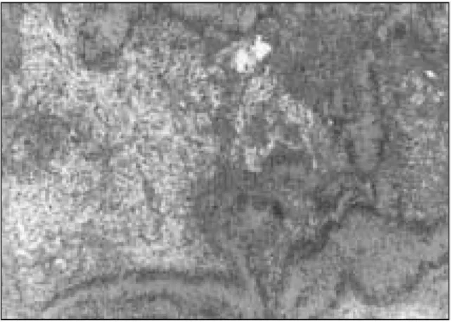
조직학적으로 신경초종은 거의 대부분 피막화되어있고, 물결무늬의 유신경 세포가 관찰되며 2가지의 특이한 조직 형 태를 띄게 되는데, 먼저 Antoni A조직에서는 책상배열과 신장 된 핵이 동종의 초자양 무세포성 섬유지대인 소위 Verocay
body에 의해 둘러싸이게 되고, Antoni B 조직에서는 소섬유가
좀더 느슨한 망상체를 이루고 공포화된 세포의 공간 구간을 갖는 난원형 핵을 보여 Verocay body는 관찰되지 않는다(Fig.11,12)
4,13,14).
Fig. 11. Histopathologic findings (H-E stain ×40) Spindle-shaped cells with palisading forming Verocay bodies of the Antoni type A tissue and zone of micro- cystic and myxoid change of the Antoni type B tissue.
Fig. 12. Histopathologic findings (H-E stain × 400)
Spindle-shaped cells of the Antoni type A tissue and
Antoni type B tissue shows Schwann cells dispersed in
a loose and random fashion with a meshwork of
delicate reticulin fibers and numerous microcystic
spaces.
막화가 되어 있지는 않지만 잘 경계지어지며 myxoid 또는 col-
lagen stroma 내에 방추 세포가 증식되어 있는 상태로 나타나고,
핵의 형태는 타원형의 파상의 경계를 갖고 또한 신경섬유종증 의 한 증상으로 나타나기도 하기 때문에 이들과의 감별이 필 요한데, 신경 섬유종증은 전체 경우의 약 5~16%에서 악성 전 환의 가능성이 있고, 특징적으로 다발성 신경 섬유종, 피부 착 색(cafe、au lait spot), 골내 병소를 가지는 상염색체 우성 질환으 로 단독으로 1.5cm이상 직경의 cafe、au lait spot을 6개 이상 가질 경우 신경섬유종증으로 진단이 가능하다7).
즉, 신경초종은 신경섬유종 및 신경섬유종증에 비해 더 많은 유조직 구조, 피막의 존재 및 Verocay body를 포함하는 Antoni A 조직과 Antoni B 조직으로 구분할 수 있고 또한 낮은 재발율과 악성 이환율을 갖는다4,10,19)
. 이러한 임상 및 조직학적 특성은 비
슷한 기원을 갖는 신경섬유종 및 신경섬유종증과의 감별을 가 능하게 한다.본 증례에서도 역시 신경초종의 특징적 양상인 Verocay body 를 포함하는 Antoni A 및 Antoni B조직을 관찰할 수 있었다.
Robertson 등
15)은 두경부에 발생한 31증례의 악성 신경초종중 18증례에서 하치조 신경과 연관되어 발생함을 보고하였다.
조직학적으로 악성 신경초종이 경우에 따라 유골 형성과 치밀 한 섬유 조직을 포함해서 골화성 섬유종으로 오진할 수 있고 섬유모세포성 육종과도 감별이 어려워 이를 구분하는 특징은 악성 신경초종의 신경 기원 즉, 수술 도중 종양의 신경 부착을 확인하는 것이다16)
.
Eversole 등
17)은 구강 내 19증례의 신경성 육종 중 6증례에서중심성임을 보고했고 하악이 상악보다 그리고 여성에서 더 호 발함을 보고했다. Shirasuna 등18)은 몇몇의 경우에서 방사선학 적으로 불명확한 변연을 갖는 방사선 투과상과 하악관 혹은 이공 확대의 소견을 보고하였다.
치료방법은 외과적 적출술로 악성으로의 전이 및 재발이 드 물어 예후는 매우 양호하며, 방사선 치료는 신경초종이 방사 선 저항성이 강하고 악성으로의 전이 가능성이 있어 시행하지
않는다1,10,20)
.
종양의 외과적 적출 시 통상 하치조 신경은 종양과 경계가 명확하고 피막이 잘 형성되어 있기 때문에 주위조직으로부터 쉽게 분리되어 대부분의 환자에서 술후 일시적인 지각 마비 증상을 호소하지만4,5,9,10,11,13)
, Shimura 등
21)은 우측 하악지와 하악 체에 이르는 광범위한 종양의 적출시 제 1대구치 부위에서 오 훼돌기까지 광범위한 변연골 절제술 후 우측 하순의 감각소실 을 보고하였으며, Llewelyn과 Sugar 등14)은 종양 적출 후 골결손 부위에 장골 이식술을 시행하였다.본 증례에서는 하치조 신경이 종양과 분리되지 않아 신경을 포함한 종양 적출술을 시행하였고 동시에 병소가 정중부에서 좌측 하악지에 이르는 광범위한 상태여서 병리학적 골절의 예
Ⅳ. 결 론
저자 등은 28세 남환의 하악지에서 하악체를 거쳐 하치조 신 경을 따라 정중부까지 광범위하게 포함되고, 협부 및 하순의 이신경을 따라 결절성으로 발생한 주변성 및 중심성 신경초종 환자에서 종양 적출술 및 골 이식술로 치아 발거없이 양호한 결과를 얻었기에 문헌 고찰과 함께 보고하는 바이다.
참고문헌