• 검색 결과가 없습니다.

Osteogenic differentiation of bone marrow derived stem cells in gelatin-hydroxyapatite nanocomposite

N/A
N/A
Protected

Academic year: 2021

Share "Osteogenic differentiation of bone marrow derived stem cells in gelatin-hydroxyapatite nanocomposite"

Copied!
6
0
0

로드 중.... (전체 텍스트 보기)

전체 글

(1)

Introduction

Bone is generally considered to be a nanocomposite of min- erals and proteins. Calcified tissue is regarded as a biological, chemical bonded composite between hydroxyapatite (HA) and type-I collagen. The preparation of a nanocomposite of HA- collagen that is similar to the nanostructure of real bone have been reported.

1-3)

The largest problems with type-I collagen, which makes it difficult to follow up on well controlled pro- cessing, are its cost and limited commercial sources of this material. Type-I collagen can be replaced with a gelatin pre- cursor. The commercial sources of gelatin show good water

solubility and well-defined physical and chemical properties.
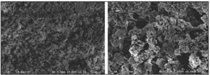
4,5)

Recently, HA-gelatin composite powders were made by the precipitation of HA. These composites showed nano-sized HA powders dispersed in a gelatin network. The preparation of Gel-HA nanocomposite was previously reported by Chang et

al.

4-11)

In this study, the Gel-HA nanocomposite similar to the

one made by Dr. Chang was used.

Bone marrow has a multipotent population of cells that are capable of differentiating into a number of mesodermal lin- eages; adipocytes, osteoblasts, and other mesodermal path- ways. Bone marrow stromal cells (BMSCs) also support the proliferation and differentiation of hematopoietic stem cells.
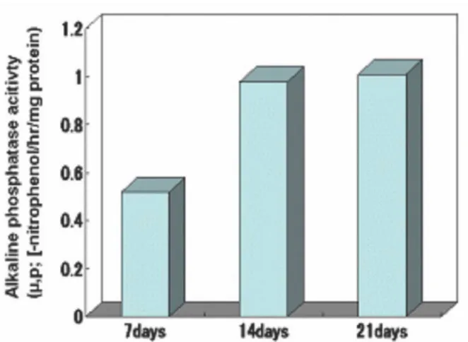
12)

This study examined the osteogenic differentiation and pro- liferation of rabbit bone marrow stem cells in a Gel-HA nanocomposite and elucidated the possibility of osteoinductivi- ty of a Gel-HA nanocomposite seeded with BMSCs as a bone tissue engineering scaffold.

*Corresponding author Uk-Kyu Kim

Dept.OMFS. Surgery, School of Dentistry, Pusan National University 1-10, Ami-dong, Seo-gu,602-739, Busan, Republic of Korea, Tel: 82-51-240-2803

E-mail: kuksjs@pusan.ac.kr

Osteogenic differentiation of bone marrow derived stem cells in gelatin- hydroxyapatite nanocomposite

Hyun-Jun Jeon

1

, Young-Sup Hwang

1

, Uk-Kyu Kim

1

*, Dae-Seok Hwang

1

, Kwang-Ho Lee

1

, Myung-Cheol Chang

2

1

School of Dentistry, Pusan National University, Korea,

2

School of Materials Science and Chemical Engineering, Kusan National University, Korea

Purpose: Gelatin-hydroxyapatite nanocomposite is similar to inorganic nanostructure of bone. To make a scaffold with osteoinductivity, bone mar- row derived stem cells from rabbit femur were impinged into the nanocomposite. This vitro study was to test osteogenic differentiation of the stem cells in the nanocomposite, which was made by authors.

Material & Methods: Gel-HA nanocomposite with 10g of HA, 3 g of Gel has been made by co-precipitation process. Bone marrow was obtained from femur of New Zealand White rabbits and osteogenic differentiation was induced by culturing of the BMSCs in an osteogenic medium. The BMSCs were seeded into the Gel-HA nanocomposite scaffold using a stirring seeding method. The scaffolds with the cells were examined by scan- ning electron microscopy (SEM), colorimetry assay, biochemical assay with alkaline phosphatase (ALP) diagnostic kit, osteocalcin ELISA kit.

Results: Gel-HA nanocomposite scaffolds were fabricated with relatively homogenous microscale pores (20-40 μm). The BMSCs were obtained from bone marrow of rabbit femurs and confirmed with flow cytometry, Alizarin red staining. Attachment and proliferation of BMSCs in Gel-HA nanocomposite scaffold could be identified by SEM, ALP activity and osteocalcin content of BMSCs.

Conclusion: The Gel-HA nanocomposite scaffold with micropores could be fabricated and could support BMSCs seeding, osteogenic differentia- tion.

Key words

Gelatin (Gel), Hydroxyapatite(HA), Nanocomposite, Osteogenic differentiation, Bone Marrow derived Stem Cell (BMSC)

Abstract

(2)

J Korean Assoc Oral Maxillofac Surg. Vol. 35 No. 1, 2009

MATERIAL AND METHOD 1. Scaffold fabrication

The co-precipitation process of a Gel-HA nanocomposite has been reported in detail elsewhere.

4-11)

In this study the molecu- lar weight (MW) of tropocollagen (about 310,000 ± 50,000) was used as the basis for the batch calculations due to the high variability in the MW of gelatin (50,000 to 250,000 Da) according to the processing conditions. The starting materials were CaCO

3

(Alkaline analysis grade, Aldrich, USA), H

3

PO

4

(AP grade, Aldrich, USA) and GEL (Unflavored, Canada).

The amount of Ca (OH)

2

and H

3

PO

4

was calculated to make 10 g of HA. A homogeneous suspension, which included Ca(OH)

2

dispersed in 2 liters of DI water, and an aqueous solution of H

3

PO

4

with 3g of GEL was added gradually to the reaction vessel through peristaltic pumps. After the co-precipitation reaction, the total volume was adjusted to 4 liters. The temper- ature and pH of the reaction solution in the vessel was main- tained at 38℃ and 8.0, respectively. After the reaction, the resulting slurry was aged at 38℃ for 24 h.

2. Isolation and culture of stem cell

Bone marrow was obtained from the femur of New Zealand White rabbits by aspiration and flushing out with a 16 gauze needle and a 10 ml syringe containing 1 ml of heparin (3000U/ml). After placing the bone marrow in a 50 ml tube containing 5 ml of α-modified Eagle’s medium (α-MEM), containing 10% fetal bovine serum (FBS), it was centrifuged at 1200×g for 10 min to obtain a pellet. The pellet was filtered through a 75-㎛ nylon mesh to remove the cellular debris and incubated overnight at 37℃/ 5% CO

2

in a control medium(α- MEM; Gibco/Invitrogen Corp, Grand Island, NY) containing 10% FBS(Gibco/Invitrogen), 1% of penicillin- streptomycin(Gibco/Invitrogen). After incubation, the plate was washed extensively with phosphate-buffered saline (PBS) to remove the residual nonadherent red blood cells. The result- ing cell population was maintained at 37℃/5% CO

2

in the con- trol media. After expansion to passage 2, the BMSCs were washed with PBS and collected via trypsinization (trysin- EDTA solution; 0.05% trysin Sigma, USA) at 37℃ for 2 min.

3. Confirmation of osteogenic potential of BMSC

Flow cytometry was performed to determine the surface pro- tein expression on the undifferentiated BMSC. Adipogenic dif- ferentiation was induced by culturing the MSC for 2 weeks in

an adipogenic Medium (10% FBS, 1μM dexamethasone, 100 μ g/mL 3-isobutyl-1-methylxanthine, 5μg/mL insulin, and 60 μM indomethacin in α-MEM) and assessed using an Oil Red O stain as an indicator of intracellular lipid accumulation.

12)

In order to obtain the quantitative data, 1 ml of isopropyl alcohol was added to the stained culture dish.

Osteogenic differentiation was induced by culturing the MSC for a minimum of 3 weeks in an osteogenic Medium (10% FBS, 0.1mM dexamethasone, 10 μM α-glycerophos- phate, and 50 μg/mL ascorbic acid in α-MEM). The level of extracellular matrix calcification was examined by alizarin red staining.

13)

Osteogenic differentiation was quantified by mea- suring the alizarin red stained area in 6 well dishes by image analysis.

4. Cell seeding into scaffold

The gelatin-hydroxyapatite nanocomposte scaffolds were prepared to dimensions of 5 mm×5 mm×3 mm, and steril- ized three times in 70% ethanol for 2 min and pre-soaked in the culture medium for 5 min.

The BMSCs were seeded into the gelatin-hydroxyapatite nanocomposite scaffold using a stirring seeding method.

Briefly, the scaffolds were hung over wire and threads, and placed in a sterilized flask containing 300ml of a osteogenic medium (10% FBS, 0.1mM dexamethason, 10mM β-glyc- erophosphate, and 50μg/mL ascorbic acid in α-MEM) with the cells at a concentration of 5×10

4

cells/ml. The cell-containing medium was then stirred with a magnetic stirrer at 150 rpm speed for 6 hours

14)

5. Cell culture of cell-seeded scaffold

After an attachment period of 6 hours, each of the cell-seed- ed scaffolds were removed and washed three times with PBS.

The viable cells attached to the scaffolds were quantified using an MTS assay. The absorbance was measured at 490nm using a microplate reader. The other cells on the cell-seeded scaf- folds were transferred into new wells and cultured in α-MEM for 3 weeks.

6. Scanning electron microscopy observation

After culturing for 3 weeks, the scaffolds with the cells were primary fixed with a 2.5% glutaraldehyde solution in PBS.

After rinsing with PBS, the scaffolds with the cells were sec-

ondary fixed with 1% osmium tetroxide. They were subse-

quently dehydrated with a graded series of ethanol solutions

(3)

dehydrated samples were immersed in HMDS (H00326-500, Lancaster) and dried. After sputter coating with gold/palladium (HITACHI, ion-sputter E-1010), the samples were examined by scanning electron microscopy (SEM, S3500N, HITACHI, Japan).

7. Colorimetry assay

The assays measuring the mitochondrial activity are a safe, simple, rapid method for measuring the level of cellular prolif- eration. This was performed by measuring the activity of mito- chondrial dehydrogenases of viable cells, which reduces 3-(4, 5-dimethylthiazol-2-yl) - 5-(3-carboxy-methoxyphenyl) -2-(4- sulfophenyl)-2H-tetrazolium [MTS] to a water-soluble for- mazan product in the presence of phenazine methosulfate. The amount of formazan generated is proportional to the total mito- chondria activity per cell in the culture. The amount of for- mazan product was measured at a wavelength of 490 nm.

The MTS assay was carried out by combining a MTS solu- tion (CellTiter 96R AQueous Assay, Promega). Briefly, the scaffolds were washed with phosphate-buffered saline (PBS), transferred into a new 24-well cell culture plate (Costar, Corning, NY) containing 1 mL culture media and 200 mL of MTS solution, and incubated for 2 h at 37℃. Subsequently, 250 mL of a 10% sodium dodecyl sulfate (SDS) solution was added, and the resulting solution was incubated at room tem- perature for 5 min. The absorbance at 490 nm of each well was measured using a spectrophotometric plate reader (TECAN, Crailsheim, Germany). The number of cells was determined using a standard curve that was established using a known number of cells counted on a Coulter counter. The scaffolds were analyzed at 6 hours, 7 days, 14 days and 21 days after.

8. Biochemical assay

Osteogenic differentiation potential of the bone marrow- derived stromal cells was assessed using the alkaline phos- phatase activity and osteocalcin content. At each defined time point, the plated cells were fixed with 10% paraformaldehyde and stained with alkaline phosphatase to evaluate the level of osteogenic differentiation of bone marrow derived stem cells.

The alkaline phosphatase activity within the cytoplasm was quantified using an alkaline phosphatase diagnostics kit (Sigma, USA). Osteocalcin, which is a late bone turnover marker only secreted by terminally differentiated osteoblasts, was quantified using an osteocalcin enzyme-linked immunosorbent assay kit (Quidel Co., San Diego, USA). All

er’s protocols. The scaffolds were analyzed at 7 days, 14 days and 21 days.

RESULTS

1. Characterization of gelatin-HA nanocomposite scaffold

Fig. 1 shows scanning electron micrographs of the gelatin-HA nanocomposite, which have an irregular pore of scaffold. The morphology of the gelatin-HA nanocomposite was described by Chang through TEM observations of the ED patterns.

11)

Various morphologies were revealed according to the denaturing condi- tions of the Gel precursors in an aqueous solution of H

3

PO

4

. The organic-inorganic interaction and resulting morphology can serve as a guide to develop a microcircuit design in a biomass.

Gel-HA nanocomposite scaffolds were fabricated with relatively homogenous microscale pores (20-40μm).

2. Confimation of osteogenic potential

The mesenchymal stem cell population obtained from the rabbit bone marrow was cultured. The BMSC were maintained in α-MEM supplemented with 10% FBS. The cells expanded easily in vitro and exhibited a fibroblast-like morphology, sim- ilar to that of the MSC obtained from other tissues. Flow cytometry was performed to determine the surface protein expression on the undifferentiated BMSC. CD45 and CD44 were used. Figure 2 shows representative histograms. The undifferentiated BMSCs tested positive to CD44 and the mes- enchymal stem cell markers, and negative to CD45 and the hematopoietic markers.

13)

The multilineage capacity of the BMSC was examined by

differentiation toward adipogenic and osteogenic lineages

using lineage specific induction factors. In order to determine

if ADSC undergo adipogenesis, the cells were cultured in a

medium containing these agents (adipogenic medium, AM)

and stained with Oil Red-O. The ADSC cultured in AM were

Fig 1. Scanning electron microscopy findings of the gelatin-HA

nanocomposite scaffold (50um, 10um).

(4)

reproducibly induced toward the adipogenic lineage as early as 2 weeks after induction (Fig. 3). A significant proportion of the cells contained multiple, intracellular lipidfilled droplets that accumulated Oil Red-O. No lipid droplets were observed in the undifferentiated ADSC. Osteogenic differentiation was con- firmed by measuring the level of calcification of the extracellu- la matrix (ECM) in the MSC using Alizarin red staining.

Calcification appears as red regions within the cell monolayer.

Consistent with osteogenesis, several red regions, which are indicative of a calcified ECM, were observed in the BMSC treated for 3 weeks in osteogenic medium. No calcification was observed in the undifferentiated MSC (Fig. 3).

3. Attachment and proliferation of MSC in gelatin-HA nanocomposite scaffold

A stirring method (150 rpm, 6 h) was used at a constant cell density in medium (5×10

4

cells/ml) to assist the cells to attach uniformly to the three-dimensional scaffolds.

Fig. 4 shows the SEM morphology of the cells grown on the gelatin-HA nanocomposite scaffolds. The cells grew favorably with the cell membranes spreading out actively on the surface.

The cell viability on the scaffolds after culturing for 6 hours, 1, 2, 3 weeks were quantified using an MTS assay, as shown in

Fig. 5. For all the scaffolds, the cells proliferated to higher degrees with increasing culturing period, which suggests good cell viability on all scaffolds.

4. Osteogenic differentiation of MSC in gelatin-HA nanocomposite scaffold

Fig. 6 shows the alkaline phosphatase (ALP) activity of the MSC cultured in the gelatin-HA nanocomposite scaffold 1, 2, 3 weeks after the stirring method. The ALP levels on the scaf- folds increased with increasing culturing period.

Fig.7 shows the osteocalcin (OC) content of MSC cultured in gelatin-HA nanocomposite scaffold 1, 2, 3 weeks after the stir- ring method. The level of OC production increased with increasing culturing period.

DISCUSSION

Human bone tissue is a biologically and chemically bonded composite of inorganic apatite nanocrystals embedded in an organic matrix of collagen and noncollagenous proteins.

3)

Generally, hydroxyapatite is the main component of bone min- eral. This study used the gelatin-hydroxyapatite nanocomposite scaffold reported by Chang.

4-11)

This scaffold was made using a co-precipitation technique of HA nano-sized particles in a gelatine matrix. The amount of gelatin in a constant volume

J Korean Assoc Oral Maxillofac Surg. Vol. 35 No. 1, 2009

Fig 2. Expression of the surface markers in MSC , Expression of the mesenchymal stem cell marker in the bone marrow derived stem cell was identified by flow cytometry (CD44, CD45).

Fig 3. Mesenchymal stem cell induced with the adipogenic medi- um(AM) and osteogenic medium(OM). The Bone marrow-derived MSC were cultured for 2 weeks in AM(right), 3 weeks in OM(left) and stained with Oil REd O, Alizarin S to identify the differentiation

Fig 4. Scanning electron micrographs of the MSC attached to the gelatin-HA nanocomposite scaffold.

Fig 5. Proliferation of the MSC on nanocomposite scaffolds at 6

hours, 7, 14 and 21days after culturing, as assessed by the MTS

method.

(5)

batch greatly influenced the nucleation and the development of HA nanocrystals. The co-precipitated gelatin-HA nanocom- posites showed chemical bond formation between the HA nanocrystals and gelatin macromolecules and had a self-orga- nized structure along the gelatin fibrils

15-17)

.

Porous structures on the scaffold are important because they determine where the cells will initially be distributed during the seeding process, where the scaffold will eventually guide tissue formation, and how well the tissue will survive in vitro or in vivo depending on the availability of nutrients or blood vessel formation.

18-19)

The scaffold used in this study did not have a homogenous pore size but showed stem cells seeding. It is difficult to fabricate a scaffold with the proper and homoge- nous pores through the co-precipitation of HA particles within a gelatin matrix. This will form the basis of future studies.

These results show that the adult rabbit bone marrow is a suitable and feasible source of a large number of MSC, and that the bone marrow derived stem cells can be easily induced to differentiate into an osteogenic lineage. Furthermore, it was demonstrated that the gelatin-HA nanocomposite scaffold is

nature of the cells derived from the bone marrow of adult rab- bits was demonstrated by their capacity of extensive prolifera- tion and ability to differentiate into several different lineages.

The capacity of the bone marrow derived stem cell to differen- tiate towards an osteogenic lineage and deposit calcium has been identified by several methods. Based on these results, the increase in the alkaline phosphatase and osteocalcin levels, which are early markers of cells oriented towards osteogenic production, can explain why adult MSC exposed to osteogenic medium differentiate towards an osteoblastic lineage

20-27)

.

In this study, the stirring method was used for cell seeding and proliferation on the scaffold

14,19,24)

. This method allows effi- cient transfer of oxygen and nutrients to the cells because the culture medium is circulated. In addition, the stirring method creates mechanical stimulation to the cells. Mechanical strain and fluid shear stress stimulates the cells to produce prostaglandins, alkaline phosphatase and collagen type I, while they increase the level of osteoblast proliferation and mineral- ization. The ALP activity and osteocalcin content of the MSC as osteogenic differentiation markers were increased as the cell culture passages were processed. This suggests that the gelatin- HA nanocomposites seeded with BMSCs can bio-mimetically have high potential as a hard tissue scaffold.

CONCLUSION

HA particles were co-precipitated on gelatin matrix in order to create better environmental conditions for gelatin-HA nanocomposite scaffolds in stem cell seeding. BMSCs were extracted from a rabbit femur, cultured and seeded into the gelatin-HA scaffold using the stirring method. The osteogenic differentiation on the scaffold was confirmed by a MTS assay and a biochemical assay for ALP and OC. Mesenchymal stem cells from rabbit bone marrow differentiated towards an osteogenic lineage, representing a suitable cell source for bone formation. The gelatin-HA nanocomposite scaffolds could be fabricated by the co-precipitation of hydroxyapatite within a gelatin matrix. This scaffold supported BMSC seeding and dif- ferentiation. The findings are expected to provide fundamental information to design gelatin-nanocomposite scaffolds suitable for bone tissue engineering.

REFERENCES

1. Zerwekh JE, Kourosh S, Schienbergt R.: Fibrillar collagen.biphasic calcium phosphate composite as a bone graft substitute for spinal fu- sion. J Orthop Res 1992;10: 562-72.

2. Liu L, Zhang L, Ren B, Wang F, Zhang Q. : Preparation and charac- Fig 6. Alkaline phosphatase(ALP) activity of the MSCs on the

nanocomposite scaffolds at 1, 2, 3 weeks after culturing.

Fig 7. Ostecalcin(OC) production of the MSC on the nanocomposite

scaffolds at 1, 2, 3 weeks after culturing

(6)

J Korean Assoc Oral Maxillofac Surg. Vol. 35 No. 1, 2009

terization of collagen-hydroxyapatite composite used for bone tissue engineering scaffold. Artif Cells Blood Substit ImmobilBiotechnol 2003; 31:435-48.

3. Sachlos E, Gotora D, Czernuszka JT.: Collagen scaffolds reinforced with biomimetic composite nano-sized carbonate-substituted hy- droxyapatite crystals and shaped by rapid prototyping to contain in- ternal microchannels. Tissue Eng 2006; 12:2479-87.

4. Chang MC, Ikoma T, Tanaka J.: Cross-linkage of hydroxyapatite- gelatin nanocomposite using EDGE. J Mater Sci 2004; 39: 5547 - 5550.

5. Kim HW, Knowles JC, Kim HE.: Porous scaffolds of gelatin- hy- droxyapatite nanocomposites obtained by biomimetic approach:

Characterization and antibiotic drug release. J Biomed Mater Res B Appl Biomater 2005; 74: 686-698.

6. Chang MC, Ko CC, Douglas WH.: Modification of hydroxyapatite- gelatin composite by polyvinylalcohol. J Mater Sci 2005; 40: 505 - 509.

7. Chang MC, Ko CC, Douglas WH.: Preparation of hydroxyapatite - gelatin nanocomposite. Biomaterials 2003; 24: 2853-2862.

8. Chang MC.: Molecular orbital calculation on the configuration of hydroxyl group in hexagonal hydroxyapatite. J Kor Ceramic Society 2005; 42: 304-307.

9. Chang MC, Douglas WH, Tanaka J.: Organic-inorganic interaction and the growth mechanism of hydroxyapatite crystals in gelatin matrics between 37 and 80�C. J Mater Sci:Mater Med 2006; 17:

387-396.

10. Chang MC.: Morphology development of HAp crystallites in Gel Matrix. J Kor Ceramic Society 2007; 44: 133-136.

11. Chang MC, Ko CC, Douglas WH.: Conformational change of hydroxyaptite/gelatin nanocomposite by glutaradehyde. Biomaterials 2003; 23: 3087-3094.

12. Preece A: A manual for histologic technicians. Boston. MA.Little, Bowrn and Co, 1972: 145-147.

13. Lee RH, Kim BC, Choi IS: Characterizaiton and expression analysis of mesenchymal stem cells from human bone marrow and adipose Tissue. Cell Physiol Biochem 2004; 14: 311-324.

14. Kim HW, Kim HE, Salih V.: Stimulation of osteoblast responses to biomimetic nanocomposites of gelatin-hydroxyapatite for tissue enginnering scaffolds. Biomaterials 2005; 26: 5221-5230.

15. Yaylaoglu MB, Korkusuz P, Ors U, Korkusuz F, Hasirci V.:

Development of a calcium phosphate.gelatin composite as a bone

substitute and its use in drug release. Biomaterials1999; 20:711-719.

16. Fukunaka Y, Iwanaga K, Morimoto K, Kakemi M, Tabata Y.:

Controlled release of plasmid DNA from cationized gelatin hydrogels based on hydrogel degradation. J Control Rel 2002; 80:333-43.

17. Kim HW, Knowles JC, Kim HE.: Hydroxyapatite and gelatin com- posite foams processed via novel freeze-drying and crosslinking for use as temporary hard tissue scaffolds. J Biomed Mater Res A. 2005;

72:136-45.

18. Shen FH, Zeng Q, Choi L: Osteogenic differentiation of adipose- derived stromal cells treated with GDF-5 cultured on a novel three- dimensional sintered microsphere matrix. Spine 2006; 6: 615-623.

19. Donzelli E, Salvade A, Mimo P: Mesenchymal stem cells cultured on a collagen scaffold: in vitro osteogenic differentiation. Arch Oral Biol 2007; 52: 64-73.

20. Kimura Y. Biodegradable polymers. In; Tsuruta T, editor: Biomedical applications in polymeric materials. Boca Raton: CRC press, 1993:

163-165.

21. Wei G, Ma PX.: Structure and properties of nano- hydroxyapatite/polymer composite scaffolds for bone tissue engi- neering. Biomaterials 2004; 25:4749-57.

22. Kenley RA, Yim K, Abrams J, Ron E, Turek T, Marden LJ:

Biotechnology and bone graft substitutes. Pharm Res 1993;10: 1393- 401.

23. Khan SN, Cammisa Jr FP, Sandhu HS, Diwan AD, Girardi FP, Lane JM.: The biology of bone grafting. J Am Acad Orthop Surg, 2005;

13: 77-86.

24. Pittenger MF, Mackay AM, Beck SC, Jaiswal RK, Douglas R, Mosca JD: Multilineage potential of adult human mesenchymal stem cells.

Science 1999; 284:143-7

25. Jaiswal N, Haynesworth SE, Caplan AI, Bruder SP.: Osteogenic dif- ferentiation of purified, culture-expanded human mesenchymal stem cells in vitro. J Cell Biochem 1997; 64: 295-312.

26. Yang S, Leong K-F, Du Z, Chua C-K.: The design of scaffolds for use in tissue engineering. Part I.traditional factors. Tissue Eng 2001;

7:679-89.

27. El-Amin SF, Lu HH, Burems J, Mitchell J, Tuan RS, Laurencin CT.:

Extracellular matrix production by human osteoblasts cultured on biodegradable polymers applicable for tissue engineering.

Biomaterials 2003; 4:1213-21.

수치

Fig. 1 shows scanning electron micrographs of the gelatin-HA nanocomposite, which have an irregular pore of scaffold
Fig 7. Ostecalcin(OC) production of the MSC on the nanocomposite scaffolds at 1, 2, 3 weeks after culturing

참조

관련 문서

_____ culture appears to be attractive (도시의) to the

1 John Owen, Justification by Faith Alone, in The Works of John Owen, ed. John Bolt, trans. Scott Clark, "Do This and Live: Christ's Active Obedience as the

The proliferations of MC3T3-E1 cell seeded on pristine PCL scaffold and surface modified scaffolds for 1, 3, and 5 days... ALP activity of the MC3T3-E1 cell seeded

mRNA expression of osteonectin, Runx2 and BSP(bone sialoprotein) in MC3T3-E1 cells cultured for 24 hours on 4 different titanium surfaces.. The mRNA were analyzed

3 Mean percentages of bone implant contact ratio in the control group and experimental groups at 6 and 12 weeks after placement of the

Determining optimal surface roughness of TiO₂blasted titanium implant material for attachment, proliferation and differentiation of cells derived from

Approved clinical use of bone marrow stem cells for myocardial infarction treatment... Cardiac

Comparison of cell attachment on the bone grafting materials synthesized as the defined mixing ratio of Hydroxyapatite (HA) and β-tricalcium phosphate (β-TCP) by DAPI staining