13 To the editor,
The coronavirus disease 2019 (COVID-19) is highly contagious, and spread globally following an outbreak in China in 2019. Although the severe acute respiratory syndrome coronavirus 2 (SARS-CoV-2) virus primarily targets the respiratory system, we herein report on COVID-19- associated-encephalopathy which manifests as multiple enhanced lesions in brain magnetic resonance imaging (MRI).
A 67-year-old-male was hospitalised for COVID-19-associated-pneumonia on April 2, 2020.
His first symptoms consisted of myalgia and a cough. COVID-19 was diagnosed based on the detection of SARS-CoV-2 viral nucleic acid through a nasopharyngeal swab specimen using real-time polymerase-chain-reaction assay. Chest computerised tomography showed patchy areas of consolidation in the peribronchial and subpleural areas of both lungs, which was consistent with COVID-19-associated-pneumonia. He had diabetes, chronic kidney disease without dialysis, and alcoholic liver disease. The patient was not a smoker, but a heavy drinker. Lopinavir/ritonavir and antibiotic therapy were initiated. On the 5th day of illness, the patient was intubated due to desaturation. On the 14th day, multi-organ failure was diagnosed, and continuous-renal-replacement-therapy and extracorporeal-membrane- oxygenation were administered. On the 25th day, he was released from quarantine due to the negative results of three consecutive COVID-19-tests. He had stupor, but diffusion, and MRI showed no abnormal findings. The response was not improved until 42nd day, although sedative was not used.
On the 42nd day, he showed decreased response (semi-coma) due to pain and intermittent apnoea. His pupils were 3-mm bilaterally and prompt. He had a blood pressure of 149/90 mmHg, body temperature of 37°C, heart rate of 120 beats/min, respiratory rate of 25 breaths/
min, saturation of 100% under mechanical ventilation, haemoglobin of 9.3 g/dL, C-reactive protein of 11.83 mg/L, erythrocyte-sedimentation rate of 46 mm/hr, and creatinine of 3.1 mg/
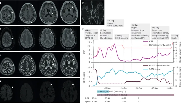
dL, which was elevated. The other routine laboratory findings measured as normal. Brain MRI showed multifocal gyriform and nodular enhancing lesions scattered in the cortex and deep grey matter in the left cerebral hemisphere on a contrast-enhanced T1-weighted-image and high signal intensities and hemorrhages in the corresponding areas of the enhanced lesions on a T2-weighted-image. MR-angiography detected no abnormality in the large intra/
extracranial vessels (Fig. 1).
Dement Neurocogn Disord. 2021 Apr;20(2):13-15 https://doi.org/10.12779/dnd.2021.20.2.13 pISSN 1738-1495·eISSN 2384-0757
Letter to the Editor
Received: Oct 21, 2020 Revised: Feb 26, 2021 Accepted: Mar 23, 2021 Correspondence to Young Hee Jung
Department of Neurology, Myongji Hospital, College of Medicine, Hanyang University, 55 Hwasu-ro 14beon-gil, Deokyang-gu, Goyang 10475, Korea.
E-mail: [email protected]
© 2021 Korean Dementia Association This is an Open Access article distributed under the terms of the Creative Commons Attribution Non-Commercial License (https://
creativecommons.org/licenses/by-nc/4.0/) which permits unrestricted non-commercial use, distribution, and reproduction in any medium, provided the original work is properly cited.
ORCID iDs Yu Min Kang
https://orcid.org/0000-0002-4368-9878 Noh Hyuck Park
https://orcid.org/0000-0003-4716-3491 Min Jae Seong
https://orcid.org/0000-0002-8731-5245 Hyun Jeong Han
https://orcid.org/0000-0002-1129-6340 Young Hee Jung
https://orcid.org/0000-0002-8945-2200 Funding
This work was supported by a fund by Research of Korea Centers for Disease Control and Prevention (2021ER100500).
Conflict of Interest
The authors have no financial conflicts of interest.
Yu Min Kang ,1 Noh Hyuck Park ,2 Min Jae Seong ,3 Hyun Jeong Han ,3 Young Hee Jung 3
1 Department of Infectious disease, Myongji Hospital, College of Medicine, Hanyang University, Goyang, Korea
2Department of Radiology, Myongji Hospital, College of Medicine, Hanyang University, Goyang, Korea
3Department of Neurology, Myongji Hospital, College of Medicine, Hanyang University, Goyang, Korea
Multiple Enhanced Lesions in the Brain MRI of a Patient with COVID-19
https://dnd.or.kr
Author Contributions
Conceptualization: Jung YH; Data curation:
Kang YM, Park NH, Seong MJ, Jung YH; Formal analysis: Park NH; Investigation: Park NH, Seong MJ, Han HJ, Jung YH; Supervision:
Jung YH; Validation: Jung YH; Visualization:
Jung YH; Writing - original draft: Kang YM, Park NH, Seong MJ, Jung YH; Writing - review
& editing: Kang YM, Park NH, Seong MJ, Han HJ, Jung YH.
To evaluate the cardiac source of emboli, transthoracic echocardiography was performed.
There was no evidence of embolic sources such as infective endocarditis. To evaluate vasculitis, additional laboratory tests were performed. Serologic testing revealed negative fluorescent antinuclear and p-type antineutrophil cytoplasmic antibodies through a serum immunofluorescence test. Enzyme-linked immunosorbent assay revealed negative myeloperoxidase and peroxidase-3 antibodies. Cerebrospinal fluid studies on the 48th day showed an increased red blood cell level count (234/μL). Otherwise, the composition of cerebral fluids and the opening pressure were normal. No specific rheumatologic diagnoses were made because the patient did not present any systemic manifestations such as skin lesions, arthritis, or signs of small vessel vasculitis except for the brain MRI finding. Steroid pulse was not considered due to his severe pneumonia, and intravenous immunoglobulin was not administered due to his unstable vital signs. On the 52nd day, he died due to the progression of pneumonia and shock, which did not respond to vasopressors.
The radiologic findings in our patient might suggest that small intracranial vessels were involved in COVID-19-associated vasculitis in that punctate and patchy enhancements have been reported as characteristic of vasculitis.1-3 In addition, scattered enhanced lesions with hemorrhages were not uncommon findings on the MRIs of multiple embolic infarctions.
14 https://doi.org/10.12779/dnd.2021.20.2.13
COVID-19 Associated Encephalopathy
https://dnd.or.kr
+5 Day Desaturation Intubation ICU admission
+14 Day Stupor CRRT, ECMO start
+20 Day
ECMO weaning +53 Day
Death +25 Day
Stupor Released from quarantine, No abnormal finding in diffusion MRI
+42 Day Semi-coma Intermittent apnea, Multiple enhancing lesions in brain MRI +1 Day
Myalgia, cough Diagnosis of COVID-19
25 15 5 10 20
0 12345678
CRP Clinical severity score
20 15 5 10
0 0
5 10 15
SOFA score Glascow coma scale
+20 Day
+10 Day +30 Day +40 Day +50 Day
Lopinavir/ritonavir (Day 2–day 11) Antibiotics therapy (Day 2–day 53) RdRP
E gene 30.33 30.08
26.65 30.08
35.27 35.35
0 0
A
B
C
D
E
F
CRP
Clinical severity score
Glascow coma scale SOFA score
Fig. 1. Radiologic findings and the clinical course of the patient. (A) Axial CE-T1WI show multifocal scattered enhanced lesions in the cortex of the left frontoparietal lobe, the left caudate nucleus, and the left thalamus. (B) Axial gradient echo images show multifocal microhemorrhages in the corresponding areas of the enhanced lesions as seen on CE-T1WI. (C) Axial T2WI show multifocal high signal intensity lesions (arrowheads) in the corresponding areas of the enhanced lesions seen on CE-T1WI. Perilesional oedema can be observed. (D) An axial diffusion-weighted image shows no evidence of abnormal diffusion restriction in the enhanced lesions. (E) Brain magnetic resonance angiography shows no abnormal findings in the intra- or extracranial large vessels. (F) The clinical course of the patient, clinical severity score (1 = no limit of activity; 2 = limit of activity but no O2; 3 = O2 with nasal prong; 4 = O2 with facial mask; 5 = non- invasive ventilation; 6 = invasive ventilation; 7 = multi-organ failure; 8 = death).
COVID-19: coronavirus disease 2019, ICU: intensive care unit, CRRT: continuous renal replacement therapy, ECMO: extracorporeal membrane oxygenation, MRI:
magnetic resonance imaging, RdRP: Ct value of RNA-dependent RNA polymerase of SARS-CoV-2, E gene: Ct value of envelope small membrane protein of SARS- CoV-2, CRP: C-reactive protein, SOFA: sequential organ failure assessment, CE-T1WI: contrast-enhanced T1-weighted images, T2WI: T2-weighted images, Ct:
cycle threshold, SARS-CoV-2: severe acute respiratory syndrome coronavirus 2.
COVID-19-associated neurologic complications involving the central nervous system (CNS) have also been reported, including acute hemorrhagic necrotizing encephalopathy and CNS- vasculitis.2-5 The previous CNS-vasculitis case was considered as a result of the endothelial injury caused by the binding of SARS-CoV-2 to angiotensin converting enzyme-2. However, a delayed immune reaction, and not SARS-CoV-2 infection, may have caused multiple brain lesions in our case. The patient's consciousness deteriorated approximately a month later, and MRI showed a finding resembling CNS-vasculitis, although the patient was released from quarantine after he tested negative for COVID-19 on the 25th day.
Furthermore, the asymmetric pattern that characterised the case of our patient was different from the symmetric pattern of the brain lesions on the MRI in a previously reported case of acute hemorrhagic necrotizing encephalopathy. In addition, we suspected the possibility of subacute stroke was caused by multiple emboli migrating to left middle cerebral artery territory. However, diffusion-MRI on the 25th day and 42nd day showed no abnormal findings.
The limitation is that diagnostic uncertainty remains. Brain imaging is the only evidence to support CNS-vasculitis and the diagnostic criteria of primary angiitis of the CNS were not satisfied. The possibility of cerebral infarct cannot be excluded, despite no evidence of cardiac embolism and consecutive, negative diffusion-MRI. Brain-biopsy, detailed neurologic exams and studies including other vasculitis markers, cytokines and complement fraction should be followed to confirm the diagnosis in future studies.
In conclusion, a neurologic complication with multiple enhanced lesions in brain MRIs may be present in patients with COVID-19.
REFERENCES
1. Hodel J, Darchis C, Outteryck O, Verclytte S, Deramecourt V, Lacour A, et al. Punctate pattern: a promising imaging marker for the diagnosis of natalizumab-associated PML. Neurology 2016;86:1516-1523.
PUBMED | CROSSREF
2. Hanafi R, Roger PA, Perin B, Kuchcinski G, Deleval N, Dallery F, et al. COVID-19 neurologic complication with CNS vasculitis-like pattern. AJNR Am J Neuroradiol 2020;41:1384-1387.
PUBMED | CROSSREF
3. Vaschetto R, Cena T, Sainaghi PP, Meneghetti G, Bazzano S, Vecchio D, et al. Cerebral nervous system vasculitis in a COVID-19 patient with pneumonia. J Clin Neurosci 2020;79:71-73.
PUBMED | CROSSREF
4. Poyiadji N, Shahin G, Noujaim D, Stone M, Patel S, Griffith B. COVID-19–associated acute hemorrhagic necrotizing encephalopathy: CT and MRI features. Radiology 2020;296:E119-E120.
CROSSREF
5. Varatharaj A, Thomas N, Ellul MA, Davies NWS, Pollak TA, Tenorio EL, et al. Neurological and neuropsychiatric complications of COVID-19 in 153 patients: a UK-wide surveillance study. Lancet Psychiatry 2020;7:875-882.
PUBMED | CROSSREF
15 https://doi.org/10.12779/dnd.2021.20.2.13
COVID-19 Associated Encephalopathy
https://dnd.or.kr