ABSTRACT
Background: Coronavirus disease 2019 (COVID-19) is caused by severe acute respiratory syndrome coronavirus 2 (SARS-CoV-2) infection. This disease, which is quickly spreading worldwide, has high potential for infection and causes rapid progression of lung lesions, resulting in a high mortality rate. This study aimed to investigate the effects of SARS-CoV-2 infection on renal function in patients with COVID-19.
Methods: From February 21 to April 24, 2020, 66 patients diagnosed with COVID-19 at Chungnam National University Hospital were analyzed; all patients underwent routine urinalysis and were tested for serum creatinine, urine protein to creatinine ratio (PCR), and urine albumin to creatinine ratio (ACR).
Results: Acute kidney injury (AKI) occurred in 3 (4.5%) of the 66 patients, and 1 patient with AKI stage 3 underwent hemodialysis. Upon follow-up, all 3 patients recovered normal renal function. Compared with patients with mild COVID-19, AKI (n = 3) occurred in patients with severe COVID-19, of whom both urine PCR and ACR were markedly increased.
Conclusion: The incidence of AKI was not high in COVID-19 patients. The lower mortality rate in SARS-CoV-2 infection compared with previous Middle East respiratory syndrome and SARS-CoV infections is thought to be associated with a low incidence of dysfunction in organs other than the lungs.
Keywords: Coronavirus Disease 2019; COVID-19; Severe Acute Respiratory Syndrome Coronavirus 2; SARS-CoV-2; Acute Kidney Injury; Proteinuria
INTRODUCTION
Novel coronavirus disease is a newly discovered contagious disease caused by severe acute respiratory syndrome coronavirus 2 (SARS-CoV-2), primarily manifesting as an acute respiratory illness accompanied by interstitial and alveolar pneumonia; however, it can also affect multiple organs, such as kidneys and heart, the digestive tract, the blood, and the nervous system.1 This disease was reported in December 2019 in Wuhan, Hubei province,
Original Article
Received: May 11, 2020 Accepted: Jun 30, 2020 Address for Correspondence:
Jae Wan Jeon, MD, PhD
Department of Internal Medicine, Chungnam National University School of Medicine, 266 Munhwa-ro, Jung-gu, Daejeon 35015, Republic of Korea.
E-mail: [email protected]
© 2020 The Korean Academy of Medical Sciences.
This is an Open Access article distributed under the terms of the Creative Commons Attribution Non-Commercial License (https://
creativecommons.org/licenses/by-nc/4.0/) which permits unrestricted non-commercial use, distribution, and reproduction in any medium, provided the original work is properly cited.
ORCID iDs Ki Ryang Na
https://orcid.org/0000-0002-0136-176X Hae Ri Kim
https://orcid.org/0000-0001-8766-6064 Youngrok Ham
https://orcid.org/0000-0001-9140-6900 Dae Eun Choi
https://orcid.org/0000-0003-2870-3958 Kang Wook Lee
https://orcid.org/0000-0003-3407-1205 Jae Young Moon
https://orcid.org/0000-0001-8724-6289 Yeon-Sook Kim
https://orcid.org/0000-0003-1142-5488
Ki Ryang Na ,1,2 Hae Ri Kim ,3 Youngrok Ham ,1,2 Dae Eun Choi ,1,2
Kang Wook Lee ,1,2 Jae Young Moon ,1,4 Yeon-Sook Kim ,1,5 Shinhye Cheon ,1,5 Kyung Mok Sohn ,1,5 Jungok Kim ,1,6 Sungmin Kim ,1,6 Hyeongseok Jeong ,5 and Jae Wan Jeon 1,3
1Department of Internal Medicine, Chungnam National University School of Medicine, Daejeon, Korea
2Department of Nephrology, Chungnam National University Hospital, Daejeon, Korea
3Department of Nephrology, Chungnam National University Sejong Hospital, Sejong, Korea
4 Department of Pulmonary and Critical Care Medicine, Chungnam National University Sejong Hospital, Sejong, Korea
5Department of Infectious Disease, Chungnam National University Hospital, Daejeon, Korea
6Department of Infectious Disease, Chungnam National University Sejong Hospital, Sejong, Korea
Acute Kidney Injury and Kidney Damage in COVID-19 Patients
Nephrology
Shinhye Cheon
https://orcid.org/0000-0002-1783-121X Kyung Mok Sohn
https://orcid.org/0000-0002-3237-044X Jungok Kim
https://orcid.org/0000-0002-0694-1579 Sungmin Kim
https://orcid.org/0000-0003-3518-966X Hyeongseok Jeong
https://orcid.org/0000-0002-4539-079X Jae Wan Jeon
https://orcid.org/0000-0003-4256-3672 Funding
This research was supported by Basic Science Research Program through the National Research Foundation of Korea (NRF) funded by the Ministry of Education (NRF- 2018R1D1A1B07050193).
Disclosure
The authors have no potential conflicts of interest to disclose.
Author Contributions
Conceptualization: Choi DE, Kim YS, Jeon JW. Data curation: Kim HR, Ham Y, Cheon S, Jeon JW. Formal analysis: Sohn KM, Jeon JW. Investigation: Kim J, Kim S. Methodology:
Lee KW, Jeon JW. Resources: Jeong H.
Supervision: Na KR, Moon JY, Kim YS. Writing - original draft: Na KR, Jeon JW. Writing - review
& editing: Na KR, Jeon JW.
Eventually, on January 30, 2020, the World Health Organization (WHO) declared the outbreak a Public Health Emergency of International Concern.7 On February 11, 2020, WHO named the disease coronavirus disease 2019 (COVID-19).8
In Korea, as of April 25, 2020, 10,718 patients have been diagnosed with COVID-19, and 240 patients have already died.9 Worldwide, about 2.82 million patients have been diagnosed with the disease, and about 199,455 patients have died.10 These numbers are difficult to compare due to differences in the medical systems of each country. Nevertheless, the mortality rate of COVID-19 is high (2.2% in Korea and 7.0% worldwide). In this study, the clinical data of 66 patients diagnosed with COVID-19 were analyzed, and the effects of SARS-CoV-2 infection on renal function and its complications were explored.
METHODS
Study population
We enrolled 68 patients with COVID-19 who were hospitalized at Chungnam National University Hospital (Daejeon, Korea) from February 1 to April 24, 2020. All patients were tested for serum creatinine (SCr) and underwent routine urinalysis. The inclusion criteria were: 1) Age of at least 18 years, 2) Having undergone urine protein to creatinine ratio (PCR) and urine albumin to creatinine ratio (ACR) testing, and 3) Estimated glomerular filtration rate (eGFR) > 60 mL/min/1.73 m2 at the time of visit. Of 68 patients, one was pediatric, one was excluded due to a SCr level of 4.3 mg/dL (eGFR, 14.8 mL/min/1.73 m2), and 66 were retained for the study.
Measurements and definitions
The patients' medical records were reviewed. Data were collected, including age, gender, initial and follow-up SCr and eGFR (chronic kidney disease [CKD]-epidemiology collaboration), routine urinalysis with microscopy, urine PCR, urine ACR, underlying disease (diabetes mellitus [DM], hypertension, CKD, and cardiovascular disease), and whether mechanical ventilation, extracorporeal membrane oxygenation (ECMO), or renal replacement therapy was implemented.
Both nasopharyngeal (using a swab) and sputum (secretion) samples were collected from all patients and tested by real-time reverse transcription polymerase chain reaction (RT-PCR) using the PowerChek 2019-nCoV real-time polymerase chain reaction kit (KogeneBiotech Co., Ltd., Seoul, Korea). When positive was found in real-time RT-PCR, COVID-19 was diagnosed.
Acute kidney injury (AKI) was identified according to the guidelines of Kidney Disease:
Improving Global Outcomes (KDIGO). It is defined as any of the following: an increase in SCr of ≥ 0.3 mg/dL within 48 hours; an increase in SCr of ≥ 1.5 times the baseline, which is known or presumed to have occurred within the past 7 days; or urine volume < 0.5 mL/kg/hr for 6 hours. AKI is staged for severity according to the criteria presented in Table 1.11 The degree of urine PCR and ACR was classified according to the KDIGO 2012 Clinical Practice Guideline for the Evaluation and Management of CKD (Table 2).12
Statistical analysis
Continuous variables were analyzed using Student's t-test and categorical covariates using Pearson's χ2 test and Fisher's exact test (used with limited data). Continuous variables are expressed as means and standard deviation and discrete variables as percentages (%). The differences in the duration of follow-up according to group were evaluated using the Kruskal- Wallis test. All analyses were conducted using SPSS Statistics version 20.0 (IBM Corp., Armonk, NY, USA), and P values of less than 0.05 were considered statistically significant (SPSS version 20.0; SPSS Inc., Chicago, IL, USA).
Ethics statement
The Institutional Review Board (IRB) of Chungnam National University Hospital (IRB No.2020-04-030) approved this study. The informed consent was waived. We conducted this study in compliance with the principles of the Declaration of Helsinki.
RESULTS
Baseline characteristics
A total of 66 patients, 35 (53.0%) of whom were male, were analyzed, and the mean age was 45.6 years. The mean initial SCr level was 0.65 ± 0.18 mg/dL, and the mean initial eGFR was 113.1 ± 17.4 mL/min/1.73 m2. The prevalence of DM, hypertension, and cardiovascular disease was 15.2%, 18.2%, and 3.0%, respectively (Table 3).
AKI
During the observation period, three (4.5%) patients were found to have AKI, according to the criteria defined in this study. Three patients had AKI stage 1, AKI stage 2, and AKI stage 3 each. Hemodialysis was performed in the patient with AKI stage 3 due to renal function deterioration. However, renal function improved, hemodialysis was discontinued, and renal function returned to normal. Two patients with AKI stages 1 and 2 each regained normal renal function.
Table 1. Staging of acute kidney injury
Stage SCr Urine output
1 1.5–1.9 folds baseline < 0.5 mL/kg/hr for 6–12 hours
OR
≥ 0.3 mg/dL increase
2 2.0–2.9 folds baseline < 0.5 mL/kg/hr for ≥ 12 hours
3 3 folds baseline < 0.3 mL/kg/hr for ≥ 24 hours
OR OR
Increase in SCr to ≥ 4.0 mg/dL Anuria for ≥ 12 hours OR
Initiation of renal replacement therapy SCr = serum creatinine.
Table 2. Relationship among categories for albuminuria and proteinuria
Categories Normal to mildly increased Moderately increased Severely increased
ACR, mg/g < 30 30–300 > 300
PCR, mg/g < 150 150–500 > 500
Protein reagent strip Negative to trace Trace to + + or greater
ACR = albumin to creatinine ratio, PCR = protein to creatinine ratio.
For the three AKI patients, it was confirmed that nephrotoxic agents were more likely to have been the cause than other causes of AKI. All three patients used vancomycin, and AKI developed after using vancomycin.
Clinical presentation of the three AKI patients
The first patient, a 62-year-old-man, had no other underlying disease than schizophrenia, and his SCr was 0.81 mg/dL at the first examination after hospitalization. Chest computed tomography (non-contrast) examination during the first visit showed multifocal
consolidation with ground-glass opacity and reticular opacity in all 5 lobes of both lungs.
The patient took lopinavir/ritonavir for 19 days starting from the day of hospitalization.
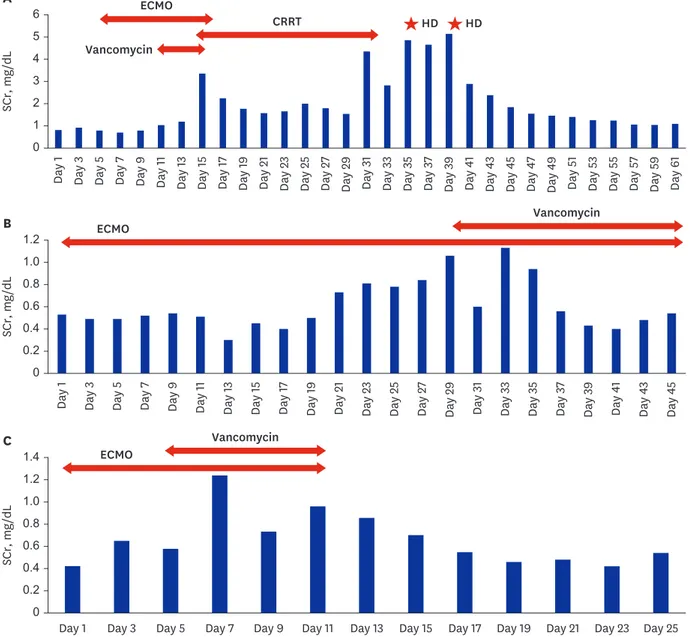
Mechanical ventilation was performed the next day, and ECMO was commenced 4 days later and maintained for 17 days. Blood cultures were performed when fever was identified, and vancomycin was added on the 10th day of hospitalization. Before using vancomycin, his SCr level was 1.0 mg/dL, but his renal function worsened after using vancomycin. His SCr gradually deteriorated to 1.19 mg/dL after 2days, 2.26 mg/dL after 3 days, and 3.35 mg/dL (peak) after 4 days of using vancomycin. The urine output decreased too (< 400 mL/day), so vancomycin was discontinued and continuous renal replacement therapy (CRRT) was started on day 14 of hospitalization and maintained for 18 days. The patient's laboratory data were closely followed up after CRRT was discontinued. The urine output was well maintained, but his SCr was continuously elevated (peak SCr: 5.14 mg/dL) and hemodialysis was performed twice. Afterwards, the SCr values improved and remained in the normal range without hemodialysis. The last recorded SCr value for this patient was 1.09 mg/dL.
The Second patient, a 78-year-old-woman, had hypertension as underlying disease and was on medication. Although she had no history of DM, her serum glucose was over 200 mg/dL at the time of visit. The HbA1c level measured after admission was 6.2%. On the first day of hospitalization, she was intubated and mechanical ventilation was maintained. An inotropic agent (norepinephrine) was used as blood pressure decreased. ECMO was commenced 1 day after the admission. Initial SCr was 0.53 mg/dL upon hospitalization. Piperacillin/
tazobactam and lopinavir/ritonavir were used from the first day of hospitalization.
Piperacillin/tazobactam was maintained for 4 weeks, and lopinavir/ritonavir was used for 9 days. Trimethoprim/sulfamethoxazole was used after 17 days of admission, and vancomycin was used after 29 days of admission. The SCr level before using vancomycin was 0.60 mg/
dL, SCr 0.61 mg/dL 1 day after, SCr 1.10 2 days after, and SCr 1.13 mg/dL (peak) after 3 days of vancomycin use indicated AKI (stage 1). Since then, though vancomycin was used continuously, SCr improved to baseline Cr level. The last recorded SCr value was 0.54 mg/dL.
Table 3. The characteristics of all the analyzed patients
Characteristics Values
Age, yr 45.6 ± 18.5
Male, sex 35 (53.0)
SCr, mg/dL 0.65 ± 0.18
Initial eGFR (CKD-EPI), mL/min/1.73 m2 113.1 ± 17.4 Comorbidities (n = 66)
Diabetes 10 (15.2)
Hypertension 12 (18.2)
Dyslipidemia 5 (7.6)
Cardiovascular disease 2 (3.0)
Data are presented as mean ± standard deviation or number (%).
SCr = serum creatinine, eGFR = estimated glomerular filtration rate, CKD-EPI = chronic kidney disease-epidemiology collaboration.
The third patient, a 64-year-old-woman, had surgery for thyroid cancer 8 years ago and had no other underlying disease. Her SCr was 0.41 mg/dL at the time of admission. Immediately after admission, the patient was intubated, after which mechanical ventilation and ECMO were performed. Mechanical ventilation was maintained for 12 days, and ECMO was maintained for 11 days. During hospitalization, the patient took lopinavir/ritonavir for 12 days. Vancomycin was commenced 4 days after admission. SCr level before vancomycin use was 0.56 mg/dL, SCr a day after the commencement of vancomycin use was 1.11 mg/dL, and SCr 2 days after the commencement of vancomycin was 1.20 mg/dL (peak); indicating AKI (stage 2). After that, although vancomycin was maintained for one week, SCr improved to baseline Cr level. The last recorded SCr level was 0.54 mg/dL. The three patients' laboratory data and clinical events are shown in Fig. 1.
SCr, mg/dL
0 1 3 6
4
2 5
Day 1 Day 3 Day 5 Day 7 Day 9 Day 11 Day 13 Day 15 Day 17 Day 19 Day 21 Day 23 Day 25 Day 27 Day 29 Day 31 Day 33 Day 35 Day 37 Day 39 Day 41 Day 43 Day 45 Day 47 Day 49 Day 51 Day 53 Day 55 Day 57 Day 59 Day 61
A ECMO
CRRT Vancomycin
HD HD
Day 1 Day 3 Day 5 Day 7 Day 9 Day 11 Day 13 Day 15 Day 17 Day 19 Day 21 Day 23 Day 25 Day 27 Day 29 Day 31 Day 33 Day 35 Day 37 Day 39 Day 41 Day 43 Day 45
B ECMO Vancomycin
SCr, mg/dL
0 0.2 0.6 1.2
0.8
0.4 1.0
C
SCr, mg/dL
0 0.2 0.6 1.2
0.8 1.4
0.4 1.0
Day 1 Day 3 Day 5 Day 7 Day 9 Day 11 Day 13 Day 15 Day 17 Day 19 Day 21 Day 23 Day 25 Vancomycin
ECMO
Fig. 1. Changes in SCr and clinical events in three patients with acute kidney injury. (A) Sixty-two-year-old-man. (B) Seventy-eight-year-old-woman. (C) Sixty- four-year-old-woman.
Proteinuria and albuminuria
In all 66 patients, routine urinalysis with a dipstick test was performed at the time of or during hospitalization. Trace and + albumin on a dipstick test was found in nine (13.6%) patients (trace in one patient, 1+ in two patients, and 2+ in six patients).
All patients were randomly tested for urine PCR and ACR more than once. In 20 (30.3%) out of 66 patients, urine PCR was higher than the normal range, 11 (16.7%) had severely increased proteinuria, and 9 (13.6%) had moderately increased proteinuria. Of the 11 patients with severely increased proteinuria, 10 had increased albuminuria, and one patient had no albuminuria. None of the nine patients with moderately increased proteinuria had albuminuria. Ten of 66 patients showed hematuria with red blood cell (RBC) counts > 3/
HPF on urine microscopy.13 Eight of them were patients with severely increased proteinuria, and two were young women whose hematuria was not clinically meaningful due to the overlapping menstrual period.
The recovery of 20 patients with elevated urine PCR was compared. For patients with moderately increased proteinuria, on follow-up, six out of nine had not improved, and three had recovered to a normal range. For patients with severely increased proteinuria, on follow- up, 1 out of 11 had not improved, 4 had improved (a reduction of over 50% compared with their highest urine PCR levels), and 6 had recovered to a normal range.
Comparison of kidney damage according to COVID-19 severity
Among COVID-19 patients, the clinical classification was done according to the National Health Commission of China guidelines (version 7) into mild, moderate, and severe cases.14 There were 21 mild cases, 37 moderate cases, and 8 severe cases. A total of three patients with AKI were included in the severe cases (P = 0.001). There was a high frequency of both the urine PCR and ACR in the severe cases, and proteinuria and albuminuria were also more severe in the severe COVID-19 group (P < 0.001) (Table 4).
No patients died of COVID-19 during the study period.
DISCUSSION
The initial step of SARS-CoV-2 infection is its entry into the human cells. SARS-CoV-2 and SARS-CoV share a common ancestor resembling the bat coronavirus HKU9-1.15 These Table 4. AKI and urine PCR and ACR analysis according to the severity of coronavirus disease 2019
Variables Mild (n = 21) Moderate (n = 37) Severe (n = 8) P value
AKI (n = 3, 4.5%) 0 (0.0) 0 (0.0) 3 (37.5) 0.001a
Albuminuria dipstick (n = 9, 13.6%) 0 (0.0) 2 (5.4) 7 (87.5) < 0.001a
PCR, mg/g (n = 20) < 0.001a
< 150 18 (85.7) 28 (75.7) 0 (0.0)
150–500 3 (14.3) 6 (16.2) 0 (0.0)
> 500 0 (0.0) 3 (8.1) 8 (100)
ACR, mg/g (n = 10) < 0.001a
< 30 21 (100) 34 (91.9) 1 (12.5)
30–300 0 (0.0) 2 (5.4) 2 (25.0)
> 300 0 (0.0) 1 (2.7) 5 (62.5)
Data are presented as number (%).
AKI = acute kidney injury, PCR = protein to creatinine ratio, ACR = albumin to creatinine ratio.
aUsing Fisher's exact test.
coronaviruses have very similar spike protein 3D structures considered to have a strong binding affinity to the human cell receptor, angiotensin-converting enzyme 2 (ACE2).
Therefore, cells expressing ACE2 may be target cells and are thus susceptible to SARS-CoV-2 infection; such cells include alveolar type II cells of the lungs.16 Thus, we believe that the pattern of ACE2 expression in different organs and tissues could reveal the potential risk for SARS-CoV-2 infection because the target cells expressing ACE2 may permit the entry, multiplication, spread, and pathogenesis of coronavirus. Previously, the RNA and protein expressions of ACE2 were investigated using bulk samples from the heart, lungs, kidneys, and other organs.17 Therefore, SARS-CoV-2 can affect not only the lungs but also other organs, as well as cause organ failure in other organs, including the kidney.
Chu et al.18 reported that of the 536 SARS patients analyzed, 36 (6.7%) had AKI, and in a study that analyzed 30 patients with Middle East respiratory syndrome coronavirus (MERS- CoV) infection, AKI occurred in eight (26.7%).19 In our study, 3 (4.5%) out of 66 patients had AKI, and they all recovered to normal SCr levels. Compared with previous human coronaviruses, such as MERS-CoV and SARS-CoV, SARS-CoV-2 infections were found to have a lower incidence of AKI. This is similar to the results of the analysis of 119 patients who were recently reported to have COVID-19 infection (AKI occurred in 0 out of 116 patients).20 In a meta-analysis of COVID-19 patients reported by Hu et al.,21 the results were similar to those of AKI occurring in 2.1% of patients.
In 20 patients, the urine PCR increased, and in 10 patients, the urine ACR increased. When the urine ACR is normal and the urine PCR is increased, renal tubular damage can be estimated in relation to infection rather than glomerular disease or glomerular damage.22,23 Compared with a previous study on MERS-CoV infection,19 SARS-CoV-2 infection exhibited less proteinuria (30.3%), suggesting that AKI and renal tubular damage caused by SARS- CoV-2 is less severe compared with previous coronavirus (MERS-CoV) infections (proteinuria occurred in 60% of patients). Our study revealed results similar to those reported recently on SARS-CoV-2 infection. In a study recently published by Wang et al.,20 dipstick tests were conducted on 111 SARS-CoV-2-infected patients (except for those with CKD), and 8 (7.2%) patients had trace or + albumin. This result is similar to that of the 9 (13.6%) out of 66 patients, in our study, whose dipstick tests revealed trace or + albumin.
This study had some limitations. The study population was selected from a single institution.
All three patients with AKI underwent mechanical ventilation and ECMO, and it was difficult to distinguish whether AKI was caused by SARS-CoV-2 infection or was associated with treatments, such as ECMO and vancomycin.24,25 Therefore, it is possible that the incidence of AKI related to COVID-19 was overestimated. In fact, all three patients used both ECMO and vancomycin, and AKI developed after vancomycin use.26 Therefore, it is thought that AKI is more likely to be associated with the treatment than with SARS-CoV-2 infection, and the likelihood of AKI being associated with SARS-CoV-2 infection may be lower. In addition, when tubular damage, namely, acute tubular necrosis, occurred, a follow-up of more than 2–4 weeks after improvement of infection was considered sufficient27,28; however, due to insufficient follow-up, it was difficult to evaluate whether there was an improvement in proteinuria. Finally, it was difficult to tell whether it was proteinuria caused by kidney damage or functional proteinuria caused by fever and infection. In this study, tests for hematuria such as RBC count and dysmorphic RBC were not performed. However, hematuria was also found in eight out of 11 patients with severely increased proteinuria. As such, it can be assumed that the possibility of the proteinuria being due to kidney
In conclusion, COVID-19, which is caused by SARS-CoV-2 infection, is thought to have less effect on the kidneys than the lungs, where it leads to rapidly progressing lung lesions. In our study, there was a lower percentage of patients with AKI (4.5%) and moderately to severely increased proteinuria (30.3%) than in previous human coronavirus infections. Compared with the mortality rates of MERS-CoV and SARS-CoV infections, which are 34.4% and 9.6%, respectively, the mortality rate of SARS-CoV-2 infection is lower, which may be due to the less organ dysfunction it causes overall despite its effect on the lungs. Moreover, the highly infectious nature of SARS-CoV-2 infection, compared with MERS-CoV and SARS- CoV infections, and the rapid progression of lung lesions it causes make future research on vaccines and therapeutics of utmost importance.
REFERENCES
1. Naicker S, Yang CW, Hwang SJ, Liu BC, Chen JH, Jha V. The novel coronavirus 2019 epidemic and kidneys.
Kidney Int 2020;97(5):824-8.
PUBMED | CROSSREF
2. Huang C, Wang Y, Li X, Ren L, Zhao J, Hu Y, et al. Clinical features of patients infected with 2019 novel coronavirus in Wuhan, China. Lancet 2020;395(10223):497-506.
PUBMED | CROSSREF
3. Li Q, Guan X, Wu P, Wang X, Zhou L, Tong Y, et al. Early transmission dynamics in Wuhan, China, of novel coronavirus-infected pneumonia. N Engl J Med 2020;382(13):1199-207.
PUBMED | CROSSREF
4. Phan LT, Nguyen TV, Luong QC, Nguyen TV, Nguyen HT, Le HQ, et al. Importation and human-to-human transmission of a novel coronavirus in Vietnam. N Engl J Med 2020;382(9):872-4.
PUBMED | CROSSREF
5. Holshue ML, DeBolt C, Lindquist S, Lofy KH, Wiesman J, Bruce H, et al. First case of 2019 novel coronavirus in the United States. N Engl J Med 2020;382(10):929-36.
PUBMED | CROSSREF
6. Giovanetti M, Benvenuto D, Angeletti S, Ciccozzi M. The first two cases of 2019-nCoV in Italy: where they come from? J Med Virol 2020;92(5):518-21.
PUBMED | CROSSREF
7. World Health Organization. Statement on the second meeting of the international health regulations (2005) emergency committee regarding the outbreak of novel coronavirus (2019-nCoV). https://www.
who.int/news-room/detail/30-01-2020-statement-on-the-second-meeting-of-the-international-health- regulations-(2005)-emergency-committee-regarding-the-outbreak-of-novel-coronavirus-(2019-ncov).
Update 2020. Accessed January 30, 2020.
8. World Health Organization. WHO director-general's remarks at the media briefing on 2019-nCoV on 11 February 2020. https://www.who.int/dg/speeches/detail/who-director-general-s-remarks-at-the-media- briefing-on-2019-ncov-on-11-february-2020. Update 2020. Accessed February 11, 2020.
9. Ministry of Health & Welfare (KR). COVID-19. http://ncov.mohw.go.kr. Updated 2020. Accessed April 25, 2020.
10. CoronaBoard. COVID-19 dashboard. https://coronaboard.kr. Updated 2020. Accessed April 25, 2020.
11. Kidney Disease: Improving Global Outcomes (KDIGO) Acute Kidney Injury Work Group. KDIGO clinical practice guideline for acute kidney injury. Kidney Int Suppl 2012;2(1):1-138.
12. Kidney Disease: Improving Global Outcomes (KDIGO) CKD Work Group. KDIGO 2012 clinical practice guideline for the evaluation and management of chronic kidney disease. Kidney Int Suppl 2013;3(1):1-150.
13. Davis R, Jones JS, Barocas DA, Castle EP, Lang EK, Leveillee RJ, et al. Diagnosis, evaluation and follow-up of asymptomatic microhematuria (AMH) in adults: AUA guideline. J Urol 2012;188(6 Suppl):2473-81.
PUBMED | CROSSREF
14. Wei PF. Diagnosis and treatment protocol for novel coronavirus pneumonia (trial version 7). Chin Med J (Engl) 2020;133(9):1087-95.
PUBMED | CROSSREF
15. Xu X, Chen P, Wang J, Feng J, Zhou H, Li X, et al. Evolution of the novel coronavirus from the ongoing Wuhan outbreak and modeling of its spike protein for risk of human transmission. Sci China Life Sci 2020;63(3):457-60.
PUBMED | CROSSREF
16. Zhou P, Yang XL, Wang XG, Hu B, Zhang L, Zhang W, et al. A pneumonia outbreak associated with a new coronavirus of probable bat origin. Nature 2020;579(7798):270-3.
PUBMED | CROSSREF
17. Zou X, Chen K, Zou J, Han P, Hao J, Han Z. Single-cell RNA-seq data analysis on the receptor ACE2 expression reveals the potential risk of different human organs vulnerable to 2019-nCoV infection. Front Med 2020;14(2):185-92.
PUBMED | CROSSREF
18. Chu KH, Tsang WK, Tang CS, Lam MF, Lai FM, To KF, et al. Acute renal impairment in coronavirus- associated severe acute respiratory syndrome. Kidney Int 2005;67(2):698-705.
PUBMED | CROSSREF
19. Cha RH, Joh JS, Jeong I, Lee JY, Shin HS, Kim G, et al. Renal complications and their prognosis in Korean patients with Middle East respiratory syndrome-coronavirus from the central MERS-CoV designated hospital. J Korean Med Sci 2015;30(12):1807-14.
PUBMED | CROSSREF
20. Wang L, Li X, Chen H, Yan S, Li D, Li Y, et al. Coronavirus disease 19 infection does not result in acute kidney injury: an analysis of 116 hospitalized patients from Wuhan, China. Am J Nephrol 2020;51(5):343-8.
PUBMED | CROSSREF
21. Hu Y, Sun J, Dai Z, Deng H, Li X, Huang Q, et al. Prevalence and severity of corona virus disease 2019 (COVID-19): a systematic review and meta-analysis. J Clin Virol 2020;127:104371.
PUBMED | CROSSREF
22. Birn H, Christensen EI. Renal albumin absorption in physiology and pathology. Kidney Int 2006;69(3):440-9.
PUBMED | CROSSREF
23. D'Amico G, Bazzi C. Pathophysiology of proteinuria. Kidney Int 2003;63(3):809-25.
PUBMED | CROSSREF
24. Vinclair C, De Montmollin E, Sonneville R, Reuter J, Lebut J, Cally R, et al. Factors associated with major adverse kidney events in patients who underwent veno-arterial extracorporeal membrane oxygenation.
Ann Intensive Care 2020;10(1):44.
PUBMED | CROSSREF
25. Hsu CK, Wu IW, Chen YT, Tsai TY, Tsai FC, Fang JT, et al. Acute kidney disease stage predicts outcome of patients on extracorporeal membrane oxygenation support. PLoS One 2020;15(4):e0231505.
PUBMED | CROSSREF
26. Sinha Ray A, Haikal A, Hammoud KA, Yu AS. Vancomycin and the risk of AKI: a systematic review and meta-analysis. Clin J Am Soc Nephrol 2016;11(12):2132-40.
PUBMED | CROSSREF
27. Deurdulian C, Tchelepi H. Imaging-based monitoring of the renal graft. In: Orlando G, Remuzzi G, Williams DF, editors. Kidney Transplantation, Bioengineering, and Regeneration. London: Academic Press; 2017, 373-402.
28. Cereda M, Neligan P, Shashaty MGS, Horak J. Renal diseases. In: Fleisher LA, editor. Anesthesia and Uncommon Disease. 6th ed. Philadelphia, PA: Saunders; 2012, 225-50.