R E V I E W Open Access
Being a front-line dentist during the Covid- 19 pandemic: a literature review
Hamid Reza Fallahi
1,2, Seied Omid Keyhan
3,4,5*, Dana Zandian
1,2, Seong-Gon Kim
6,7*and Behzad Cheshmi
8Abstract
Coronavirus is an enveloped virus with positive-sense single-stranded RNA. Coronavirus infection in humans mainly affects the upper respiratory tract and to a lesser extent the gastrointestinal tract. Clinical symptoms of coronavirus infections can range from relatively mild (similar to the common cold) to severe (bronchitis, pneumonia, and renal involvement). The disease caused by the 2019 novel coronavirus (2019-nCoV) was called Covid-19 by the World Health Organization in February 2020. Face-to-face communication and consistent exposure to body fluids such as blood and saliva predispose dental care workers at serious risk for 2019-nCoV infection. As demonstrated by the recent coronavirus outbreak, information is not enough. During dental practice, blood and saliva can be scattered.
Accordingly, dental practice can be a potential risk for dental staff, and there is a high risk of cross-infection. This article addresses all information collected to date on the virus, in accordance with the guidelines of international health care institutions, and provides a comprehensive protocol for managing possible exposure to patients or those suspected of having coronavirus.
Keywords: Coronavirus, Covid-19, 2019-nCoV, Dental care, Dentistry
Background
Since the first reported case in Wuhan, China, in December 2019, coronavirus disease-19 (Covid-19) has widely spread to Japan, Korea, Iran, and many European countries [1].
The World Health Organization (WHO) declared a pan- demic in March 2020. As saliva is a main tool of spread, dentists are in danger of contracting Covid-19. Although the exact nature of this disease must be clarified in detailed studies, current knowledge of coronavirus infection should be shared without any restrictions.
This article was written by an Iranian team of oral and maxillofacial surgeons. As Iran has many Covid-19 patients, they have significant experience with this disease. Maxillo- facial Plastic and Reconstructive Surgeryis an open-access journal, and this type of important information can be shared via our publication platform without restrictions.
Coronaviruses
Coronaviruses are enveloped viruses with a positive- sense single-stranded RNA genome. Their helical sym- metry nucleocapsid is approximately 26–32 kb in size, making it the largest investigated genome among RNA viruses [2,3]. Coronaviruses have a fundamental resem- blance in their organization and genome expression [4].
Previously, it was thought that coronaviruses only cause enzootic infections in a number of animals, including certain birds and mammals, but recent findings indicate that a variety of these viruses, including antigenic groups 1 (229E and NL63), antigenic groups 2 (OC43), and HKU1, can infect humans [5,6]. These viruses often lead to upper respiratory tract infection, frequently resulting in common cold symptoms. Three specific strains of these viruses that are of zoonotic origin, including severe acute respiratory syndrome coronavirus (SARS-CoV), Middle East respiratory syndrome coronavirus (MERS- CoV), and 2019 novel coronavirus (2019-nCoV), have recently caused lethal infections in humans [4,6,7].
Coronavirus infections in humans mainly affect the upper respiratory tract and to a lesser extent the gastrointestinal
© The Author(s). 2020 Open Access This article is licensed under a Creative Commons Attribution 4.0 International License, which permits use, sharing, adaptation, distribution and reproduction in any medium or format, as long as you give appropriate credit to the original author(s) and the source, provide a link to the Creative Commons licence, and indicate if changes were made. The images or other third party material in this article are included in the article's Creative Commons licence, unless indicated otherwise in a credit line to the material. If material is not included in the article's Creative Commons licence and your intended use is not permitted by statutory regulation or exceeds the permitted use, you will need to obtain permission directly from the copyright holder. To view a copy of this licence, visithttp://creativecommons.org/licenses/by/4.0/.
* Correspondence:[email protected];[email protected]
3CMFRC, National Advance Center for Craniomaxillofacial Reconstruction, Tehran, Iran
6Department of Oral and Maxillofacial Surgery, College of Dentistry, Gangneung-Wonju National University, Gangneung 28644, Republic of Korea Full list of author information is available at the end of the article
tract. Manifestations of coronavirus infections can range from relatively mild (similar to the common cold) to severe (bronchitis, pneumonia, and renal in- volvement) [8] (Table 1).
Pathogenesis
The ability to infect humans is mainly due to the infection of peridomestic animals, which are considered intermedi- ate hosts, nurturing recombination and mutation events as well as the development of genetic diversity among cor- onaviruses [29]. Studies have suggested that the spike glycoprotein (S glycoprotein) plays an important role in host range restriction by attaching virions to the host cell membrane [30]. Generally, coronaviruses primarily repli- cate in the respiratory and intestinal epithelial cells and subsequently cause cytopathic alterations [31].
Covid-19
Since December 2019, numerous unexplained cases of pneumonia have been reported in China. The disease caused by 2019-nCoV was called Covid-19 by the WHO in February 2020 [32]. Limiting the exposure of suspi- cious cases to the rest of society could be an effective strategy in the early outbreak phases. However, the sub- sequent worldwide virus spread and person-to-person transmission made the situation more complex and un- controllable [33].
No detailed studies have been conducted to expound the pathogenicity of 2019-nCoV on a molecular scale.
However, exploratory data established via whole-genome sequencing and subsequent bioinformatics analyses re- vealed that 2019-nCoV is phylogenetically related to SARS-CoV that was isolated for the first time in Chinese horseshoe bats between 2015 and 2017 [34–36].
Clinical manifestations
To a large extent, the clinical similarities of 2019-nCoV infection with SARS-CoV infection are substantial. The incubation period of 2019-nCov has been estimated to
be 1–14 days, and it has been shown that asymptomatic individuals may also be involved in the spread of this virus [26–28]. Since the possibility of the transmission from asymptomatic carriers has been raised currently, checking body temperature only may not be enough to screen asymptomatic carriers. According to a recent report, temperature-based screening in the airport can detect only 46% of 2019-nCoV carriers and the others were found during the self-isolation period after immi- gration [29]. To suppress the disease spread, a wide range of laboratory tests for immigrants and general population seems to be necessary. However, the infec- tion rate from asymptomatic carriers has not been clari- fied until now. The primary non-specific reported symptoms of 2019-nCoV infection at the prodromal phase are malaise, fever, and dry cough. The most com- monly reported signs and symptoms are fever (98%), cough (76%), dyspnea (55%), and myalgia or fatigue (44%) [25, 26]. Unlike patients with other human cor- onavirus infections (such as SARS-CoV), upper respira- tory tract and intestinal manifestations such as sore throat, rhinorrhea, and diarrhea in those with 2019- nCoV infection are infrequent [15,25,26] (Fig.1).
Patient characteristics
The patient mean age is generally between 49 and 61 years. Studies have shown that males are more likely to have this infection [25, 26, 37]. The lack of serious illness in youngsters is a characteristic of SARS-CoV infection, which is similarly observed in 2019-nCoV infection [32]. Increased exposures to 2019-nCoV due to occupational requirements, for instance health care workers, maybe another factor contributing to the higher risk of infection.
Diagnosis
Following the outbreak, the full 2019-nCoV genomic sequence was released in public databases [38]. This facilitates the way for further PCR assays for virus
Table 1 Comparison of clinical symptoms and incubation time of human coronaviruses [4]
Human coronaviruses Clinical symptoms Incubation period Refs.
229E General malaise, headache, nasal discharge, sneezing, sore throat, fever and cough
(10–20% of patients) 2–5 days [9–11]
OC43 General malaise, headache, nasal discharge, sneezing, sore throat, fever and cough
(10–20% of patients) 2–5 days [9,11–13]
NL63 Cough, rhinorrhea, tachypnea, fever, hypoxia, obstructive laryngitis (croup) 2–4 days [14–19]
HKU1 Fever, running nose, cough, dyspnea 2–4 days [20–22]
SARS-CoV Fever, myalgia, headache, malaise, chills, non-productive cough, dyspnea, respiratory
distress, diarrhea (30–40% of patients) 2–11 days [14–19]
MERS-CoV Fever, cough, chills, sore throat, myalgia, arthralgia, dyspnea, pneumonia, diarrhea and vomiting (one third of patients), acute renal impairment
2–13 days [15,23,24]
2019-nCoV Malaise, fever, dry cough, cough, dyspnea, myalgia, fatigue 1–14 days [25–28]
detection. The WHO recommendations for outpatient cases and patients with more critical conditions respect- ively include rapid collection and nucleic acid amplification testing (NAAT) of respiratory samples including nasopha- ryngeal and oropharyngeal swabs as well as sputum and/or endotracheal aspirate or bronchoalveolar lavage [39].
Table 2 presents recommended instructions that all practitioners in the field of dental care, including dentists, assistants, and others, should consider when treating pa- tients or those suspected of having coronavirus.
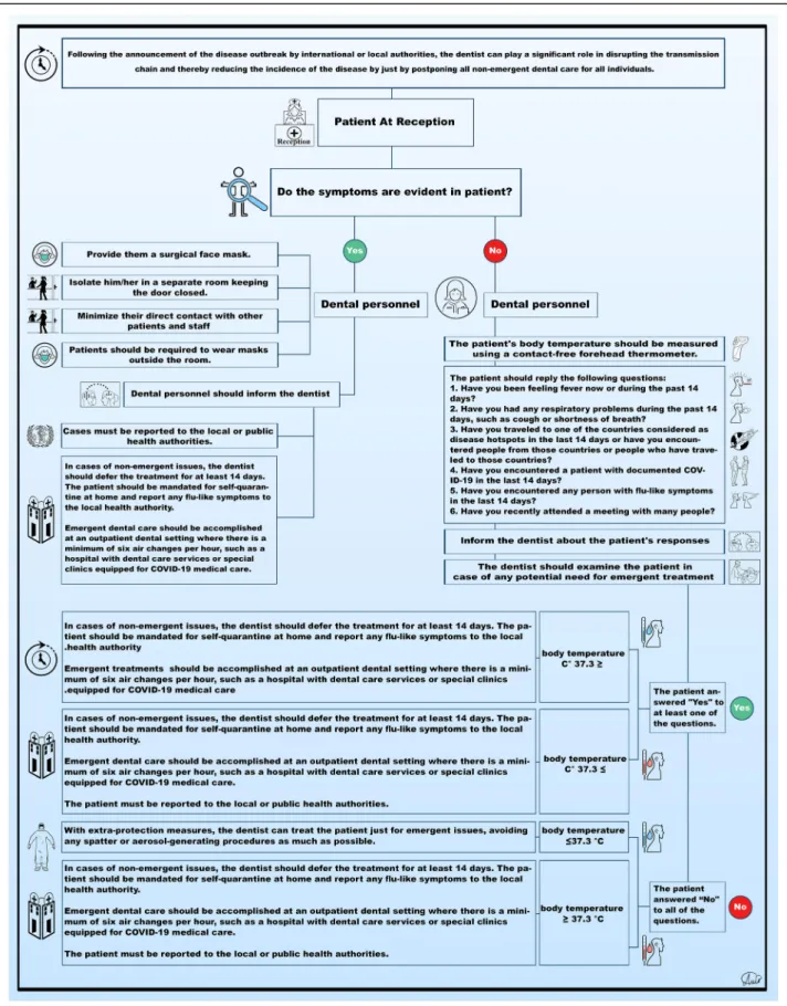
Protocol
Figure 2 shows a protocol that can organize our ap- proach to managing suspected or infected patients. The purpose of this protocol is to protect the entire dental care team, prevent any cross-infection in the office, in- form health authorities active in the field of controlling and managing the disease, and ultimately provide the optimal medical and dental care for patients affected by the virus according to the CDC and the ADA guidelines.
Transmission dynamics
The two main routes known for 2019-nCoV transmis- sion include (1) direct transmission (through coughing, sneezing, and inhalation of droplets) and (2) contact trans- mission (through contact with nasal, oral, and ocular mu- cosa) [43]. Typical clinical manifestations of Covid-2019 do not comprise ocular symptoms. However, conjunctival sample analysis has revealed that the transmission of 2019-nCoV is not limited to the respiratory tract route [26], but ocular exposure is also an effective method of virus transmission [44]. Moreover, studies have revealed that via direct/indirect contact or course and/or droplets, respiratory viruses such as 2019-nCoV may be trans- mitted from human to human. Studies have also shown direct and indirect transmission of 2019-nCoV through saliva [45].
For a comprehensive understanding of the transmission dynamics of 2019-nCoV, it is also important to know that this virus is also transmissible through asymptomatic pa- tients [34]. The remarkable feature of 2019-nCoV is that its RNA is detectable via quantitative reverse transcriptase polymerase chain reaction (qRT-PCR) in stool samples after the first week of infection [46]. However, the aerosol and fecal-oral transmission routes, which carry more pub- lic concern, still need further investigation and confirm- ation [47] (Fig.3).
Transmission dynamics in dentistry practice
New evidence suggests that 2019-nCoV may be trans- mitted directly from human to human via respiratory secretion containing droplets [44, 48]. Virus transmis- sion through contact and fomites is also likely [44, 48].
To et al. [44] reported that using the viral culture
Fig. 1 Signs and symptoms of Covid-19
method, they detected 2019-nCoV in infected individ- uals’ saliva samples. In addition, 2019-nCov invades the cells through the angiotensin-converting enzyme 2 (ACE2) receptor in the same way as the SARS corona- virus [49]. Since 2019-nCoV effectively uses ACE2 receptor for cell invasion, it can promote human-to- human transmission [50]. ACE2+ cells are abundantly present all over the respiratory tract. ACE2+ epithelial cells present in the salivary glands were considered one of the main targets of SARS coronavirus infection. Simi- larly, 2019-nCoV may also use the same mechanism to induce infection, although definitive judgment regarding this issue needs further study [51].
Due to close face-to-face contact with patients and fre- quent utilization of sharp devices, dental personnel are repeatedly exposed to respiratory tract secretions, blood, saliva, and other contaminated body fluids and are al- ways at risk for 2019-nCoV infection. 2019-nCoV trans- mission in dental settings occurs through four major routes: (1) direct exposure to respiratory secretions con- taining droplets, blood, saliva, or other patient materials;
(2) indirect contact with contaminated surfaces and/or instruments; (3) inhalation of suspending airborne vi- ruses; and (4) mucosal (nasal, oral, and conjunctival) contact with infection-containing droplets and aerosols
that are propelled by coughing and talking without a mask [51–55] (Fig.4).
The most important concern in dental clinics is the transmission of 2019-nCoV via droplets and aerosol be- cause, despite all of the precautions taken, it is almost impossible to reduce droplet and aerosol production to zero during dental procedures [54]. Dental handpieces utilize high-speed gas to rotate with running water, which leads to the generation of a considerable amount of droplets and aerosol mixed with patients’ saliva and/
or blood [55]. Therefore, it can be deduced that 2019- nCoV is capable of transmitting through dental practice;
this transmission can be from patients to clinic staff or other patients at the clinic [56].
Research has shown that coronaviruses can remain on metal, glass, and plastic surfaces for several days [52,57].
Therefore, as surfaces in dental clinics serve as venues for droplets and aerosol mixed with patients’ saliva and/
or blood, they can effectively help spread infection. Cor- onaviruses can actively maintain their virulence at room temperature from 2 h up to 9 days. Their activity at 50%
humidity was significantly higher than 30%. Therefore, in the dental environment, it seems that keeping surfaces clean and dry will play a significant role in preventing 2019-nCoV transmission [52].
Table 2 Standard precautions based on CDC and ADA guidelines for dentists on the coronavirus disease [40–42]
Postponing Following the announcement of disease outbreak by international or local authorities, dentists can play a significant role in disrupting the transmission chain, thereby reducing the incidence of the disease by simply postponing all non-emergency dental care for all patients.
Where to treat All dental care should be provided in an outpatient dental setting with a minimum of six air changes per hour, such as a hospital with dental care services or customized clinics equipped for Covid-19 patients.
Symptoms and history Primary non-specific reported symptoms of 2019-nCoV infection at the prodromal phase are malaise, fever, and dry cough. The most commonly reported signs and symptoms are fever (98%), cough (76%), dyspnea (55%), and myalgia or fatigue (44%).
They also may have traveled to one of the countries considered disease hotspots in the prior 14 days or have encountered people from those countries or people who have traveled to those countries.
Some patients may be asymptomatic or have unexpected symptoms such as diarrhea.
How long? Since it is not possible to know the etiology of each patient’s illness, it is crucial to follow the guidelines and precautions at all times during the disease outbreak.
Preparations and arrangement
Be alert, identify patients with respiratory illnesses, and provide them a disposable surgical face mask. Isolate them in a room with the door closed. Limit their direct contact with others. Isolated patients must wear masks outside their room.
Isolate suspected patients before and during care to minimize their direct contact with other patients and staff and immediately report any cases to local and state public health authorities.
Transmission prevention consideration
To prevent 2019-nCoV transmission, dental practices should adhere to the infection control protocol, including hand hygiene, providing tissues and no-touch receptacles, and providing face masks for coughing patients.
Dental health care personnel should wear white coats, gowns, head caps, goggles, face shields, masks, latex gloves, and impermeable shoe covers to prevent exposure.
Disposable masks should be substituted between patients or even during treatment if they get wet.
Guidelines updates Since Covid-19 recommendations may change rapidly with increasing information about the disease, the ADA recommends checking for updates on the CDC’s coronavirus infection control web page for health care professionals.
Health care workers The CDC strongly recommends that all health care staff, including dentists and personnel, should receive the flu vaccine and that staff with influenza must not report to work.
Fig. 2 Protocol for the management of patients during the Covid-19 pandemic
Infection control
Since the fecal-oral route is considered one of the 2019- nCoV transmission routes, attention to hand hygiene before, during, and after dental practice is important. Den- tists should exercise extreme caution to avoid contact with their own facial mucosal surfaces including their eyes, mouth, and nose. Since transmission of airborne droplet is considered one of the main routes of infection spread,
application of personal protective equipment such as masks, protective goggles, gowns, helmet, gloves, caps, face shields, and shoe covers is strongly recommended for all health care personnel.
Covid-19 patients should not be treated in a regular dental care setting without special considerations. Unex- pected circumstances may occur when the dentist cannot delay treatment or refer the patient to the appropriate
Fig. 3 Common transmission routes of the 2019-nCoV
Fig. 4 2019-nCoV transmission dynamics in dental care settings
medical institution. Under such circumstances, special protective clothing such as hazardous materials (hazmat) suits are required. If hazmat suits are not available, white coats, gowns, head caps, protective eyewear, face shields, masks, latex gloves, and virus-proof shoe covers should be used [56].
Mouth rinses
The effect of chlorhexidine, which is commonly used for pre-procedural mouth washing in dental practice, has not yet been demonstrated to be capable of eliminating 2019- nCoV. However, oxidative agents containing mouth rinses with 1% hydrogen peroxide or 0.2% povidone-iodine are recommended. Pre-procedural use of mouthwash, espe- cially in cases of inability to use a rubber dam, can signifi- cantly reduce the microbial load of oral cavity fluids [58].
Rubber dam isolation
Using rubber dams due to the creation of a barrier in the oral cavity effectively reduces the generation of drop- lets and aerosol mixed with patient saliva and/or blood in 1 m diameter of the surgical field by 70% [59]. Follow- ing the placement of the dam, extra high-volume suction is also required for maximum prevention of aerosol and spatter from spreading [60]. If it is not possible to use rubber dams for any reason, manual tools such as Cari- solvs or hand scalers are preferable.
Anti-retraction handpiece
Throughout the COVID-19 pandemic, the use of any dental handpieces that do not have an anti-retraction function should be avoided. For emergency treatment, anti-retraction handpieces designed with anti-retractive valves can play an effective role in preventing the diffu- sion and dispersion of droplets and aerosol [60,61].
Appropriate disinfectants
Since there is still little information available regarding 2019-nCoV, relatively similar genetic features between 2019-nCoV and SARS-CoV indicate that the novel cor- onavirus can be vulnerable to disinfectants such as sodium hypochlorite (1000 ppm or 0.1% for surfaces and 10,000 ppm or 1% for blood spills), 0.5% hydrogen per- oxide, 62–71% ethanol, and phenolic and quaternary ammonium compounds if utilized in accordance with the manufacturer’s instructions. Studies show that other biocidal agents such as 0.05–0.2% benzalkonium chlor- ide or 0.02% chlorhexidine digluconate probably have lower efficiency. In addition to the type of disinfectant, paying attention to other factors such as the duration of use, dilution rate, and especially the expiration time fol- lowing the preparation of the solution according to the manufacturer’s instructions is also crucial.
Management of medical waste
Prior to any inappropriate accumulation, dental office waste should be routinely transported to the institution’s temporary storage facility. Reusable tools and equipment must be properly pre-treated, cleaned, sterilized, and properly stored until the next use. Dental waste resulting from the treatment of suspected or confirmed 2019- nCoV patients is considered medically infectious waste that must be strictly disposed of in accordance with the official instructions using double-layer yellow medical waste package bags and“gooseneck” ligation.
Conclusion
Following the announcement of the disease outbreak by international or local authorities, dentists can play a sig- nificant role in disrupting the transmission chain, thereby reducing the incidence of disease by simply postponing all non-emergency dental care for all patients. Dental profes- sionals must be fully aware of 2019-nCoV spreading mo- dalities, how to identify patients with this infection, and, most importantly, self-protection considerations. The ef- fect of chlorhexidine, which is commonly used for pre- procedural mouth washing in dental practice, has not yet been demonstrated to be capable of eliminating 2019- nCoV. However, the prescription of oxidative agents con- taining mouth rinses such as 1% hydrogen peroxide or 0.2% povidone is recommended. A higher rate of virus ex- posure because of occupational commitments in health care workers is considered a key factor associated with the increased risk of infection.
Abbreviations
Covid-19:Coronavirus disease-19; WHO: World Health Organization; SARS- CoV: Severe acute respiratory syndrome coronavirus; MERS-CoV: Middle East respiratory syndrome coronavirus; 2019-nCoV: 2019 novel coronavirus; qRT- PCR: Quantitative reverse transcriptase polymerase chain reaction;
ACE2: Angiotensin-converting enzyme 2; NAAT: Nucleic acid amplification testing
Acknowledgements
Prof. Esmaeil Yazdi passed away due to coronavirus infection few days ago.
He was one of the Iranian Society of OMFS founder 60 years ago. The authors send sincere condolence to his family and colleagues. The authors also appreciate his great contribution on the progress of maxillofacial surgery.
Authors’ contributions
The article was written by HRF and BC. DZ collected the data. Illustrations were drawn by SOK and BC. SOK and KSG corrected the article and performed the critical review. The authors read and approved the final manuscript.
Funding Not applicable.
Availability of data and materials Not applicable.
Ethics approval and consent to participate Not applicable.
Consent for publication Not applicable.
Competing interests
The authors declare that the authors have no competing interests as defined by Nature Research or other interests that might be perceived to influence the results and/or discussion reported in this paper.
Author details
1School of Advanced Technologies in Medicine, Shahid Beheshti University of Medical Sciences, Tehran, Iran.2Dental Research Center, Research Institute of Dental Sciences, Shahid Beheshti University of Medical Sciences, Tehran, Iran.3CMFRC, National Advance Center for Craniomaxillofacial
Reconstruction, Tehran, Iran.4Craniomaxillofacial Research Center, Tehran University of Medical Sciences, Tehran, Iran.5Bahar building, Shahid Nuranian Alley, Saadat Abad, Isfahan, Iran.6Department of Oral and Maxillofacial Surgery, College of Dentistry, Gangneung-Wonju National University, Gangneung 28644, Republic of Korea.7Department of Oral and Maxillofacial Surgery, College of Dentistry, Gangneung-Wonju National University, 7 Jukhyun-gil, Gangneung 25457, Republic of Korea.8Faculty of Dentistry, Boroujerd Islamic Azad University, Boroujerd, Iran.
Received: 16 March 2020 Accepted: 2 April 2020
References
1. Lai CC, Liu YH, Wang CY, Wang YH, Hsueh SC, Yen MY et al Asymptomatic carrier state, acute respiratory disease, and pneumonia due to severe acute respiratory syndrome coronavirus 2 (SARS-CoV-2): facts and myths. J Microbiol Immunol Infecthttps://doi.org/10.1016/j.jmii.2020.02.012 2. Sexton NR, Smith EC, Blanc H, Vignuzzi M, Peersen OB, Denison MR (2016)
Homology-based identification of a mutation in the coronavirus RNA- dependent RNA polymerase that confers resistance to multiple mutagens. J Virol 90(16):7415–7428
3. Weiss SR, Navas-Martin S (2005) Coronavirus pathogenesis and the emerging pathogen severe acute respiratory syndrome coronavirus.
Microbiol Mol Biol Rev 69(4):635–664
4. Su S, Wong G, Shi W, Liu J, Lai AC, Zhou J et al (2016) Epidemiology, genetic recombination, and pathogenesis of coronaviruses. Trends Microbiol 24(6):490–502
5. Schoeman D, Fielding BC (2019) Coronavirus envelope protein: current knowledge. Virol J 16(1):69
6. World Health Organization (2020) Coronavirus.https://www.who.int/health- topics/coronavirus. .
7. Habibzadeh P, Stoneman EK (2020) The novel coronavirus: a bird’s eye view.
Int J Occup Environ Med 11(2):65–71
8. Wevers BA, van der Hoek L (2009) Recently discovered human coronaviruses. Clin Lab Med 29(4):715–724
9. Monto AS (1974) Medical reviews. Coronaviruses. Yale J Biol Med 47(4):234–251 10. Pene F, Merlat A, Vabret A, Rozenberg F, Buzyn A, Dreyfus F et al (2003)
Coronavirus 229E-related pneumonia in immunocompromised patients. Clin Infect Dis 37(7):929–932
11. Papa A, Papadimitriou E, de Souza Luna LK, Al Masri M, Souliou E, Eboriadou M et al (2007) Coronaviruses in children, Greece. Emerg Infect Dis 13(6):947–949 12. Cabeça TK, Granato C, Bellei N (2013) Epidemiological and clinical features
of human coronavirus infections among different subsets of patients.
Influenza Other Respir Viruses 7(6):1040–1047
13. Dijkman R, Jebbink MF, Koekkoek SM, Deijs M, Jónsdóttir HR, Molenkamp R et al (2013) Isolation and characterization of current human coronavirus strains in primary human epithelial cell cultures reveal differences in target cell tropism. J Virol 87(11):6081–6090
14. Peiris J, Lai S, Poon L, Guan Y, Yam L, Lim W et al (2003) Coronavirus as a possible cause of severe acute respiratory syndrome. Lancet 361(9366):
1319–1325
15. Assiri A, Al-Tawfiq JA, Al-Rabeeah AA, Al-Rabiah FA, Al-Hajjar S, Al-Barrak A et al (2013) Epidemiological, demographic, and clinical characteristics of 47 cases of Middle East respiratory syndrome coronavirus disease from Saudi Arabia: a descriptive study. Lancet Infect Dis 13(9):752–761
16. Ricketti A, Vemaleo J (2010) Severe acute respiratory syndrome (SARS).
Infect Dis Clin N Am 24(1):175–202
17. Gu J, Gong E, Zhang B, Zheng J, Gao Z, Zhong Y et al (2005) Multiple organ infection and the pathogenesis of SARS. J Exp Med 202(3):415–424 18. Tsang KW, Ho PL, Ooi GC, Yee WK, Wang T, Chan-Yeung M et al (2003) A
cluster of cases of severe acute respiratory syndrome in Hong Kong. N Engl J Med 348(20):1977–1985
19. Peiris JSM, Chu C-M, Cheng VC-C, Chan K, Hung I, Poon LL et al (2003) Clinical progression and viral load in a community outbreak of coronavirus-associated SARS pneumonia: a prospective study. Lancet 361(9371):1767–1772 20. Moës E, Vijgen L, Keyaerts E, Zlateva K, Li S, Maes P et al (2005) A novel
pancoronavirus RT-PCR assay: frequent detection of human coronavirus NL63 in children hospitalized with respiratory tract infections in Belgium.
BMC Infect Dis 5(1):6
21. Bastien N, Anderson K, Hart L, Caeseele PV, Brandt K, Milley D et al (2005) Human coronavirus NL63 infection in Canada. J Infect Dis 191(4):503–506 22. van der Hoek L, Sure K, Ihorst G, Stang A, Pyrc K, Jebbink MF et al (2005)
Croup is associated with the novel coronavirus NL63. PLoS Med 2(8):e240 23. Al-Tawfiq JA, Hinedi K, Ghandour J, Khairalla H, Musleh S, Ujayli A et al
(2014) Middle East respiratory syndrome coronavirus: a case-control study of hospitalized patients. Clin Infect Dis 59(2):160–165
24. Arabi YM, Arifi AA, Balkhy HH, Najm H, Aldawood AS, Ghabashi A et al (2014) Clinical course and outcomes of critically ill patients with Middle East respiratory syndrome coronavirus infection. Ann Intern Med 160(6):389–397 25. Chen N, Zhou M, Dong X, Qu J, Gong F, Han Y et al (2020) Epidemiological and clinical characteristics of 99 cases of 2019 novel coronavirus pneumonia in Wuhan, China: a descriptive study. Lancet 395(10223):507–513
26. Huang C, Wang Y, Li X, Ren L, Zhao J, Hu Y et al (2020) Clinical features of patients infected with 2019 novel coronavirus in Wuhan, China. Lancet 395(10223):497–506
27. Guan W, Ni Z, Hu Y, Liang W, Ou C, He J et al Clinical characteristics of coronavirus disease 2019 in China. N Engl J Medhttps://doi.org/10.1056/
NEJMoa2002032
28. Backer JA, Klinkenberg D, Wallinga J Incubation period of 2019 novel coronavirus (2019-nCoV) infections among travellers from Wuhan, China, 20–28 January 2020. Euro Surveillhttps://doi.org/10.2807/1560-7917.ES.2020.
25.5.2000062
29. Paules CI, Marston HD, Fauci AS (2020) Coronavirus infections—more than just the common cold. JAMA. 323(8):707–708
30. de Wilde AH, Snijder EJ, Kikkert M, van Hemert MJ (2017) Host factors in coronavirus replication. In: Roles of Host Gene and Non-coding RNA Expression in Virus Infection. Springer, pp 1–42
31. Fields BN, Knipe DM, Howley PM (1996) Fields virology. Lippincott-Raven 32. Sun P, Lu X, Xu C, Sun W, Pan B (2020) Understanding of COVID-19 based
on current evidence. J Med Virol
33. Rothe C, Schunk M, Sothmann P, Bretzel G, Froeschl G, Wallrauch C et al (2020) Transmission of 2019-nCoV infection from an asymptomatic contact in Germany. N Engl J Med
34. Chan JF-W, Yuan S, Kok K-H, To KK-W, Chu H, Yang J et al (2020) A familial cluster of pneumonia associated with the 2019 novel coronavirus indicating person-to-person transmission: a study of a family cluster. Lancet 395(10223):514–523
35. Hu D, Zhu C, Ai L, He T, Wang Y, Ye F et al (2018) Genomic characterization and infectivity of a novel SARS-like coronavirus in Chinese bats. Emerg Microbes Infect 7(1):1–10
36. Hu T, Li G, Zuo Y, Zhou X (2007) Risk of hepatitis B virus transmission via dental handpieces and evaluation of an antisuction device for prevention of transmission. Infect Control Hosp Epidemiol 28(1):80–82
37. Peiris JS, Yuen KY, Osterhaus AD, Stöhr K (2003) The severe acute respiratory syndrome. N Engl J Med 349(25):2431–2441
38. Cohen J (2020) Chinese researchers reveal draft genome of virus implicated in Wuhan pneumonia outbreak
39. Organization WH. Laboratory testing of 2019 novel coronavirus ( 2019- nCoV) in suspected human cases: interim guidance, 17 January 2020. 2020.
40. ADA releases coronavirus handout for dentists based on CDC guidelines:
American Dental Association; 2020. Available from:https://www.ada.org/en/
publications/ada-news/2020-archive/february/ada-releases-coronavirus- handout-for-dentists-based-on-cdc-guidelines.
41. CDC reminds clinicians to use standard precautions, recommends isolating patients with coronavirus symptoms: American Dental Association; 2020.
Available from:https://www.ada.org/en/publications/ada-news/2020- archive/february/cdc-recommends-isolating-patients-with-coronavirus- symptoms.
42. Infectious Diseases: 2019 Novel Coronavirus: American Dental Association;
2020. Available from:https://success.ada.org/en/practicemanagement/
patients/infectious-diseases-2019-novel-coronavirus?_ga=2.107628096.39854 0419.1583353764-477095557.1583353762.
43. Lu C-w, X-f L, Z-f J (2020) 2019-nCoV transmission through the ocular surface must not be ignored. Lancet
44. To KK-W, Tsang OTY, Chik-Yan Yip C, Chan K-H, Wu T-C, Chan J et al (2020) Consistent detection of 2019 novel coronavirus in saliva. Clin Infect Dis 45. Belser JA, Rota PA, Tumpey TM (2013) Ocular tropism of respiratory viruses.
Microbiol Mol Biol Rev 77(1):144–156
46. Holshue ML, DeBolt C, Lindquist S, Lofy KH, Wiesman J, Bruce H et al (2020) First case of 2019 novel coronavirus in the United States. N Engl J Med 47. Peng X, Xu X, Li Y et al (2020) Transmission routes of 2019-nCoV and
controls in dental practice. Int J Oral Sci
48. Rodríguez-Morales AJ, MacGregor K, Kanagarajah S, Patel D, Schlagenhauf P (2020) Going global-Travel and the 2019 novel coronavirus. Travel Med Infect Dis:101578
49. Letko MC, Munster V (2020) Functional assessment of cell entry and receptor usage for lineage Bβ-coronaviruses, including 2019-nCoV. bioRxiv 50. Zhou P, Yang X-L, Wang X-G, Hu B, Zhang L, Zhang W et al (2020) A
pneumonia outbreak associated with a new coronavirus of probable bat origin. Nature.:1–4
51. Liu L, Wei Q, Alvarez X, Wang H, Du Y, Zhu H et al (2011) Epithelial cells lining salivary gland ducts are early target cells of severe acute respiratory syndrome coronavirus infection in the upper respiratory tracts of rhesus macaques. J Virol 85(8):4025–4030
52. Kampf G, Todt D, Pfaender S, Steinmann E (2020) Persistence of coronaviruses on inanimate surfaces and its inactivation with biocidal agents. J Hosp Infect
53. Chen J (2020) Pathogenicity and Transmissibility of 2019-nCoV—A Quick Overview and Comparison with Other Emerging Viruses. Microbes Infect 54. Cleveland JL, Gray SK, Harte JA, Robison VA, Moorman AC, Gooch BF (2016)
Transmission of blood-borne pathogens in US dental health care settings:
2016 update. J Am Dent Assoc 147(9):729–738
55. Harrel SK, Molinari J (2004) Aerosols and splatter in dentistry: a brief review of the literature and infection control implications. J Am Dent Assoc 135(4):
429–437
56. Peng X, Xu X, Li Y, Cheng L, Zhou X, Ren B (2020) Transmission routes of 2019-nCoV and controls in dental practice. Int J Oral Sci 12(1):1–6 57. Otter J, Donskey C, Yezli S, Douthwaite S, Goldenberg S, Weber D (2016)
Transmission of SARS and MERS coronaviruses and influenza virus in healthcare settings: the possible role of dry surface contamination. J Hosp Infect 92(3):235–250
58. Xian Peng XX, Li Y, Cheng L, Zhou X, Ren B (2020) Transmission routes of 2019-nCoV and controls in dental practice. Int J Oral Sci Vol
59. Committee GOoNH. Office of State Administration of Traditional Chinese Medicine. Notice on the issuance of a program for the diagnosis and treatment of novel coronavirus (2019-nCoV) infected pneumonia (Trial Version 3). 2020. 2020.
60. Samaranayake L, Reid J, Evans D (1989) The efficacy of rubber dam isolation in reducing atmospheric bacterial contamination. ASDC J Dentistry Children 56(6):442–444
61. Samaranayake LP, Peiris M (2004) Severe acute respiratory syndrome and dentistry: a retrospective view. J Am Dent Assoc 135(9):1292–1302
Publisher ’s Note
Springer Nature remains neutral with regard to jurisdictional claims in published maps and institutional affiliations.
![Table 1 Comparison of clinical symptoms and incubation time of human coronaviruses [4]](https://thumb-ap.123doks.com/thumbv2/123dokinfo/5103779.327448/2.892.82.826.871.1098/table-comparison-clinical-symptoms-incubation-time-human-coronaviruses.webp)

![Table 2 Standard precautions based on CDC and ADA guidelines for dentists on the coronavirus disease [40 – 42]](https://thumb-ap.123doks.com/thumbv2/123dokinfo/5103779.327448/4.892.80.810.145.630/table-standard-precautions-based-guidelines-dentists-coronavirus-disease.webp)