Perinatology Vol. 27, No. 3, September, 2016 http://dx.doi.org/10.14734/PN.2016.27.3.168 pISSN 2508-4887 • eISSN 2508-4895
Hyun-A Kim, MD, Chae-Ku Jo, MD, Myo-Jing Kim, MD
Department of Pediatrics, Dong-A University College of Medicine, Busan, Korea
Purpose: Few studies have investigated the neurological complications of rotavirus infection in newborns. This study reports on clinical characteristics of newborns with seizures during rotavirus infection period and the neurological complications through case reports of infants who experienced seizures during rotavirus infection and demonstrated deep white matter injury on brain magnetic resonance imaging (MRI).
Methods: The study retrospectively investigated the medical records of newborn patients with positive rotavirus results who admitted to the neonatal intensive care unit of Dong-a University Hospital between January 2010 and May 2015. We analyzed the clinical characteristics of patients with seizures compared to without seizures among the patients of positive rotavirus results. The clinical characteristics of seizure patients according to the stool rotavirus antigen test results (positive vs. negative) and MRI results (normal vs. abnormal) were compared and analyzed.
Results: Of the 144 infants with positive rotavirus results, 17 infants (11.8%) had the seizures.
These 17 patients showed no other symptom except seizures, positive rotavirus results predo- minantly at 4-6 days after birth, and more abnormal MRI results compared to without seizures.
These results investigated in the seizure infants with positive rotavirus and infants of abnormal MRI results with positive rotavirus in same trend. In 15 newborns who showed positive stool test results and abnormal MRI results, seizure symptoms occurred at 4-6 days after birth, mostly following a pattern of clonic seizures. On MRI, all these newborns showed cerebral deep white matter injury in areas including the corpus callosum (CC) and internal capsule. Six out of 15 newborns underwent follow-up MRI scans, and five of them showed porencephalic white matter disease or periventricular leukomalacia.
Conclusion: In cases when a newborn shows seizures and cerebral deep white matter injury on diffusion-weighted MRI, rotavirus infection should be strongly suspected, and the patient should be subjected to long-term follow-up for neurological development.
Key Words: Newborns, Seizures, Rotavirus, Brain magnetic resonance imaging
서론
로타바이러스는 영유아에서 발열, 구토, 설사를 주증상으로 하는 급성 위장관염의 가장 흔한 원인이다.1-4 신생아의 로타바이러스 감염은 무증상을 나타내거나 복부팽만, 수유장애 등의 비특 이적 증상을 나타내는 경우가 많으며, 황달, 무호흡 등 위장관계 외 증상을 보이기도 한다.5-8 최근 연구들은 로타바이러스 위장관염 환아의 2-5%정도에서 급성 위장관염에 동반된 양성 경련, 뇌 증, 뇌염(cerebritis) 등 신경학적 합병증이 발생한다고 보고하였다.9,10
하지만 이러한 신경학적 합병증에 관한 연구들은 주로 영유아 및 소아를 대상으로 하고 있으 며, 신생아를 대상으로 한 연구결과는 많지 않다. 최근 신생아에서 로타바이러스 감염과 관련하 Received: 8 July 2016
Revised: 19 September 2016 Accepted: 21 September 2016 Correspondence to
Myo-Jing Kim, MD
Department of Pediatrics, Dong-A University College of Medicine, 1,3-ga, Dongdaesin-dong, Seo-gu, Busan 602-714, Korea
Tel: +82-51-240-2589 Fax: +82-51-242-2765 E-mail: [email protected]
Copyright© 2016 by The Korean Society of Perinatology
This is an Open Access article distributed under the terms of the Creative Com mons Attribution Non-Commercial License (http://creativecommons.org/license/
by-nc/4.0/), which permits unrestricted non-commercial use, distribution, and
Neonatal Seizures Accompanied by Cere
bral White Matter Injury Associated with
Rotavirus Infection
병증에 대한 관심을 불러일으키고 있다.11 본 연구는 로타바이러스 감염 기간 동안 경련 증상을 보였던 환아와 경련이 없었던 환아들 을 비교하여 경련 증상 발생과 관련된 특징들을 알고자 하였다. 또 한 경련을 보이면서 확산 강조 뇌 자기공명영상(brain diffusion- weighted magnetic resonance imaging) 검사에서 대뇌 심부 백 질 손상(cerebral deep white matter injury)을 보인 신생아 증례 들을 통해 신생아 로타바이러스 감염과 관련된 신경계 증상의 특 징을 보고하고자 한다.
대상 및 방법
2010년 1월부터 2015년 5월까지 동아대학교병원 신생아 중환 자실에서 시행한 로타바이러스 항원 검사에서 양성을 보인 144명 의 환아들을 대상으로 경련 증상을 보인 환아(17명)와 경련 증상 이 없었던 환아(127명)로 구분하여 임상적인 특징을 의무기록지 를 이용하여 후향적으로 조사하였다. 또한 같은 연구 기간 동안 신 생아 경련을 주소로 내원하여 확산 강조 뇌 자기공명영상 검사와 대변 로타바이러스 항원 검사를 모두 시행한 32명 중 주산기 가사 로 진단된 1명을 제외한 31명을 대상으로 각 검사 결과에 따른 임 상적인 특징을 비교 분석하였다. 대상 환아들은 경련이 의료진에 의해 확인되어 항경련제 치료를 받은 경우로 하였다.
대상 환아들의 임상적인 특징에 대해 알기 위해 재태주수, 성별, 출생체중, 분만방법, 1분/ 5분 아프가 점수, 임상 증상, 경련 발생시 기, 경련 형태, 뇌 자기공명영상 이상 소견 등을 조사하였다.
뇌 자기공명영상은 1.5 T or 3.0 T 시스템(GE Medical Systems, Milwaukee, WI, USA)을 이용하여 경련 증상 발현 후 2-5일 사이 에 시행하였다. T1 강조영상, T2 강조영상, 액체감쇠역전회복영상 (fluid-attenuated inversion recovery images, FLAIR images), 그리 고 확산강조영상(diffusion-weighted images, DWI)을 포함하였다.
로타바이러스 검사는 외부 병원에서 출생하여 전원된 환자의 경 우 입원 당일 채취한 대변으로, 신생아 중환자실에 입원해 있던 환 아의 경우는 의심되는 임상증상이 있거나 다른 환자 발생에 따른 선별 검사 시행을 위해 채취한 대변으로 효소결합면역흡착측정법 (enzyme-linked immunosorbent assay, ELISA)으로 시행하였다.
통계치는 평균±표준편차로 표기하였으며, IBM SPSS Statistics 22 프로그램(IBM Corporation, Armonk, NY, USA)을 이용하여 분 석하였다. 모든 통계적 검정은 양측 검정을 하였으며, P값이 0.05 미만인 경우에만 통계적 유의성이 있는 것으로 판단하였다. 독립 집단인 두 군 사이의 연속형 변수의 평균 분석은 Mann-Whitney U
test를 이용하였으며, 범주형 변수의 빈도와 분율의 비교는 Fisher’s exact test를 이용하였다.
결과
1. 대상 환아들의 임상적 특징
연구 기간 동안 로타바이러스 항원 검사에서 양성이었던 144명 중 17명(11.8%)에서 경련 증상을 보였다. 경련 증상을 보인 환아 들은 모두 외부 병원에서 출생 후 전원된 환아들이었다. NICU 입원 중 선별 검사에서 로타바이러스 양성을 보인 환아는 34명이었으나 경련을 보인 경우는 없었다. 경련 증상을 보인 환아들은 경련이 없 었던 환아들에 비해 경련 이외의 다른 임상 증상이 거의 없었으며, 로타바이러스 항원 검출 시기가 경련이 없었던 환아들에 비해 통계 적으로 의미 있게 빨랐다(4.8±0.6일 vs. 14.3±17.2일, P=0.03). 경 련 증상을 보인 17명의 환아 중 15명에서 비정상 뇌 자기공명영상 결과를 보여, 경련이 없었던 환아들에 비해 의미 있게 신경계 영상 검사에서 이상이 많았다(88.2% vs. 5.3%, P<0.01)(Table 1). 같은 연구 기간 동안 신생아 경련을 주소로 내원하여 확산 강조 뇌 자기 공명영상 검사와 대변 로타바이러스 항원 검사를 모두 시행한 31 명 중 1명의 환아에서 경련이 발생하기 전 설사 증상이 있었으나, 나머지 30명은 경련 외에 다른 증상을 보이지 않았다. 검사실 검사 에서 1명의 환아에서 저혈당증 (38 mg/dL)을 보였고, 1명의 환아
Table 1. Comparison of the Clinical Characteristics between Infants with Seizures and without Seizures in Rotavirus Group
Rotavirus+
Seizure (+) (n=17)
Rotavirus+
Seizure (-)
(n=127) P value
Gestational age (weeks) 37.6±1.0 35.8±4.2 0.09
Male, n (%) 12 (70.6) 67 (52.8) 0.11
Birth weight (kg) 3.1±0.4 2.7±0.9 0.06
Cesarean section, n (%) 7 (41.2) 73 (57.5) 0.42 Apgar score (1’/5’) 8.3±0.8/9.1±0.9 7.4±1.9/9.2±0.9 0.12/0.48 Age at positive rotavirus (days) 4.8±0.6 14.3±17.2 0.03
NICU screening, n (%) 0 34 (26.8) 0.01
Vomiting, n (%) 0 14 (11.0) 0.37
Poor feeding, n (%) 0 32 (25.2) 0.02
Diarrhea, n (%) 1 (5.9) 20 (15.7) 0.13
Fever, n (%) 0 19 (15.0) 0.13
Abnormal MRI, n (%) 15/17 (88.2) 1/19 (5.3) <0.01 Values are expressed as number (percent) or mean±standard deviation Abbreviations: 1’, one minute; 5’, five minutes; NICU, neonatal intensive care unit;
MRI, magnetic resonance imaging
에서 뇌척수액 검사에서 백혈구 상승(40/μL)을 보였으나 출혈성 천자된 검체였다. 대상 환아 모두에서 시행된 혈액 배양 검사 및 뇌 척수액 배양 검사에서 음성이었다. 이들 31명 중 대변 로타바이러 스 항원 양성인 환아(Group A)는 17명 이었고(후기 조산아 35+5 주, 36+6주 포함), 대변 로타바이러스 항원 음성인 환아(Group B) 는 14명 이었다(후기 조산아 36+2주 포함). 두 군간의 임상적인 특 징의 비교에서 Group A군의 재태연령이 Group B군에 비해 의미 있게 어렸고(38.2±1.0주 vs. 39.2±1.2주, P<0.01), Group A군의 증상 발생이 출생 후 4-6일 사이에 의미 있게 집중되어 발생하였다 (94.1% vs. 14.3%, P<0.01). 비정상 뇌 자기공명영상 결과를 보인 환 아들은 24명이었고, Group A군에서 Group B군에 비해 높은 경향 을 보였으나 통계적인 의미는 없었다(88.2% vs. 64.3%) (Table 2).
2. 비정상 확산 강조 뇌 자기공명영상 결과를 보인 환아들 의 특징
확산 강조 뇌 자기공명영상 검사와 대변 로타바이러스 항원 검사 를 모두 시행한 대상 환아 31명 중 24명이 확산 강조 뇌 자기공명영 상에서 비정상 소견을 보여 대상 환아의 77.4%가 비정상 소견을 보 였다. 이들 환아들의 경우 로타바이러스 검사 양성율이 확산 강조 뇌 자기공명영상 정상 소견을 보인 환아들에 비해 높은 경향을 보 였으나 통계적으로 유의하지는 않았다(62.5% vs. 28.5%)(Table 3).
이들 환아들을 대변 로타바이러스 항원 검사에서 양성(15명)인 경우와 음성(9명)인 경우로 나눠서 임상적인 특징을 비교하였다.
대변 로타바이러스 항원 양성인 환아들의 경우 평균 재태연령이 더 어렸다(38.0±0.9주 vs. 39.4±0.8 P<0.01). 특징적으로 확산 강조
뇌 자기공명영상에서 비정상 소견을 보이면서 대변 로타바이러스 항원 양성인 환아들의 경련 발생시기가 생후 4-6일 사이에 집중되 어 있었다(100% vs. 0%, P<0.01)(Table 4).
3. 대변 로타바이러스 항원 양성이면서 비정상 확산 강조 뇌 자기공명영상 결과를 보인 환아들의 임상적인 특징 대상 환아 15명의 평균 재태연령은 38.0±0.9주였으며, 출생체 중은 3.2±0.5 kg 이었다. 질식분만이 8명, 제왕절개 분만이 7명이 었으며, 아프가 점수는 모두 7점과 10점로 신생아 가사는 없었다.
경련 발생 시기는 생후 4일 3명, 생후 5일 9명, 생후 6일 3명으로 생후 4일에서 6일 사이에 집중적으로 발생하였으며 생후 5일에 가 장 많이 발생하였다. 경련의 형태는 국소 간대성 경련(focal clonic seizure) 2명, 다국소 간대성 경련(multifocal clonic seizure) 8명,
Table 2. Comparison of the Clinical Characteristics between Infants with Positive Rotavirus and Negative Rotavirus in Seizure Group
Seizure+
Rotavirus (+) (n=17)
Seizure+
Rotavirus (-)
(n=14) P value Gestational age (weeks) 38.2±1.0 39.2±1.2 <0.01
Male, n (%) 13 (76.5) 6 (42.9) 0.08
Birth weight (kg) 3.2±0.5 3.1±0.4 0.54
Cesarean section, n (%) 8 (47.1) 9 (64.3) 0.47
Apgar score(1’/5’) 8.3±0.8/9.1±0.9 7.9±1.4/8.9±1.2 0.54/0.59 Age at seizure onset (days) 4.9±0.8 5.0±3.3 0.85 Seizure onset between 4 and 6
days of life, n (%)
16 (94.1) 2 (14.3) <0.01
Abnormal findings in MRI, n (%) 15 (88.2) 9 (64.3) 0.20 Abnormal findings in EEG, n (%) 5 (29.4) 2 (20.0) 0.68 Values are expressed as number (percent) or mean±standard deviation Abbreviations: 1’, one minute; 5’, five minutes MRI, magnetic resonance imaging;
EEG, electroencephalography
Table 3. Comparison of the Clinical Characteristics between Infants with Abnormal MRI and Normal MRI in Seizure Group
Seizure+
Abnormal MRI (n=24)
Seizure+
Normal MRI
(n=7) P value
Gestational age (weeks) 38.6±1.1 39.0±1.5 0.25
Male, n (%) 16 (66.7) 3 (42.9) 0.38
Birth weight (kg) 3.1±0.5 3.0±0.4 0.56
Cesarean section, n (%) 13 (54.2) 4 (57.1) 1.0
Apgar score(1’/5’) 8.0±1.2 / 8.9±1.1 8.4±0.8 / 9.3±0.8 0.38/0.53 Age at seizure onset (days) 4.8±2.2 5.3±2.4 0.95 Positive rotavirus antigen, n (%) 15 (62.5) 2 (28.5) 0.20 Values are expressed as number (percent) or mean±standard deviation Abbreviations: 1’, one minute; 5’, five minutes MRI, magnetic resonance imaging Table 4. Comparison of the Clinical Characteristics between Infants with Positive Rotavirus and Negative Rotavirus in Abnormal MRI Group
Abnormal MRI+
Rota (+) (n=15)
Abnormal MRI+
Rota (-)
(n=9) P value Gestational age (weeks) 38.0±0.9 39.4±0.8 <0.01
Male, n (%) 12 (80) 4 (44.4) 0.10
Birth weight (kg) 3.2±0.5 3.1±0.5 0.73
Cesarean section, n (%) 7 (46.7) 6 (66.7) 0.42
Apgar score(1’/5’) 8.2±0.8 / 9.0±1.0 7.5±1.6 / 8..6±1.4 0.34/0.55 Age at seizure onset (days) 5.0±0.7 4.6±3.6 0.68 Seizure onset between 4 and 6
days of life, n (%)
15 (100) 0 (0) <0.01
Abnormal findings in EEG, n (%) 4 (36.3) 0 (0) 0.26 Values are expressed as number (percent) or mean±standard deviation Abbreviations 1’, one minute; 5’, five minutes MRI, magnetic resonance imaging;
EEG, electroencephalography
고찰
본 연구에서 로타바이러스 감염 기간 동안 신생아 경련을 보인 환아들의 빈도는 11.8%였다. 경련을 보인 환아들의 경우 경련 이 외 다른 동반 증상이 거의 없었으며, 로타바이러스 검출 시기가 생 후 4.8±0.6일로 생후 4-6일 사이에 집중되어 있었고, 의미 있게 뇌 자기공명영상 결과에서 비정상 소견을 보이는 경우가 많았다. 또한 이런 결과는 신생아 경련으로 입원하여 로타바이러스가 검출된 경 우와 비정상 뇌 자기공명영상 결과를 보이면서 로타바이러스가 검 출된 경우에도 비슷한 경향을 보였다. 본 연구 결과를 통해 신생아 경련 환자의 경우 로타바이러스 감염과의 연관성과 이와 관련하여 발생하는 뇌 자기공명영상 이상 소견의 가능성에 대해 관심을 가 져야 한다는 것을 알 수 있었다.
로타바이러스 감염과 관련된 중추신경계 합병증에 대한 보고는 Salmi 등12에 의해 처음으로 보고되었다. 이후 Riedel 등13은 로타바 이러스 감염으로 인해 급성 설사를 보이는 신생아에서 서맥-무호 흡 발생을 보고하면서 로타바이러스 감염이 신생아 중추신경계 합 병증을 일으킬 수 있음을 시사한다고 하였다. Verboon-Maciolek 등14은 로타바이러스 감염기간 동안 경련을 보인 5명의 미숙아와 3 전신성 간대성 경련(generalized clonic seizure) 3명, 강직성 경련
(tonic seizure) 1명, 자전거 페달을 밟는 듯한 경련(pedaling) 1명 으로 대부분 간대성 경련(clonic seizure)의 형태로 발생하였다. 확 산강조 뇌 자기공명 영상에서 모든 경우 뇌량과 속섬유막을 포함한 심부 백질 손상을 나타내었다(Fig. 1). 15명 중 6명의 환아에서 경련 후 2주에서 3개월 이내에 추적 뇌 자기공명영상 검사를 시행하였는 데, 1명의 환아만 정상으로 회복된 소견을 보였고, 5명은 대뇌 백질 에 공뇌증 혹은 백질연화증 소견을 보였다(Fig. 2). 뇌파 검사에서 11명에서 이상 소견을 발견할 수 없었으며, 4명의 환아에서 이상 극 파 소견을 보였으며 추적 뇌파 검사는 시행하지 못하였다(Table 5).
A-1 A-2
B-1 B-2
C-1 C-2
Fig. 1. Diffusion-weighted images of the patients. Diffusion-weight- ed images of the 3 patients (A; patient 8, B; patient 9, C; patient 10) of the group of positive rotavirus and abnormal MRI. Note the high signals on deep white matter, corpus callosum and internal capsule.
A-1 A-2
B-1 B-2
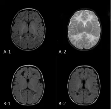
Fig. 2. Follow-up magnetic resonance image (MRI) after 2 weeks from seizure onset of patients A shows porencephaly and cerebromalacia in both frontal and parietal white matter, periventricular leukomala- cia around occipital horn (A-1, A-2; patient 8). Likewise, porencephaly in frontal and parietal white matter is demonstrated in follow-up MRI of patient B (B-1, B-2; patient 9).
명의 만삭 신생아를 대상으로 임상적 특징과 함께 영상 검사 소견 을 보고하였는데, 4명의 미숙아에서 뇌 자기공명 T2 강조영상에서 낭성 병변과 함께 대뇌 백질의 고신호강도가 관찰되었으며, 2명의 만삭 신생아의 추적 뇌 확산강조영상에서 낭성 변화를 동반한 국 소 병변이 관찰되었다. 최근, Lee 등11은 경련을 주소로 내원한 만 삭 신생아 30명을 대상으로, 뇌 자기공명 확산강조영상에서 특징 적인 소견(뇌량을 포함한 대뇌 백질에 미만성 대칭성 병변)을 보이 는 경우 로타바이러스 양성율이 의미 있게 높아 대뇌 백질의 이상 소견과 로타 바이러스 감염이 유의한 상관 관계가 있음을 보고하 였다. 이번 연구에서는 통계적으로 의미 있는 결과를 얻지는 못했 지만, 뇌 자기공명영상에서 이상 소견이 동반되었을 때 정상인 경 우에 비하여 로타바이러스 감염 비율이 높은 경향을 보였다. 특히, 로타바이러스 양성이면서 뇌 자기공명영상에서 대뇌 손상이 확인 된 환아군 모두 확산강조영상에서 뇌량과 속섬유막을 포함한 심부 대뇌 백질에 고신호강도를 보이면서 대뇌 겉질에는 병변을 보이지 않아, Lee 등11의 연구대상자들에게서 관찰된 특징적인 소견과 부 합하고, 뇌 자기공명 확산강조영상에서 이상 소견을 보인 환자군 모두 생후 4일에서 6일 사이에 경련이 발생하였다는 보고도 이번 연구 결과와 일치한다. 이번 연구의 결과 및 위의 문헌들은 로타바 이러스 감염과 신생아 경련이 상관관계가 있음을 시사하며, 특히
대뇌 백질의 손상을 동반할 수 있음을 알 수 있다.11,14
하지만 로타바이러스의 중추신경계 침범을 확진하기 위해서는, 뇌 척수액에서 로타바이러스의 항원 또는 역전사효소중합효소연쇄반 응(reverse transcription polymerase chain reaction, RT-PCR)을 통 해 리보핵산(ribonucleic acid, RNA)을 검출하는 것이 필요하다.15-17 이번 연구는 단일 기관에서 시행한 단기 예후에 대한 연구로써 대상 환자의 수가 적어 통계적으로 의미 있는 결론을 얻는데 제한 이 있었다. 그리고 로타바이러스 항원 검출이 대변 검체로만 이루 어 졌으며 혈액이나 뇌척수액 검체에서는 확인하지 못한 점, 신경 발달과 관련된 예후에 대한 자료가 부족한 점을 이번 연구의 제한 점으로 들 수 있겠다. 그리고 본 연구 결과를 통해 구체적인 인과 관계에 대해 알 수는 없었으나, 저자들의 경우 로타바이러스 감염 에 의한 염증 반응과 관련된 다양한 형태의 혈관병증이 관련 있을 것으로 고려하였다. 이번 연구 및 다른 여러 문헌들로 미루어, 생후 4-6일 사이 신생아 경련을 보이는 경우 로타바이러스 감염 여부를 확인하는 것이 필요하며, 특히 뇌 자기공명영상 검사 및 신경발달 의 추적관찰이 중요함을 알 수 있다. 향후 신생아 로타바이러스 감 염의 중추신경계 합병증의 특징과 예후에 대해서 추가적인 연구가 이루어져야 할 것으로 생각된다.
Table 5. Clinical Characteristics, Seizure Type, MRI and EEG Findings of the Group of Positive Rotavirus and Abnormal MRI Pt.
No. GA/Sex Bwt.
(g) Del. A/S (1’/5’) Onset
(days) Seizure
type Abnormal brain DWI EEG Stool rotavirus
antigen Follow-up brain MRI
1 38+4/M 3,820 NSVD 8/9 5 Clonic, F Deep white matter, CS NS + Porencephalic change in white matter
2 35+5/M 2,420 NSVD 9/10 5 Clonic, M Deep white matter NS +
3 38+3/M 3,220 NSVD 7/8 4 Clonic, M Deep white matter NS +
4 38+2/M 3,700 C/S 9/10 5 Clonic, G Deep white matter, CS, IC Multifocal sharp wave, predominant frontal
+
5 37+5/F 2,410 C/S 8/9 6 Clonic, G Deep white matter, T NS +
6 39+3/M 3,270 NSVD 8/9 5 Clonic, M Deep white matter, CS, IC Encephalopathic pattern + Cerebromalacia in white matter 7 38+4/M 3,720 C/S 8/9 5 Clonic, G Deep white matter, CS, IC, T Infrequent multifocal
sharp wave
+
8 38+0/F 2,520 C/S 9/10 5 Clonic, M Deep white matter, CS, IC, T NS + Cystic PVL, Cerebromalacia in white matter
9 37+3/M 3,100 NSVD 9/10 5 Clonic, M Deep white matter, CS, IC Multifocal sharp wave + Porencephalic change in white matter
10 36+6/M 3,200 C/S 9/10 5 Clonic, M Deep white matter NS +
11 38+0/F 2,760 NSVD 8/9 5 Clonic, M Deep white matter, CS NS +
12 38+1/M 2,970 NSVD 7/7 4 Rigidity BG NS + Normal
13 37+6/M 2,780 NSVD 9/9 4 Pedaling Deep white matter, CS ,IC, EC NS + Porencephaly in white matter
14 38+2/M 3,390 C/S 7/8 6 Clonic, M Deep white matter, CS, IC, T NS +
15 39+3/M 3,120 C/S 9/10 6 Clonic, F Deep white matter, CS, IC NS +
Abbreviations: GA, gestatioanal age; Bwt, birth weight; Del, delivery method; A/S, apgar score; DWI, diffusion-weighted imaging; EEG, electroencephalography; F, focal; M, multifocal; G, generalized; CS, corpus callosum; IC, internal capsule; EC, external capsule; T, thamus; BG, basal ganglia; NS, non-specific
References
1) Choi UY, Lee SY, Ma SH, Jang YT, Kim JY, Kim HM et al. Epidemiological changes in rotavirus gastroenteritis in children under 5 years of age after the introduction of rotavirus vaccines in Korea. Eur J Pediatr 2013;172:947-52.
2) Kim CR. Neonatal rotavirus infection. Neonatal Med 2013;20:389-401.
3) Park DK, Chung JY. The changes in the outbreak of rotavirus gastro- enteritis in children after introduction of rotavirus vaccines: a retro- spective study at a tertiary hospital. Korean J Pediatr Infect Dis 2014;
21:167-73.
4) Sohn TY, Lee CJ, Kim YJ, Kang MJ, Kim SH, Lee SY, et al. Clinical and epidemiological study of 1,165 hospitalized cases of rotaviral gastro- enteritis before and after the introduction of rotavirus vaccine, 2006- 2013. Korean J Pediatr Infect Dis 2014;21:174-180.
5) Langley JM, LeBlanc JC, Hanakowski M, Goloubeva O. The role of Clostridium difficile and viruses as causes of nosocomial diarrhea in children. Infect Control Hosp Epidemiol 2002;23:660-4.
6) Gurwith M, Wenman W, Hinde D, Feltham S, Greenberg H. A prospec- tive study of rotavirus infection in infants and young children. J Infect Dis 1981;144:218-24.
7) Santosham M, Yolken RH, Quiroz E, Dillman L, Oro G, Reeves WC, et al.
Detection of rotavirus in respiratory secretions of children with pneu- monia. J Pediatr 1983;103:583-5.
8) Park MK, Park JO, Kim CH. Comparison of clinical manifestation of rotaviral gastroenteritis between neonates and infants. Korean J
Pediatr Gastroenterol Nutr 2006;9:153-61.
9) Lynch M, Lee B, Azimi P, Gentsch J, Glaser C, Gilliam S, et al. Rotavirus and central nervous system symptoms: cause or contaminant? Case reports and review. Clin Infect Dis 2001;33:932-8.
10) Shiihara T, Watanabe M, Honma A, Kato M, Morita Y, Ichiyama T, et al.
Rotavirus associated acute encephalitis/encephalopathy and con- current cerebellitis: report of two cases. Brain Dev 2007;29:670-3.
11) Lee KY, Oh KW, Weon YC, Choi SH. Neonatal seizures accompanied by diffuse cerebral white matter lesions on diffusion-weighted imaging are associated with rotavirus infection. Eur J Paediatr Neurol 2014;
18:624-31.
12) Salmi TT, Arstila P, Koivikko A. Central nervous system involvement in patients with rotavirus gastroenteritis. Scand J Infect Dis 1978;10:29- 31.
13) Riedel F, Kroener T, Stein K, Nuesslein TG, Rieger CH. Rotavirus infection and bradycardia-apnoea-episodes in the neonates. Eur J Pediatr 1996;
155:36-40.
14) Verboon-Maciolek MA, Truttmann AC, Groenendaal F, Skranes J, Døllner H, Hunt RW, et al. Development of cystic periventricular leuko- malacia in newborn infants after rotavirus Infection. J Pediatr 2012;
160:165-8.
15) Wong CJ, Price Z, Bruckner DA. Aseptic meningitis in an infant with rotavirus gastroenteritis. Pediatr Infect Dis 1984;3:244-6.
16) Ushijima H, Bosu K, Abe T, Shinozaki T. Suspected rotavirus ence- phalitis. Arch Dis Child 1986;61:692-4.
17) Keidan I, Shif I, Keren G, Passwell JH. Rotavirus encephalopathy:
evidence of central nervous system involvement during rotavirus infection. Pediatr Infect Dis J 1992;11:773-5.