© 2013 The Korean Academy of Medical Sciences.
This is an Open Access article distributed under the terms of the Creative Commons Attribution Non-Commercial License (http://creativecommons.org/licenses/by-nc/3.0) which permits unrestricted non-commercial use, distribution, and reproduction in any medium, provided the original work is properly cited.
pISSN 1011-8934 eISSN 1598-6357
A Case of Fulminant Sclerosing Peritonitis Presented Like Acute Culture-Negative Peritonitis and Successfully Treated with
Corticosteroid Therapy
Sclerosing peritonitis is an uncommon complication of peritoneal dialysis. It is characterized by peritoneal fibrosis and sclerosis. The most common clinical presentations of sclerosing peritonitis in peritoneal dialysis patients are ultrafiltration failure and small bowel obstruction. The prognosis and response to immunosuppressive therapy of sclerosing peritonitis presenting with ultrafiltration failure or small bowel obstruction are poor. Here, we describe the case of a 28-yr-old man with end-stage renal disease on peritoneal dialysis showing fulminant sclerosing peritonitis presented like acute culture-negative peritonitis and was successfully treated with corticosteroid therapy. It is not well recognized that sclerosing peritonitis may present in this way. The correct diagnosis and corticosteroid therapy may be life-saving in a fulminant form of sclerosing peritonitis.
Key Words: Peritonitis; Peritoneal Sclerosis; Peritoneal Dialysis; Steroids Ji Yun Jung and Jong Tae Cho
Department of Internal Medicine, Dankook University College of Medicine, Cheonan, Korea Received: 16 August 2012
Accepted: 30 October 2012 Address for Correspondence:
Jong Tae Cho, MD
Department of Internal Medicine, Dankook University College of Medicine, 201 Manghyang-ro, Dongnam-gu, Cheonan 330-715, Korea
Tel: +82.41-550-3925, Fax: +82.41-556-3256 E-mail: [email protected]
http://dx.doi.org/10.3346/jkms.2013.28.4.620 • J Korean Med Sci 2013; 28: 620-623
CASE REPORT
Nephrology
INTRODUCTION
Sclerosing peritonitis (SP) is a rare form of peritoneal inflam- mation which involves both the visceral and the parietal surfac- es of the abdominal cavity. It is characterized by a fibrous thick- ening and sclerotic changes of the peritoneum and is reported to complicate peritoneal dialysis (PD) in most cases. The com- mon clinical manifestations are ultrafiltration and clearance failure in PD patients. Furthermore, small bowel obstruction due to adhesions and encapsulation with abdominal pain, an- orexia, nausea, and vomiting is frequently observed and ulti- mately entails weight loss and malnutrition. It has a poor re- sponse to immunosuppressive therapy (1, 2).
We describe the case of a 28-yr-old man with end-stage renal disease on PD showing fulminant SP presented like acute cul- ture-negative peritonitis and successfully treated with cortico- steroid therapy. On review of the literature, there have been only a few case reports where the presentation of SP has been dominated by an acute inflammatory state (3-7). There was a delay in diagnosis due to the rare mode of presentation, and also a delay in commencement of appropriate therapy. A high degree of clinical suspicion is necessary to diagnose this form of SP, and initiate corticosteroid therapy which appears particu- larly effective for fulminant SP (1, 7).
CASE DESCRIPTION
A 28-yr-old man had been maintained on continuous ambula- tory peritoneal dialysis (CAPD) for 6 yr due to end-stage renal disease of unknown etiology. He used Baxter CAPD system and dialysate with lactate as the base. In December 2011, he had first episode of uncomplicated peritonitis by methicillin-sensi- tive Staphylococcus epidermidis, which was treated with intra- peritoneal cefazolin. On April 06, 2012, he was admitted due to diffuse abdominal pain which had developed 5 days before ad- mission. At the time of admission, blood pressure was 143/77 mmHg, body temperature 36.4°C, height 155 cm, and weight 39 kg. Physical examination showed decreased bowel sound and diffuse abdominal tenderness without rebound tenderness.
Laboratory tests showed the following: hemoglobin 12.4 g/dL, hematocrit 35.6%, leukocyte count 8,540/μL with neutrophil 86.4%, platelet 219,000/μL, C-reactive protein (CRP) 5.17 mg/
dL, blood urea nitrogen 74.5 mg/dL, serum creatinine 16.59 mg/dL, glucose 184 mg/dL, cholesterol 181 mg/dL, total pro- tein 8.2 g/dL, albumin 4.0 g/dL, AST 19 U/L, ALT 23 U/L, total bilirubin 0.42 mg/dL, alkaline phosphatase 258 IU/L, CK 96 U/
L, LDH 411 U/L, amylase 7 U/L, lipase 42 U/L, and peritosol leukocyte count 66/μL with neutrophil 75%. On the fifth hospi- tal day, CRP remained high (6.05 mg/dL) and peritosol leuko- cyte count was 297/μL with neutrophil 90%. Repeat cultures
Jung JY, et al. • Case of Fulminant Sclerosing Peritonitis
http://jkms.org 621
http://dx.doi.org/10.3346/jkms.2013.28.4.620
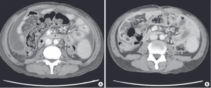
were all negative for bacteria, fungi, and mycobacteria. Despite numerous antimicrobial agents, diffuse abdominal pain per- sisted and peritosol leukocyte count remained high (594/μL with neutrophil 57%). On the eleventh hospital day, PD cathe- ter was removed, and he was switched to hemodialysis. Never- theless, his condition deteriorated. On the twenty-first hospital day, computed tomography (CT) scan of the abdomen (Fig. 1A) showed diffuse thickening of parietal peritoneum and moder- ate amount of ascites. On the twenty-third hospital day, oral prednisolone was prescribed at a dose of 40 mg/day (1.0 mg/
kg/day) on the diagnosis of fulminant SP. On the next day after corticosteroid therapy, he was getting better with decreased ab- dominal pain. On the twenty-fifth hospital day, he did not have abdominal pain. CRP decreased significantly from 19.49 mg/dL to 6.75 mg/dL (Fig. 2). On the thirty-ninth hospital day, he was discharged with normalization of CRP (0.22 mg/dL). On June 20, 2012, a follow-up CT scan (Fig. 1B) showed decreased amount
of ascites without significant change of peritoneal thickening.
In August 2012, he is still free of abdominal pain with a dose of 10 mg/day of oral prednisolone.
DISCUSSION
SP is a rare form of peritoneal inflammation which is reported to occur as an idiopathic form or secondary form in association with long-standing PD, prior abdominal surgery, recurrent peri- tonitis, and beta-blocker (practolol) treatment. The major risk factor for SP is PD treatment. In PD, the continual exposure to nonphysiologic PD solutions and intermittent peritonitis are probable precipitants for the development of SP, with the dura- tion of PD being the most relevant single factor (1, 8). Nonphys- iologic PD solutions such as high concentrations of glucose and lactate, low pH, and bioincompatible substances may induce a chronic sterile inflammation in the peritoneal membrane with upregulation of several cytokines resulting in collagen synthesis by mesothelial cells and fibroblasts (2). The underlying process of SP may be immunological. Moreover, they can directly dam- age the peritoneal membrane, and might contribute to the de- velopment of SP.
Peritoneal sclerosis has two distinct forms. Simple peritoneal sclerosis (SS form) is a mild fibrosing condition of the peritone- al membrane that appears in most patients after several years of PD. The SP form is a dramatic progression of the sclerosis af- ter an inflammatory insult, such as peritonitis. The sclerotic tis- sue is thicker than in SS, and a marked chronic inflammatory infiltrate is present (9). In a small minority, these sclerotic chan- ges are magnified so that the visceral organs are encased in a fi- brotic abdominal cocoon. The severe SP like this has been termed sclerosing encapsulating peritonitis (SEP) or encapsulating peri-
A B
Fig. 1. CT scan of the abdomen. (A) On April 26, 2012, it showed diffuse thickening of parietal peritoneum, mesenteric thickening with vascular engorgement, and moderate amount of ascites. (B) On June 20, 2012, it showed decreased amount of ascites without significant change of peritoneal and mesenteric thickening.
Fig. 2. Clinical course of the present case of fulminant sclerosing peritonitis success- fully treated with corticosteroid therapy.
CRP (mg/dL)
Hospital day
1 2 3 4 5 6 7 8 9 10 11 12 13 14 15 16 17 18 19 20 21 22 23 24 25 26 27 28 29 30 31 32 33 34 35 36 37 38 39 30
25
20
15
10
5
0
Antifungal agent Antibiotics
PD catheter removal CT
Oral prednisolone 40 mg 30 mg
Jung JY, et al. • Case of Fulminant Sclerosing Peritonitis
622 http://jkms.org http://dx.doi.org/10.3346/jkms.2013.28.4.620
toneal sclerosis (EPS) (1). Kawanishi et al. (4) proposed the stage classification and therapeutic options for EPS. Inflammatory stage with manifestations of fever, increased CRP levels, and as- cites develop several weeks or months after the removal of a PD catheter. Steroid therapy is recommended during the early stage.
If the EPS with ileus symptoms is not relieved or recurs within 1 month, steroid dose reduction and total parenteral nutrition (TPN) is recommended at this encapsulating stage. If ileus sym- ptoms remain despite the absence of inflammatory manifesta- tions, laparatomy and enterolysis is considered at this ileus stage.
With the increasing number and survival of PD patients, SP becomes an important problem. Estimated prevalence of SP is reported to be 0.5%-0.9% of PD patients (1, 8, 10, 11). SP usually occurs in patients receiving PD for more than 4-5 yr, and most SP patients have a past medical history of infectious peritonitis.
The clinical presentation of SP is variable. The common mani- festations are ultrafiltration and clearance failure in PD patients, abdominal pain, anorexia, nausea, vomiting, weight loss and malnutrition. These reflect fibrous thickening of the peritoneal membrane, inhibition of peristalsis, encapsulating sclerosis and small bowel obstruction. The uncommon manifestation is acute sterile peritonitis. This reflects an acute peritoneal inflam- mation of SP. The fulminant form of SP like this is rare, but it can occur as a second phase phenomenon immediately following acute bacterial peritonitis (7). In our case, a fulminant SP may present like or develop immediately following acute culture- negative peritonitis.
Diagnosis of SP is made by clinical and radiological methods such as CT scanning, or by surgical laparotomy with excision of sclerosed peritoneum. Histopathology is now seldom required as CT scanning with the clinical features can make a diagnosis of SP. SP should be considered in any patient on prolonged PD who develops recurrent abdominal pain or bowel obstruction in the absence of active infectious peritonitis. The combined CT imaging features of peritoneal enhancement, calcification, and thickening, bowel wall thickening, loculated fluid collections, and tethered small bowel loops in this clinical setting should be considered diagnostic of SP (1, 12). In our case, SP was diag- nosed by the CT findings of peritoneal enhancement and thick- ening, bowel wall thickening, mesenteric thickening with nod- ularity, and ascites even after PD catheter removal in the clini- cal setting of acute sterile peritonitis developed in PD patient.
There is no evidence-based therapy for SP. Treatment of sec- ondary SP in PD patient depends on the severity of symptoms at the time of presentation. It includes cessation of PD, change to hemodialysis, nutritional support, tamoxifen, immunosup- pressive therapy (9, 13-15), and surgery. Intestinal enterolysis can be performed if intestinal obstruction symptoms are not improved by steroid administration or TPN (4, 16). Anti-fibrotic agent, such as tamoxifen, may have been used without signifi- cant benefit. The benefit with immunosuppressive agents in the
previous case reports was variable according to drug chosen, clinical features, and treatment timing. Corticosteroid therapy may be effective for an acute inflammatory state of fulminant SP (3, 4, 7, 13). In our case, acute sterile peritonitis of fulminant SP was dramatically improved immediately after corticosteroid therapy.
Once EPS with ileus symptoms have appeared, mortality is extremely high due to illness related to bowel obstruction or complications of surgery. A major limitation to the success of therapy is the delayed diagnosis. A high index of suspicion is important for early intervention (1). CT scanning should be considered in the patient with the presence of clinical manifes- tations suspicious for SP.
REFERENCES
1. Kawaguchi Y, Kawanishi H, Mujais S, Topley N, Oreopoulos DG. Encap- sulating peritoneal sclerosis: definition, etiology, diagnosis, and treat- ment. International Society for Peritoneal Dialysis Ad Hoc Committee on Ultrafiltration Management in Peritoneal Dialysis. Perit Dial Int 2000;
20: S43-55.
2. Merkle M, Wörnle M. Sclerosing peritonitis: a rare but fatal complication of peritoneal inflammation. Mediators Inflamm 2012; 2012: 709673.
3. Mori Y, Matsuo S, Sutoh H, Toriyama T, Kawahara H, Hotta N. A case of a dialysis patient with sclerosing peritonitis successfully treated with cor- ticosteroid therapy alone. Am J Kidney Dis 1997; 30: 275-8.
4. Kawanishi H, Harada Y, Noriyuki T, Kawai T, Takahashi S, Moriishi M, Tsuchiya S. Treatment options for encapsulating peritoneal sclerosis based on progressive stage. Adv Perit Dial 2001; 17: 200-4.
5. Rajani R, Smyth J, Koffman CG, Abbs I, Goldsmith DJ. Differential effect of sirolimus vs prednisolone in the treatment of sclerosing encapsulating peritonitis. Nephrol Dial Transplant 2002; 17: 2278-80.
6. Evrenkaya TR, Atasoyu EM, Unver S, Basekim C, Baloglu H, Tulbek MY.
Corticosteroid and tamoxifen therapy in sclerosing encapsulating peri- tonitis in a patient on continuous ambulatory peritoneal dialysis. Nephrol Dial Transplant 2004; 19: 2423-4.
7. Courtney AE, Doherty CC. Fulminant sclerosing peritonitis immediately following acute bacterial peritonitis. Nephrol Dial Transplant 2006; 21:
532-4.
8. Rigby RJ, Hawley CM. Sclerosing peritonitis: the experience in Australia.
Nephrol Dial Transplant 1998; 13: 154-9.
9. Martins LS, Rodrigues AS, Cabrita AN, Guimaraes S. Sclerosing encap- sulating peritonitis: a case successfully treated with immunosuppression.
Perit Dial Int 1999; 19: 478-81.
10. Kawanishi H, Kawaguchi Y, Fukui H, Hara S, Imada A, Kubo H, Kin M, Nakamoto M, Ohira S, Shoji T. Encapsulating peritoneal sclerosis in Ja- pan: a prospective, controlled, multicenter study. Am J Kidney Dis 2004;
44: 729-37.
11. Kim BS, Choi HY, Ryu DR, Yoo TH, Park HC, Kang SW, Choi KH, Ha SK, Han DS, Lee HY. Clinical characteristics of dialysis related sclerosing en- capsulating peritonitis: multi-center experience in Korea. Yonsei Med J 2005; 46: 104-11.
12. Stafford-Johnson DB, Wilson TE, Francis IR, Swartz R. CT appearance
Jung JY, et al. • Case of Fulminant Sclerosing Peritonitis
http://jkms.org 623
http://dx.doi.org/10.3346/jkms.2013.28.4.620
of sclerosing peritonitis in patients on chronic ambulatory peritoneal di- alysis. J Comput Assist Tomogr 1998; 22: 295-9.
13. Fagugli RM, Selvi A, Quintaliani G, Bianchi M, Buoncristiani U. Immu- nosuppressive treatment for sclerosing peritonitis. Nephrol Dial Trans- plant 1999; 14: 1343-5.
14. Junor BJ, McMillan MA. Immunosuppression in sclerosing peritonitis.
Adv Perit Dial 1993; 9: 187-9.
15. Bhandari S, Wilkinson A, Sellars L. Sclerosing peritonitis: value of im- munosuppression prior to surgery. Nephrol Dial Transplant 1994; 9:
436-7.
16. Kawanishi H, Harada Y, Sakikubo E, Moriishi M, Nagai T, Tsuchiya S.
Surgical treatment for sclerosing encapsulating peritonitis. Adv Perit Dial 2000; 16: 252-6.