INTRODUCTION
Thyroid transcription factor-1 (TTF-1) is a 38-kDa home- odomain containing DNA-binding protein of the Nkx-2 gene family, which is expressed in the thyroid, lungs, and some re- stricted areas of diencephalon (1). During human development, it appears in early stages and thus could play an important role in cell differentiation and morphogenesis of both the thyroid and lung (1). TTF-1 activates transcription of thyroglobulin and thyroperoxidase genes in the follicular thyroid cells, but not in lung epithelial cells (2-5). Conversely, the role of TTF-1 in lung tissues involves regulation of the surfactant and Clara cell secretory protein gene expression, but not in follicular thy- roid cells (6-8).
Since the expression of TTF-1 is an early event during lung differentiation, the evaluation of TTF-1 expression in lung tumors may have an impact in clinical research and in the man- agement of these neoplasms (9). Also, tissue-specific transcrip- tion factors contribute to the transcriptional activation of genes expressed in particular cell types, and thus the study of tran-
scription factors like TTF-1 in human lung tumors could be of benefit in our understanding of the molecular events of their neoplastic transformation (10, 11).
In lung cancers, several reports described the differences in TTF-1 expression between histologic types (11-17). They re- ported that TTF-1 was expressed in 62.5-90% of adenocarci- nomas and 89-100% of small cell carcinoms, whereas it was not found or found at a very low frequency in squamous car- cinomas (0-27%) and large cell carcinomas (0-25%). Most re- ports have emphasized the usefulness of TTF-1 in differential diagnosis of lung cancer from nonpulmonary cancers, where- as relatively few and contradictory data are available on the prognostic implication of TTF-1 immunoreactivity because its upregulation has been considered as a favorable or unfa- vorable predictor of survival in patients with non-small cell lung carcinoma (NSCLCs) (13, 14, 16, 18). Also, there are few studies referred to possible significance of TTF-1 expres- sion in lung carcinogenesis, despite its crucial roles in lung development and maintenance of pulmonary function by means of surfactant apoprotein A and Clara cell antigen induction.
Na-Hye Myong
Department of Pathology, Dankook University College of Medicine, Cheonan, Korea
Address for correspondence Na-Hye Myong, M.D.
Department of Pathology, Dankook University College of Medicine, San 29 An-seo dong, Cheon-an 330-714, Korea
Tel : +82.41-550-3891, Fax : +82.41-561-9127 E-mail : [email protected]
*The present research was conducted by the research fund of Dankook University in 2002.
494
Thyroid Transcription Factor-1 (TTF-1) Expression in Human Lung Carcinomas: Its Prognostic Implication and Relationship with Expressions of p53 and Ki-67 Proteins
This study was aimed to evaluate the prevalence and prognostic implication of thy- roid transcription factor-1 (TTF-1) immunoreactivity in 81 human lung carcinomas, including 65 cases of non-small cell lung carcinoma (NSCLC) and 16 cases of small cell lung carcinoma (SCLC); and also to investigate its relationship with the cell pro- liferation and regulation by immunostaining of Ki-67 and p53 proteins, respectively.
The immunohistochemical staining for TTF-1 (clone 8G7G3/1) was performed and several clinicopathologic variables and the follow-up data were obtained. The immuno- staining results for TTF-1 were semiquantitatively interpreted as negative and pos- itive. Of NSCLCs, TTF-1 is highly expressed in adenocarcinomas (76%), whereas squamous cell carcinomas revealed no immunoreactivity (0%). SCLCs showed strong TTF-1 expression (88%). In NSCLC, TTF-1 expression was inversely correlated with Ki-67 proliferative activity and independent of p53 overexpression. TTF-1 (+) group tended to show better survival than TTF-1 (-) group in NSCLC. Conclusively, these observations suggest that TTF-1 is a sensitive and specific diagnostic marker for pulmonary adenocarcinomas and SCLCs; that TTF-1 might have a good prognostic implication based on its inverse correlation with Ki-67 proliferative activity and ten- dency for better survival in NSCLC; that this cell lineage marker may play a role in the molecular pathogenesis of lung cancers at the level of transcription.
Key Words : Transcription Factors; Thyroid Transcription factor-1 (TTF-1) Expression; Human; Lung Neoplasm; Ki-67 antigen; Protein p53; Pathogenesis
Received : 3 February 2003 Accepted : 18 March 2003
Other factors for risk stratification in patients with lung cancers include the tumor proliferative potential, which is com- monly evaluated by Ki-67 antigen immunostaining, and the mutation in p53 genes, the most common genetic alteration in lung cancers (14, 15, 18). Although the independent pro- gnostic role of these prognostic factors in lung cancer patients still remains a matter of debate, there are no doubts the cell proliferation and the regulation of cell cycle are essential fac- tors for tumor growth. However, the relationships between TTF-1 and cell proliferation or p53 overexpression have been only rarely reported in human lung carcinomas, including NSCLC and small cell lung carcinoma (SCLC) (14, 15, 18).
This study was aimed at evaluating the prevalence and pro- gnostic implication of TTF-1 immunoreactivity in 65 cases of NSCLC and 16 cases of SCLC patients, and at demonstrat- ing its relationship with cell proliferation assesed by Ki-67 immunostaining and cell-cycle regulation by p53 staining.
Our findings indicate that TTF-1 is expressed almost exclu- sively by adenocarcinomas and small cell carcinomas, and thus this transcriptional protein is likely to be involved in the growth of adenocarcinomas and small cell carcinomas, with inverse correlation with Ki-67 proliferative potential; and that TTF-1 immunoreactive NSCLCs show a trend for a favorable clinical course, although the TTF-1 immunoreactivity fails to con- firm a statistically significant prognostic implication.
MATERIALS AND METHODS Patients and tissue samples
Primary tumor specimens from 65 NSCLCs and 16 SCLCs were obtained by surgical resection and bronchoscopic biopsy, respectively, between 1996 and 2001 at the Dankook Univer- sity Hospital. All hematoxylin-eosin stained slides of the tis- sue samples were reviewed, and the pathological diagnoses of the histologic grades and types were confirmed by a pathol- ogist. Histologic typing was performed according to the World Health Organization diagnostic criteria for lung carcinomas (1999). The hospital records of all 81 patients (65 NSCLCs and 16 SCLCs) were reviewed to obtain the clinicopathologic variables such as age, sex, smoking history, tumor-node-metas- tasis (TNM) stage (in NSCLC), and tumor extent (in SCLC).
The pathologic staging of NSCLC and SCLC was assessed ac- cording to the TNM Classification of AJCC staging system (1997), and the Veterans’Administration Lung Study Group (VALSG) staging system (1973), respectively. Death from lung cancer was the terminal event for survival calculations. All patients were followed for up to about 5 yr.
Immunohistochemical analysis
All specimens used in this study were 4 m-thick sections of paraffin-embedded tissue obtained at surgery and broncho-
scopic biopsy. The standard avidin-biotin-peroxidase complex method was used for immunohistochemical examination using monoclonal antibodies against TTF-1 (8G7G3/1, NeoMark- ers, CA, U.S.A.), p53 (DO7, Novocastra, Newcastle-upon- Tyne, U.K.), Ki-67 (AO47, DAKO, Carpinteria, CA, U.S.A.).
Deparaffinization of all sections were performed through a series of xylene baths, and rehydration was performed through graded ethyl alcohols. The sections were microwaved in 10 mM citrate buffer at 90℃for 10 min, and were treated with 3%
H2O2-PBS solution to reduce endogenous peroxidase activi- ty. Then, they were incubated with normal bovine serum to reduce nonspecific antibody binding and were subsequently subjected to the primry antibody reactions. The antibodies for TTF-1, p53, and Ki-67 proteins were reacted with the sections at room temperature for one hour at the same dilution (1:100).
Detection of the immunoreactive staining was carried out by avidin-biotin-peroxidase complex method using the LSAB kit (DAKO). The sections were subjected to a color reaction with 3,3-diaminobenzidine tetrahydrochloride containing 3%
H2O2in Tris buffer, and were lightly counterstained with Ma- yer’s hematoxylin.
For evaluation of the expressions of TTF-1, p53, and Ki-67 proteins, the immunostained cells were considered positive only when distinct nuclear staining was identified. The extent of the positive cells was semiquantitatively scored as follows:
0, negative reaction; 1, <5% positive tumor cells; 2, 5-50%;
3, 51-100% of tumor cells. The staining intensity was eval- uated with three categorized scores of weak 1, moderate 2, and strong 3. To exclude equivocal reactions, the staining was re- garded as positive for TTF-1 expression when the combined scores were more than 2 (≥2). Cases were considered positive for Ki-67 expression, when more than 10% of tumor cells were reactive, because the median value of Ki-67 proliferative frac- tion was about 10% in all cases. Cases were considered posi- tive for p53 overexpression, when at least 50% of tumor cells showed nuclear immunopositivity because high p53 expres- sion would be expected if p53 mutations existed (19). To obtain the survival curves, the TTF-1 nuclear staining was divided into as negative (scores 0-1) and positive (scores 2-9) groups based on the tumor nuclei with unequivocal staining.
Statistical Analysis
The comparison of TTF-1 immunoreactivity between sev- eral two-categorical clinicopathologic variables and the rela- tionships of TTF-1 expression with Ki-67 proliferative activ- ity and p53 overexpression were analysed by chi-square test (SPSS 10.0). The survival period was calculated as the time from the date of surgery to the date of death or last follow-up.
Postoperative survival curves were constructed using the Ka- plan-Meier method and then compared by the log rank test.
A p value less than 0.05 was defined as statistically significant.
RESULTS
Clinicopathologic data and correlation with TTF-1 expression
Patient profiles are shown in Tables 1 and 2. NSCLC group consisted of 49 men and 16 women, with median ages of 58.0 and 61.8 yr, respectively. The SCLC group included 14 men and 2 women with median ages of 60.6 and 62 yr, respective- ly. Overall, the median age at diagnosis was 59.9 yr (range:
31-83) in NSCLC and 60.8 yr in SCLC (range: 38-75). All 16 SCLC patients were smokers. The pathological staging of the 65 NSCLC cases revealed 25 cases of stage 1, 10 of stage 2, and 30 of stage 3. Pathological T stages were pT1 in 14 cases, pT2 in 31, pT3 in 16, and pT4 in 4. The 16 SCLC cases were staged as 8 limited and 8 extensive diseases. The histologic types of 65 NSCLCs included 30 cases of squamous cell carcinoma (SCC), 25 cases of adenocarcinoma (ADC), 3 cases of adenosquamous carcinoma (ASC), 2 cases of bronchi- oloalveolar carcinoma (BAC), 3 cases of large cell carcinoma
(LCC), and 2 cases of pleomorphic carcinoma (PLC), based on the WHO classification (1999). The adenocarcinomas and sq- uamous cell carcinomas were graded as 6 cases of grade 1 (well differentiated), 36 of grade 2 (moderately differentiated), and 13 of grade 3 (poorly differentiated). Clinicopathologic vari- ables such as age, smoking history, tumor size, and histolog- ic grades in NSCLCs were not significantly associated with TTF-1 expression, although TTF-1 expression tend to be higher in non- smokers and well-differentiated tumors. How- ever, TTF-1 was expressed significantly higher in women than in men (p<0.05) and in ADCs than SCCs (p<0.001).
In SCLCs, any significant difference in TTF-1 expression was not found between two categorical groups of the clinco- pathologic variables such as age, sex, and stage.
TTF-1 expression in normal lung, NSCLCs, and SCLCs
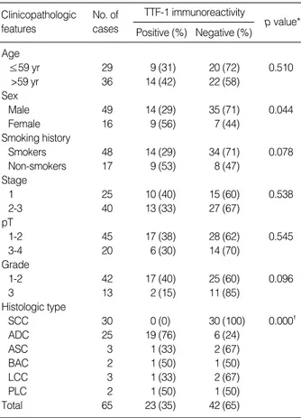
In non-neoplastic lung, nuclear TTF-1 immunoreactivity was limited in alveolar and bronchiolar epithelial cells; inter- stitial cells, lymphocytes, alveolar macrophages did not stain for TTF-1 (Fig. 1). No differences between non-neoplastic and malignant cells were observed in the staining pattern with TTF-1 antibody.
Overall, TTF-1 immunoreactivity was found in 23 (35%) of 65 NSCLCs, including 19 (76%) of 25 ADCs, 1 (33%) of 3 ASCs, 1 (50%) of 2 BACs, 1 (33%) of 3 LCCs, and 1 (50%) of 2 PLCs. The 30 squamous cell carcinomas were complete- ly devoid of TTF-1 immunoreactivity (0%). No differences in the nuclear immunoreactivity were noted among the dif- ferent tumor types, and there was no preferential distribution of TTF-1-reactive cells in perivascular, central, or peripheral viable areas of the neoplasms. Among the different histolog- ic types of NSCLCs, ADCs showed TTF-1 expression in the majority of the cases (76%), which was significantly higher than SCCs (0%) (p<0.001) (Fig. 2). TTF-1 expression did not vary with the morphologic subtypes of ADCs, which showed acinar and solid patterns. One of two BACs revealed TTF-1
*chi-square test, �: p value between the percentages of TTF-1 immunore- activity in squamous cell carcinoma versus adenocarcinoma, SCC: squa- mous cell carcinoma, ADC: adenocarcinoma, ASC: adenosquamous car- cinoma, BAC: bronchioloalveolar carcinoma, LCC: large cell carcinoma, PLC: pleomorphic carcinoma.
Clinicopathologic features
No. of cases
TTF-1 immunoreactivity Positive (%) Negative (%)
p value*
Age
≤59 yr 29 9 (31) 20 (72) 0.510
>59 yr 36 14 (42) 22 (58)
Sex
Male 49 14 (29) 35 (71) 0.044
Female 16 9 (56) 7 (44)
Smoking history
Smokers 48 14 (29) 34 (71) 0.078
Non-smokers 17 9 (53) 8 (47)
Stage
1 25 10 (40) 15 (60) 0.538
2-3 40 13 (33) 27 (67)
pT
1-2 45 17 (38) 28 (62) 0.545
3-4 20 6 (30) 14 (70)
Grade
1-2 42 17 (40) 25 (60) 0.096
3 13 2 (15) 11 (85)
Histologic type
SCC 30 0 (0) 30 (100) 0.000�
ADC 25 19 (76) 6 (24)
ASC 3 1 (33) 2 (67)
BAC 2 1 (50) 1 (50)
LCC 3 1 (33) 2 (67)
PLC 2 1 (50) 1 (50)
Total 65 23 (35) 42 (65)
Table 1.Correlations between TTF-1 expression and clinico- pathologic features in non-small cell lung carcinomas
*chi-square test, �16 cases of SCLC were staged as limited and exten- sive diseases.
Clinicopathologic features
No. of cases
TTF-1 immunoreactivity Positive (%) Negative (%)
p value*
Age
≤59 yr 7 6 (86) 1 (14) 0.849
>59 yr 9 8 (89) 1 (11)
Sex
Male 14 12 (86) 2 (14) 0.568
Female 2 2 (100) 0 (0)
Stage�
Limited disease 8 8 (100) 0 (0) 0.131
Extensive disease 8 6 (75) 2 (25)
Total 16 14 (88) 2 (12)
Table 2.Correlation between TTF-1 immunoreactivity and clini- copathologic features in 16 small cell lung carcinomas (SCLCs)
expression not in mucinous type but in the non-mucinous type (Fig. 3).
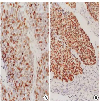
SCLCs examined in this study showed high frequency of TTF-1 expression (88%), which was higher than adenocarci- nomas (76%) (Fig. 4).
Association of TTF-1 expression with p53 and Ki-67 proteins in 65 NSCLCs
The statistical analysis for the relationships was shown in Tables 3 and 4. Significant inverse correlation was found be- tween TTF-1 expression and proliferative activity evaluated by Ki-67 protein (p<0.01). However, there was no significant
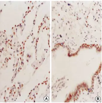
Fig. 3.A bronchioloalveolar carcinoma of non-mucinous type re- vealed high immunoreactivity for thyroid transcription factor-1 (TTF- 1) (×200).
Fig. 4.Pulmonary small cell carcinoma showing high nuclear ex- pression for thyroid transcription factor-1 (TTF-1) (×400).
Fig. 1.Nuclear immunoreactivity for thyroid transcription factor-1 (TTF-1) in normal alveolar (A) and bronchiolar (B) epithelial cells (×400).
A B
Fig. 2.Immunohistochemical finding of thyroid transcription factor- 1 (TTF-1) in moderately differentiated adenocarcinoma, showing diffuse and strong nuclear immunoreactivity for TTF-1 (×400).
correlation between TTF-1 expression and the overexpression of p53 protein, a cell cycle regulator. Two representative cases are shown for the immunostaining results for Ki-67 prolif- erative activity and p53 overexpression in Fig. 5.
Due to the small number of cases, those relationships were not analyzed in 16 SCLCs.
Survival data and its comparison between TTF-1 (+) and TTF-1 (-) groups
Complete follow-up information was available on 36 pati-
ents of 65 NSCLCs and 6 of 16 SCLCs, with a median follow- up duration of 29.7 months for living patients (range 12-66 months) in NSCLCs and 45 (range 24-66 months) in SCLCs.
At the time of analysis, 26 (72.2%) and 2 (33%) patients were alive in NSCLCs and SCLCs, respectively. When all of 65 NSCLC patients analyzed for postsurgical survival were exam- ined for the difference in survival time between TTF-1 (+) and TTF-1 (-) groups by Kaplan-Meier method and then log rank test, the group with a positive immunoreactivity for TTF-1 tended to reveal the better survival, compared with the group with a negative immunostaining for that (Fig. 6). When we also obtained the survival curves for the lower (stage 1, 2) and higher stage (stage 3) and compared them by log rank test, the patients with stage 3 revealed significantly lower survival time compared with those with stage 1, 2 (p=0.0077) (data not shown).
Due to the limited number of SCLC patients, the statistical analysis for the survival data between TTF-1 (+) and TTF-1 (-) groups was not performed.
DISCUSSION
Tissue-specific gene expression is mediated largely by tran- scritional factors, and a master regulatory gene is thus a poten- tial marker of cellular lineage (15, 20). TTF-1 is an example of such a lineage marker, which plays a crucial role in normal peripheral lung function and morphogenesis (15).
In the present study, normal lung tissue revealed a very lim- ited expression for TTF-1 in the terminal airways such as alveol- ar and bronchiolar epithelial cells. The staining was quite uni- form, and TTF-1 appeared to be useful for labeling the com- mited cells or lineage of the terminal respiratory unit. This result also indicates that TTF-1 could be used as a molecular marker of differentiation for neoplasms arising from these cell
Fig. 5.A case of squamous cell carcinoma showing moderately brisk mitotic activity evaluated by Ki-67 immunostaining (A, ×400) and diffuse nuclear immunopositivity for p53 protein (B, ×200).
A B
Cummulative Survival
Time (months)
TTF-1(+) n=23
TTF-1(-) n=42 1.1
1.0 .9 .8 .7 .6
.5-10 0 10 20 30 40 50 60 70
Fig. 6.Kaplan-Meier survival curves of patients with non-small cell lung carcinoma (NSCLC). Comparison between TTF-1 positive and TTF-1 negative groups shows a tendency for better survival in the TTF-1 positive group (log rank test; p=0.0956).
*: chi-square test.
TTF-1 expression No. of cases
Ki-67 immunoreactivity Positive (%) Negative (%)
p value*
Positive 23 9 (39) 14 (61) 0.003
Negative 42 32 (76) 10 (24)
*: chi-square test.
TTF-1 expression No. of cases
p53 overexpression Positive (%) Negative (%)
p value*
Positive 23 10 (43) 13 (57) 0.215
Negative 42 25 (60) 17 (40)
Table 3.Correlation between TTF-1 expression and Ki-67 pro- liferative activity in non-small cell lung carcinomas
Table 4.Correlation between TTF-1 expression and p53 over- expression in non-small cell lung carcinomas
types (9).
In the lung cancer group, TTF-1 expression was found in 23 NSCLCs of non-squamous cell type (35%) and 14 SCLCs (88%). Of the 23 NSCLCs, the majority (19 cases, 83%) was ordinary type of adenocarcinoma and the remaining consisted of 1 non-mucinous type of BAC, 1 ASC, 1 LCC, and 1 PLC.
In a recent report (15), adenocarcinomas with terminal res- piratory unit (TRU) morphology revealed significantly high- er TTF-1 expression than those with non-TRU morphology.
They suggested that TTF-1 expression could be used as a lin- eage marker for TRU type adenocarcinomas, and could play a role in the molecular pathogenesis of those adenocarcino- mas. Although the adenocarcinomas were not classified into TRU and non-TRU type, the present study also suggested that TTF-1 could be a useful marker of adenocarcinomas in the diagnosis of poorly differentiated NSCLCs because there was a highly significant difference in TTF-1 expression between two major groups of NSCLCs, ADC and SCC (76% versus 0%, p=0.000). Also, TTF-1 expression in this study was sig- nificantly higher in the female than in male, and tend to be higher in non-smokers than in smokers. These results seem to be associated with the fact that pulmonary ADCs tend to be more prevalent in female non-smokers than SCCs. In our study, all 30 cases of SCCs revealed no TTF-1 immunoreac- tivity (0%). The rare occurrence or lack of TTF-1 immunore- activity in SCC or metaplastic epithelium suggests that this protein is not involved in the differentiation of squamous cells.
This fact is supported by a few studies showing that the lung- specific genes activated by TTF-1 are not expressed in SCCs (8, 17, 21). Pelosi et al. (14) also proposed a possibility of the synthesized TTF-1 with an aberrant function in SCCs.
SCLC has also been reported to express TTF-1 in most cases (89-100%) (9, 11, 12, 22), and our data confirmed the high frequency of TTF-1 expression in SCLC (14 of 16, 88%). Al- though this high frequency of TTF-1 expression in SCLC ap- pears to conflict with the previously mentioned usefulness of TTF-1 as the cell lineage marker for adenocarcinoma, Yatabe et al. (15) explained it as follows; first, small cell carcinomas frequently produce TTF-1 aberrantly like ectopic hormones;
second, SCLC is a type of neuroendocrine carcinoma and can make TTF-1 because TTF-1 is reported to be expressed in the the diencephalon area (1); and third, TTF-1 expression may be atavistic in these undifferentiated cells of SCLC.
Taken together, the present study suggests that TTF-1 may play an important role as the transcriptional factor in the molec- ular pathogenesis of adenocarcinomas and small cell carcinomas.
Most of the previous studies on TTF-1 expression focused on its usefulness as the immunohistochemical marker for diag- nosis of lung tumors and for differential diagnosis of primary pulmonary adenocarcinomas from extrapulmonary adenocar- cinomas metastatic to the lung (23). Few studies investigated the prognostic role of TTF-1 in NSCLC and showed contra- dictory data on the relationship between TTF-1 expression and survival (13, 14, 16, 18). In the current study, we found
that the patients with TTF-1 expression tend to survive longer than TTF-1 (-) group, although the difference was not statis- tically significant (log rank test, p=0.096). Potential sources of the conflicting results of TTF-1 expression and survival in the literatures have been analyzed, including the scoring sys- tem of immunohistochemical staining for TTF-1, the stain- ing procedure, and statistical analysis of data (14, 16).
Although the independent prognostic role of p53 mutation and Ki-67 proliferative activity is still debatable in NSCLC, the importance of p53 mutation and cell proliferation in the pulmonary carcinogenesis seems to be obvious (14, 24). p53 is considered as the cell cycle regulator as it blocks progression through the cell cycle in cells that have sustained DNA dam- age (24). In general, p53 mutation or overexpression is con- sidered as an indicator of poor prognosis. Also, Ki-67 is fre- quently used to evaluate the prognostic value of proliferative activity of lung cancers, and high Ki-67 expression was hypothe- sized to be correlated with a poor clinical outcome (25). How- ever, the study on the relationship between TTF-1 expression and p53 overexpression or proliferative activity is very rare.
Yatabe et al. (15) reported that p53 overexpression showed the negative correlation with TTF-1-positive adenocarcinoma.
Puglisi et al. (18) performed a combined analysis of TTF-1 and MIB-1 proliferative activity in NSCLCs, showing no cor- relation between them but the poorest survival in the TTF- 1-positive/MIB-1-positive group. The presence of a relation- ship between TTF-1 expression and cell proliferation has been reported on the basis of data showing that reduced prolifer- ation rate of thyroid cells is induced by decrease in TTF-1 concentration by antisense procedures (26). Also, it has been suggested that homeodomain-containing nuclear proteins functionally similar to TTF-1 have positive effects on cell growth (27) as well as antiapoptotic effects demonstrated by loss of function experiment (28).
However, our data unexpectedly showed inverse relationship between the TTF-1 expression and Ki-67 proliferative activ- ity in NSCLCs. This contradictory result seems to be difficult to be interpreted because of paucity of the similar study except for a study by Pelosi et al. (14). The investigation showed TTF- 1 immunoreactivity correlated inversely with the tumor pro- liferative fraction assessed by Ki-67 immunostaining. But as an interpretation for the result, they commented only that the relationship of TTF-1 expression with cell proliferation of lung cancer remains to be unveiled. Therefore, our result for the inverse relationship between TTF-1 and proliferative activity in NSCLCs remains to be proved further.
In conclusion, we confirmed that TTF-1 appears to be a rea- sonably sensitive and highly specific diagnostic marker for pul- monary adenocarcinomas and small cell carcinomas; that TTF- 1 may be used a good prognostic marker based on its negative correlation with Ki-67 proliferative activity and tendency for better survival; that the cell lineage marker of the lung may in part participate in the molecular pathogenesis of lung can- cers at the level of transcription.
REFERNCES
1. Lazzaro D, Price M, de Felice M, Di Lauro R. The transcription fac- tor TTF-1 is expressed at the onset of thyroid and lung morphogen- esis and in restricted regions of the foetal brain. Development 1991;
113: 1093-104.
2. Musti AM, Ursini VM, Avvedimento EV, Zimarino R, Di Lauro R.
A cell type specific factor recognizes the rat thyroglobulin promoter.
Nucleic Acids Res 1987; 15: 8149-66.
3. Civitareale D, Lonigro R. Sinclair AJ, Di Lauro R. A thyroid-specific nuclear protein essential for tissue-specific expression of the throglobu- lin promoter. EMBO J 1989; 8: 2537-43.
4. Mizuno K, Gonzalez FJ, Kimura S. Thyroid-specific enhancer-bind- ing protein (T/EBP): cDNA cloning, functional charaterization, and structural identity with thyroid transcription factor 1 (TTF-1). Mol Cell Biol 1991; 11: 4927-33.
5. Francis-Lang H, Price M, Polycarpou-Schwarz M, Di Lauro R. Cell- type-specific expression of the rat thyroperoxidase promoter indicates common mechanisms for thyroid-specific gene expression. Mol Cell Biol 1992; 12: 576-88.
6. Bruno MD, Bohinski RJ, Huelsman KM, Whitsett JA, Korfhagen TR.
Lung cell-specific expression of the murine surfactant protein A (SP- A) gene is mediated by interaction between the SP-A promoter and thy- roid transcription factor-1. J Biol Chem 1995; 270: 6531-6.
7. Yan C, Sever Z, Whitsett JA. Upstream enhancer activity in the human surfactant protein B gene is mediated by thyroid transcription factor- 1. J Biol Chem 1995; 270: 24852-7.
8. Zhang L, Whitsett JA, Stripp BR. Regulation of Clara cell secretory protein gene transcription by thyroid transcription factor-1. Biochim Biophys Acta 1997; 1350: 359-67.
9. Fabbro D, Di Loreto C, Stamerra O, Beltrami CA, Lonigro R, Damante G. TTF-1 gene expression in human lung tumors. Eur J Cancer 1996;
32A: 512-7.
10. Kamps MP, Murre C, Sun XH, Baltimore D. A new homeobox gene contributes the DNA binding domain of the t(1;19) translocation in pre-B ALL. Cell 1990; 60: 547-55.
11. Zamecnik J, Kodet R. Value of thyroid transcription factor-1 and sur- factant apoprotein A in the differential diagnosis of pulmonary car- cinomas: a study of 109 cases. Virchows Arch 2002; 440: 353-61.
12. Di Loreto C, Di Lauro V, Puglisi F, Damante G, Fabbro D, Beltrami CA. Immunocytochemical expression of tissue specific transcription factor-1 in lung carcinoma. J Clin Pathol 1997; 50: 30-2.
13. Puglisi F, Barbone F, Damante G, Bruckbauer M, Di Lauro V, Bel- trami CA, Di Loreto C. Prognostic value of thyroid transcription fac- tor-1 in primary, resected, non-small cell lung carcinoma. Modern Pathol 1999; 12: 318-24.
14. Pelosi G, Fraggeta F, Pasini F, Maisonneuve P, Sonzogni A, Iannuc- ci A, Terzi A, Bresaola E, Valduga F, Lupo C, Viale G. Immunoreac- tivity for thyroid transcription factor-1 in stage I non-small carcino- mas of the lung. Am J Surg Pathol 2001; 25: 363-72.
15. Yatabe Y, Mitsudomi T, Takahashi T. TTF-1 expression in pulmonary adenocarcinomas. Am J Surg Pathol 2002; 26: 767-73.
16. Haque AK, Syed S, Lele SM, Freeman DH, Adegboyega PA. Immuno- histochemical study of thyroid transcription factor-1 and HER2/neu in non-small cell lung cancer: Strong thyroid transciption factor-1 predicts better survival. Appl Immunohistochem Mol Morphol 2002;
10: 103-9.
17. Kaufmann O, Dietel M. Thyroid transcription factor-1 is a the supe- rior immunohistochemical marker for pulmonary adenocarcinomas and large cell carcinomas compared to surfactant proteins A and B.
Histopathology 2000; 36: 8-16.
18. Puglisi F, Aprile G, Bruckbauer M, Barbone F, Damante G, Guerra S, Beltrami CA, Di Loreto C. Combined analysis of MIB-1 and thyroid transcription factor-1 predicts survival in non-small cell lung carci- nomas. Cancer Lett 2001; 162: 97-103.
19. Rodrigues NR, Rowan A, Smith MEF, Kerr IB, Bodmer WF, Gannon JV, Lane DP. P53 mutations in colorectal cancer. Proc Natl Acad Sci USA 1990; 87: 7555-9.
20. Weintraub H, Davis R, Tapscott S, Thayer M, Krause M, Benezra R, Blackwell TK, Turner D, Rupp R, Hollenberg S, Zhuang Y, Lasser A.
The myoD gene family: nodal point during specification of the muscle cell lineage. Science 1991; 251: 761-6.
21. Khoor A, Whitsett JA, Stahlman MT, Olson SJ, Cagle PT. Utility of surfactant protein B precursor and thyroid transcription factor 1 in differentiating adenocarcinoma of the lung from malignant mesothe- lioma. Hum Pathol 1999; 30: 695-700.
22. Folpe Al, Gown AM, Lamps LW, Garcia R, Dail DH, Zabro RJ, Schmidt RA. Thyroid transcription factor-1: Immunohistochemical evaluation in pulmonary neuroendocrine tumors. Modern Pathol 1999;
12: 5-8.
23. Lau SK, Luthringer DJ, Eisen RN. Thyroid transcription factor: A re- view. Appl Immunohistochem Mol Morphol 2002; 10: 97-102.
24. Nikinski J, Niklinska W, Laudanski J, Chyczewska E, Chyczewski L.
Prognostic molecular markers in non-small cell lung cancer. Lung Cancer 2001; 34: S53-8.
25. Tungekar MF, Gatter KC, Dunnill MS, Mason DY. Ki-67 immuno- staining and survival in operable lung cancer. Histopathology 1991; 19: 545-50.
26. Rossi DL, Acebron A, Santisteban P. Function of the homeo and paired domain proteins TTF-1 and Pax-8 in thyroid cell proliferation. J Biol Chem 1995; 270: 23139-42.
27. Sauvageau G, Thorsteinsdottir U, Eaves CJ, Lawrence HJ, Largman C, Lansdorp PM, Humphries RK. Overexpression of HOXB4 in he- matopoietic cells causes the selective expansion of more primitive pop- ulations in vitro and in vivo. Genes Dev 1995; 9: 1753-65.
28. Izon DJ, Rozenfeld S, Fong ST, Komuves L, Largman C, Lawrence HJ. Loss of function of the homeobox gene Hoxa-9 pertubes early T- cell development and induces apoptosis in primitive thymocytes. Blood 1998; 92: 383-93.