introduction
The thickness of cement mantle has a profound influence on the stability of implant fixation in total knee arthroplasty (TKA).
There are two types of bone cement mantles in the knee accord
ing to the location, one that is supposed to penetrate into the cancellous bone after TKA and the other that forms an interface between the implant and the bone16).
A thin cement mantle weakens the durability and fixation strength of a prosthesis implanted in the knee whereas a thick mantle increases the risk of thermalinduced osteonecrosis. The optimum thickness to allow for interdigitation of the cement into the cancellous bone has been suggested as 3–5 mm711). During cementing in TKA, it is also important to ensure impaction of the implant onto the bone because a thick cement mantle can reduce the gap width and boneimplant contact at the interface12). Knee implants are designed to have a 1 mm builtin cement pocket to maintain a proper mantle thickness after squeezing out of the ce
ment by impaction13) (Fig. 1). Therefore, the cement mantle at the bone cement interface has been recommended to be ≤1 mm in the proximal tibia and distal femur each to minimize its influence on the gap width, and ≥2 mm thickness should be avoided12).
Cementing techniques are essential to obtain a proper mantle thickness. Since cementing is affected by various factors including the type of cement, mixing time, temperature, and the surgeon’s experience, it is important to utilize reproducible cementing
Cement Mantle Thickness at the Bone Cement Interface in Total Knee Arthroplasty: Comparison of PS150 RP and LPSFlex Knee Implants
Dong Oh Ko, MD, Song Lee, MD, Kyung Tae Kim, MD, Jae Il Lee, MD, Jin Woo Kim, MD, and Seong Min Yi, MD
Department of Orthopedic Surgery, Seoul Sacred Heart General Hospital, Seoul, Korea
purpose: To analyze the thickness of cement mantle at the bone cement interface in knees with closed and open box designs in total knee arthroplasty (TKA).
Materials and Methods: Eighty cases of TKA were performed from October 2013 to March 2014. The average age of the patients was 68.4 years. All patients were women and they were divided into two groups: group I with a closed box implant (PS150 RP, n=40) and group II with an open box implant (LPSFlex, n=40). We measured the cement mantle thickness at the bone cement interface from the distal femur and proximal tibia. If the thickness was >1 mm, it was considered an outlier.
results: The mean cement mantle thickness at the interface was 1.4 mm in the distal femur and 0.8 mm in the proximal tibia. The value exceed 1 mm in 40 cases (50%) in the distal femur and in 6 cases (7.5%) in the proximal tibia (p<0.001). The mean cement mantle thickness measured in the distal femur was 1.7 mm in group I and 1.0 mm in group II. The value exceed 1 mm in 32 cases (80%) in group I and in 8 cases (20%) in group II (p<0.000).
conclusions: The cement mantle at the interface was thicker in the knees with the closed box implant than those with the open box implant in TKA, especially in the distal femoral area. A thick cement mantle at the interface should be avoided because it affects the gap balance. In case of using a closed box implant in TKA, cementing should be performed with extra care.
Keywords: Knee, arthroplasty, thickness, cement mantle
Knee Surgery & Related Research
Received February 15, 2016; Revised (1st) April 28, 2016;
(2nd) May 26, 2016; Accepted June 12, 2016 Correspondence to: Kyung Tae Kim, MD
Department of Orthopedic Surgery, Seoul Sacred Heart General Hospital, 259 Wangsanro, Dongdaemoongu, Seoul 02488, Korea Tel: +8229661616, Fax: +8229682394
Email: [email protected]
115
This is an Open Access article distributed under the terms of the Creative Commons Attribution NonCommercial License (http://creativecommons.org/licenses/bync/4.0/) which permits unrestricted noncommercial use, distribution, and reproduction in any medium, provided the original work is properly cited.
Copyright © 2017 KOREAN KNEE SOCIETY www.jksrr.org
techniques. We observed the cement mantle was thicker than the recommended thickness in some of our TKA patients and sus
pected the influence of the cement technique used and implant design. Therefore, we conducted this study to compare the differ
ence with the targeted cement mantle thickness and to determine the influence of implant design based on the comparison of the open box design and the closed box design.
Materials and Methods
We enrolled 80 patients who underwent TKA for degenerative arthritis of the knee between October 2013 and March 2014 in this prospective, nonrandomized study. The surgery was per
formed by the same surgeon using the same technique in all pa
tients. The indication for surgery was degenerative knee arthritis.
We included only female patients to rule out the influence of gen
der. Their mean age was 68.4 years. The exclusion criterion was severe bone defect that requires structural bone grafting or metal augmentation. Patients were divided into group I (n=40) and group II (n=40) according to the type of implant used. Group I consisted of patients who received the closed box design (PFC Sigma PS150 RP; DePuy, Warsaw, IN, USA) whereas patients in group II received the open box design (Nexgen LPSFlex; Zim
mer, Warsaw, IN, USA).
There was no statistically significant difference between the groups in terms of baseline age, body mass index, bone mineral density, limitation in range of motion, and coronal plane defor
mity (Table 1).
1. Surgical technique
The surgery was performed using the gap balancing tech
nique14). Following resection of the distal femur and the proximal tibia along the mechanical axis, the coronal lower limb alignment
and extension/flexion gap balance were assessed with a spacer block inserted. Soft tissue release was performed, if additional correction was considered necessary. With the knee in 90°
flexion, the rotational alignment of the femur in parallel to the resected tibial surface was determined using a ligament tensor and anteroposterior resection of the femur was performed. With a trial implant inserted, the lower extremity alignment, ligament balance, and gap balance in extension and flexion were assessed.
The extension angle on the sagittal plane was determined to be 0°–5°.
In all knees, the identical cementing technique was performed using a bag (40 g) of medium viscosity cement (Simplex P; Stryk
er, Mahwah, NJ, USA). Using a vacuum mixing bowl, polymer powder and monomer liquid were mixed for 1 minute until the liquid stage. At the doughy stage where the cement does not stick to a surgical glove, it was evenly applied to the bone surface and the implant including the box area. The bone surface was washed using a pulsatile lavage machine to remove residual blood or de
bris and dried with gauze. The implant was inserted in the order of tibial component, femoral component, and patellar compo
nent in one step. The cement was finger pressed to allow better Table 1. Demographics
Parameter Group I (n=40) Group II (n=40)
No. of knees 40 40
Sex (female) 40 40
Age (yr) 66.4±0.2 66.1±0.1
Body mass index (kg/m2) 25.3±0.5 24.9±0.3 Bone mineral density (g/cm2) –1.9±0.1 –1.8±0.2
Range of motion (°) 128.2±0.6 129.8±0.4
Varus deformity (°) 9.5±0.3 9.3±0.2
Values are presented as mean±standard deviation (p>0.05).
Implant
A Bone B
1 2
3
Fig. 1. (A) Cement pocket (arrow) for constant cement filling in the undersurface of an implant. (B) Composition of cement in the bony resection area: 1) cement infiltration into the cancellous bone; 2) cement mantle at the interface; and 3) cement mantle in the cement pocket.
penetration into the cancellous bone. The implant was impacted into place onto the bone surface using an impactor. Any residual cement was removed while maintaining the extension alignment until the cement hardens.
2. assessment
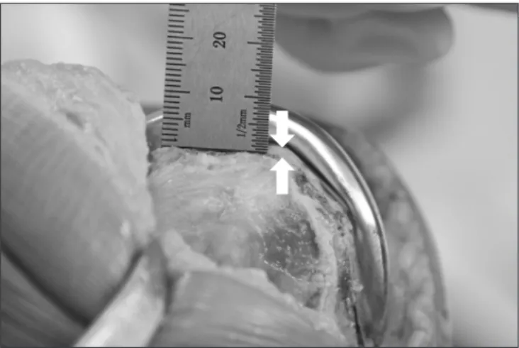
Both the open and closed box designs are not conducive to postoperative radiographic assessment of the cement mantle in the distal femur. Therefore, the mediolateral thickness of the ce
ment at the interface was measured intraoperatively after implant insertion in the distal femur and proximal tibia using a 0.5 mm graduated ruler. To minimize measurement errors, two investi
gators performed measurements twice each and used the most accurate value for analysis (Fig. 2). An outlier was defined as 1) a mantle thickness of >1 mm in the distal femur or proximal tibia or 2) a combined value of >2 mm. Statistical analysis was per
formed using the SPSS ver. 16.0 (SPSS Inc., Chicago, IL, USA).
The Student ttest and chisquare test were used to compare the cement mantle thickness between the groups. A pvalue of ≤0.05 was considered statistically significant.
results
The combined mantle thickness in the femur and tibia was 2.1±0.7 mm in 80 knees and the value was >2 mm in 16 knees (20%). The mean thickness in the distal femur was 1.3 mm in the medial area and 1.4 mm in the lateral area. The value in the proximal tibia was 0.8 mm and 0.8 mm, respectively, showing no mediolateral difference (p>0.05). The mean cement thick
ness at the interface was 1.4 mm in the distal femur and 0.8 mm in the proximal tibia. The value was ≥1 mm in 40 knees (50%)
in the distal femur, which was high compared to 6 knees (7.5%) in the proximal tibia (p<0.001). The cement thickness was sig
nificantly greater in group I patients with the closed box design (2.6±0.6 mm) than group II patients with the open box design (1.6±0.5 mm) (p<0.001). The combined value was >2 mm in 14 knees (35%) in group I and in II knees (5%) in group II, showing statistically significant intergroup difference (p<0.01). The value was greater in both the distal femur and proximal tibia in group I (1.7±0.4 mm and 0.9±0.3 mm, respectively) than group II (1.0±0.3 mm and 0.6±0.2 mm, respectively) (p<0.001). The incidence of
>1 mm thickness in the distal femur and proximal tibia was sig
nificantly higher in group I (32 [80%] and 6 [15%], respectively) than group II (8 [20%] and 0 [0%], respectively) (p<0.05) (Table 2).
discussion
In this study, we compared the thickness of the cement mantle in the distal femur and proximal tibia with the preoperatively planned target thickness in our TKA patients who had implants with two different box designs. The cement mantle thickness in the distal femur was thicker in the knees with the closed box design than in those with the open box design. Therefore, our findings suggest that particular care should be taken when using a closed box design in TKA.
Stable implant fixation for prevention of component loosening can be obtained with the use of cement that penetrates into the cancellous bone or forms a mantle at the interface between the bone and the implant. Cement penetration into the pore open
ings of the cancellous bone (interdigitation) facilitates bone to cement bonding and prevents ingress of microparticulate debris into the tibia, obviating the risk of component loosening and osteolysis5,7,8,15). Although cement is strong under compression, it between the Closed and Open Box Implants
Parameter Group I
(n=40) Closed box
Group II (n=40)
Open box pvalue Cement mantle thickness
Total (mm) 2.6±0.6 1.6±0.5 0.000
Femoral (mm) 1.7±0.4 1.0±0.3 0.000
Tibial (mm) 0.9±0.3 0.6±0.2 0.000
Total >2 mm (%) 14 (35) 2 (5) 0.002
Femoral >1 mm (%) 32 (80) 8 (20) 0.000
Tibial >1 mm (%) 6 (15) 0 (0) 0.034
Values are presented as mean±standard deviation or case.
Fig. 2. Measurement of cement mantle thickness at the interface in the distal femur.
has a low shear strength. Therefore, to obtain an optimal cement bone interface shear strength through interdigitation, a 3–5 mm cement penetration depth has been advised. Techniques using a spatula, finger pressing, and a cement gun have been suggested to improve penetration of cement into bone3,16,17). In contrast, the ideal thickness of cement mantle that bonds the prosthesis to the bone at the interface has been considered ≤1 mm12). Due to the presence of a 1 mm cement pocket in the implant, the mantle thickness can be maintained after impaction. Therefore, consid
ering its impact on the gap kinematics, the thickness should be
≤1 mm in the distal femur and proximal tibial each. However, in our patients, the mean mantle thickness was ≥1 mm in the distal femur (mean, 1.3 mm) although it was less than <1 mm in the proximal tibia (mean, 0.8). In addition, the prevalence of ≥1 mm thickness in the femur was 50% whereas it was only 7.5% in the tibia.
Factors that can influence the cement mantle thickness include cementing techniques and implant designs. Cementing tech
niques consist of the cement mixing process and implant fixation process. The former process refers to polymerization of methyl methacrylate (polymethyl methacrylate) by mixing polymer powder and monomer liquid. According to changes in morphol
ogy of the cement, it can be divided into 4 stages (wetting stage, liquid stage, doughy stage, curing stage, and setting stage12,18)).
In the doughy stage, the cement no longer adheres to a surgical glove so that it can be applied to an implant and the bone, and the implant should be inserted into the knee in this stage. The length of doughy stage is variable ranging from 3 to 7 minutes according to the type of cement, storage method, temperature, and mix
ing speed. Proper fixation of the femoral and tibial components within the optimal application time frame is critically dependent on the surgeon’s experience19,20). We mixed the polymer powder and monomer liquid for 1 full minute and started to apply it to the implant and the cut surface when it was relatively thin to se
cure sufficient time for cementing. Bone cements can be divided into three types (low, medium, and high) according to viscosity.
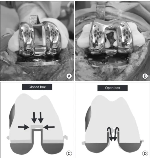
Fig. 3. (A, B) Cement leakage from the periphery of the closed box type implant and from the periphery and central box of the open box type implant. (C) Less cement leakage in the box area (arrows) can pre
vent compression of the femoral compo
nent in the case of closed box type implant.
(D) Cement can easily leak (arrows) and the implant can be compressed in the case of open box type implant.
Closed box Open box
C D
A B
ing and doughy stage, which affects the cementing technique. In TKA, high or medium viscosity cement is commonly used5,21,22). For proper implant fixation, the components should be impacted into position onto the bone surface. During this procedure, ex
cess cement that leaks between the implant and the bone surface should be removed. Then, to ensure the absence of excess cement at the interface, the component is pressed with an impactor or with an insertion of a trial bearing in knee extension under axial loading until the cement is cured.
Although we used the same cementing technique in all pa
tients, the cement mantle thickness was larger in some patients.
We think this can be attributed to the implant design. TKA can be performed either with an open box femoral component or a closed box femoral component. The open box design has a cam in the box area revealing the bone. It requires an additional time for thorough cement removal to prevent contact of residual ce
ment with the bearing post; however, it necessitates small bone resections and allows for intramedullary Kirschner wire insertion in case of periprosthetic fracture. In the closed box design, the box is covered by metal acting as a cam exhibiting the opposite advantages and disadvantages of the open box design12). We compared the cement mantle thickness between the two designs under the hypothesis that the closed box design would disrupt leaking of the excess cement between the prosthesis and the bone surface resulting in a thicker cement mantle. The combined ce
ment mantle thickness in the distal femur and the proximal tibia was larger in the closed box design group than in the open box design group (2.6±0.6 mm vs. 1.6±0.5 mm). The percentage of knees with a value of >2 mm was also higher in the closed box design group than in the open box design group (35% vs. 5%). In particular, the value in the distal femur was higher in the closed box design group than in the open box design group (1.7±0.4 mm vs. 1.0±0.3 mm) and >1 mm was significantly more preva
lent in the closed box design group than in the open box design group (80% vs. 20%). All these findings appear to corroborate our hypothesis. It is our understanding that the closed box design provides a broader contact with the bone surface than the open box design and reduces postoperative bleeding, but the excess cement contained in the box even after impaction disrupts stable fixation of the prosthesis to the bone surface6) (Fig. 3). Since there was no significant difference in the tibial component design, we did not expect the cement mantle thickness in the tibia would be different between the groups. However, the incidence of >1 mm thickness in the tibia was higher in the closed box design group than in the open box design group (15% vs. 0%). The open box
design requires wide and deep tibial reaming of the medullary cavity whereas the closed box design allows for tapered ream
ing for the stem to better fit into the cavity, resulting in increased resistance during pressfit fixation of the tibial component. We surmise that the greater resistance of the closed box design had an influence on the cement thickness between the implant and the bone surface (Fig. 4).
One of the limitations of this study is that we included only one implant for each design in the analysis. Therefore, it is question
able whether our findings can be applied to other implants of each design, and our results should be confirmed by further re
search for generalization. In addition, we did not examine various factors that can influence the cement mantle thickness other than the cementing techniques and implant designs. Although the results could be affected by the extension gap tightness variance associated with the use of a 2 mm bearing insert and errors in bone resection and thickness measurement, we did not take these factors into consideration because the surgery was performed by the same surgeon using the same technique. Last, we did not in
vestigate the clinical significance of the cement mantle thickness;
however, it was beyond the scope of this study where the impact of implant design on cement mantle thickness in TKA was exam
ined. We believe clinical outcomes should also be addressed in a future study based on a further followup.
We identified that the closed box design knee implant was as
sociated with the greater cement mantle thickness than the open box design knee implant after TKA performed using the same Fig. 4. (A) Cement filling (arrows) around the tibial stem of PS150 RP (closed box type) implant suggesting restriction of cement spread. (B) Deeper cement penetration into the medullary canal of tibia in LPSFlex (open box type) implant.
A B
surgical technique. Therefore, it is advised to take extra caution in cementing of the closed box design in TKA. In our opinion, it could be helpful to insert an implant when the cement is in a thinner stage or to use less amount of cement.
conclusions
The cement mantle thickness was greater in the distal femur than in the proximal tibia in our TKA patients (1.4 mm vs. 0.8 mm). In particular, the closed box design (PS150 RP) was associ
ated with the greater cement mantle thickness than the open box design (LPSFlex). We think that the closed box design obstructs leakage of residual cement, adversely affecting impaction of the implant. Therefore, extra care should be taken to avoid creating a thick cement mantle that affects the gap balance especially when a closed box design is used in TKA.
conflict of interest
No potential conflict of interest relevant to this article was re
ported.
references
1. Kopec M, Milbrandt JC, Duellman T, Mangan D, Allan DG.
Effect of hand packing versus cement gun pressurization on cement mantle in total knee arthroplasty. Can J Surg.
2009;52:4904.
2. Lutz MJ, Pincus PF, Whitehouse SL, Halliday BR. The ef
fect of cement gun and cement syringe use on the tibial cement mantle in total knee arthroplasty. J Arthroplasty.
2009;24:4617.
3. Schlegel UJ, Bishop NE, Püschel K, Morlock MM, Nagel K.
Comparison of different cement application techniques for tibial component fixation in TKA. Int Orthop. 2015;39:47
54.
4. Vanlommel J, Luyckx JP, Labey L, Innocenti B, De Corte R, Bellemans J. Cementing the tibial component in total knee arthroplasty: which technique is the best? J Arthroplasty.
2011;26:4926.
5. Kopec M, Milbrandt JC, Kohut N, Kern B, Allan DG. Ef
fect of bone cement viscosity and set time on mantle area in total knee arthroplasty. Am J Orthop (Belle Mead NJ).
2009;38:51922.
6. Saari T, Li MG, Wood D, Nivbrant B. Comparison of ce
menting techniques of the tibial component in total knee
replacement. Int Orthop. 2009;33:123942.
7. Norton MR, Eyres KS. Irrigation and suction technique to ensure reliable cement penetration for total knee arthro
plasty. J Arthroplasty. 2000;15:46874.
8. Walker PS, Soudry M, Ewald FC, McVickar H. Control of cement penetration in total knee arthroplasty. Clin Orthop Relat Res. 1984;(185):15564.
9. Dorr LD, Lindberg JP, ClaudeFaugere M, Malluche HH.
Factors influencing the intrusion of methylmethacrylate into human tibiae. Clin Orthop Relat Res. 1984;(183):14752.
10. Kim YH, Walker PS, Deland JT. A cement impactor for uniform cement penetration in the upper tibia. Clin Orthop Relat Res. 1984;(182):20610.
11. Fukushima H, Hashimoto Y, Yoshiya S, Kurosaka M, Matsu
da M, Kawamura S, Iwatsubo T. Conduction analysis of ce
ment interface temperature in total knee arthroplasty. Kobe J Med Sci. 2002;48:6372.
12. Cho WS, Kim MY, Youm YS. Knee joint arthroplasty. 3rd ed.
Seoul: Young Chang; 2015. p4851, 184.
13. Vertullo CJ, Davey JR. The effect of a tibial baseplate under
surface peripheral lip on cement penetration in total knee arthroplasty. J Arthroplasty. 2001;16:48792.
14. Dennis DA, Komistek RD, Kim RH, Sharma A. Gap bal
ancing versus measured resection technique for total knee arthroplasty. Clin Orthop Relat Res. 2010;468:1027.
15. Mann KA, Ayers DC, Werner FW, Nicoletta RJ, Fortino MD.
Tensile strength of the cementbone interface depends on the amount of bone interdigitated with PMMA cement. J Biomech. 1997;30:33946.
16. Cawley DT, Kelly N, McGarry JP, Shannon FJ. Cementing techniques for the tibial component in primary total knee replacement. Bone Joint J. 2013;95:295300.
17. Vaninbroukx M, Labey L, Innocenti B, Bellemans J. Cement
ing the femoral component in total knee arthroplasty: which technique is the best? Knee. 2009;16:2658.
18. Cameron HU, Mills RH, Jackson RW, Macnab I. The struc
ture of polymethylmethacrylate cement. Clin Orthop Relat Res. 1974;(100):28791.
19. Haas SS, Brauer GM, Dickson G. A characterization of poly
methylmethacrylate bone cement. J Bone Joint Surg Am.
1975;57:38091.
20. Krause WR, Krug W, Miller J. Strength of the cementbone interface. Clin Orthop Relat Res. 1982;(163):2909.
21. Silverman EJ, Landy DC, Massel DH, Kaimrajh DN, Latta LL, Robinson RP. The effect of viscosity on cement penetra
tion in total knee arthroplasty, an application of the squeeze
22. Jiang J, Lv H, Zhu J. Influence of closedbox and openbox knee prosthesis on postoperative blood loss in patients un
Jian Wai Ke Za Zhi. 2008;22:911.