Journal of Bacteriology and Virology 2009. Vol. 39, No. 3 p.165 – 171 DOI 10.4167/jbv.2009.39.3.165
Genetic Analysis of Hepatitis A Virus Isolated from Korea
Kyung-Ok Lee*, Su-Jin Jeong, Hye-Soon Seong, Kyung-Tae Kim, Yoo-Sung Hwang, Gee-Young Kim and Sun-Hwa Lee Genome Research Center, Neodin Medical Institute, Seoul, Korea
Hepatitis A virus (HAV) is one of the most important causes of acute infectious hepatitis. The aim of this study was to determine the genotypes of HAV that have been circulating in Koreans. A total of 76 sera referred to our institute for HAV genotyping from 11 Korean provinces were used for this study. Those samples were diagnosed by positive of IgM anti-HAV. HAV RNA was extracted from 150 μl of serum, and reverse transcription PCR-sequencing was used to detect and characterize HAV RNA. Primer pairs from the VP1/2A region of the HAV were used for amplification and sequencing. HAV RNA was found in 64.5% (n = 49) of the 76 patient sera with acute hepatitis A. Forty-seven strains were genotype IIIA in a total of 49 isolated strains (95.9%, 47/49); only two strains belonged to genotype IA (4.1%, 2/49). Thirty eight genotype IIIA isolates were 100% identical to consensus amino acid sequences of the reference strain AJ299467. The amino acid change of L772F was found in two IIIA strains; other IIIA isolates showed one amino acid change. Amino acid of genotype IA was compared to reference strain L20541. K801R was found in 1 strain and Q810S in both strains. The amino acid change of K801R was the first report in Koreans. Until recently HAV genotype IA has been reported as a major circulating HAV genotype in Koreans. In the present study, the predominant HAV strain in Koreans seemed to be HAV genotype IIIA.
Key Words: Hepatitis A, Genotype
INTRODUCTION
Hepatitis A virus (HAV) infection is the leading cause of clinically apparent viral hepatitis in many parts of the world, including both developed and developing countries. HAV is a hepatotropic member of the Picornaviridae family and has been classified in the genus hepatovirus. Its genome is a linear, positive-sense, single-stranded RNA, about 7.5 kb in length (1). Good sanitation practices and a clean water supply are essential to the prevention of HAV infection. In developing countries experiencing a high incidence of HAV,
the main transmission route of HAV is the fecal-oral route caused by poor sanitation, which increases the chance of ingesting contaminated food or water (2). In a number of Asian and Latin American countries, the epidemiology is presently changing from high to intermediate endemicity, due to improvements in sanitary conditions (3, 4). In Korea, due to sanitation-related improvements, epidemic outbreaks of HAV infection have become less frequent (5). However, recently HAV infection is rapidly increasing nationwide, especially in young people who have a weak immunity to HAV.
HAV strains have maintained low rates of mutation accumulation over extended periods of time (6). However recent nucleotide sequencing of selected genome regions that encode the putative VP1/2A junction region of wild-type HAV strains present in human specimens has demonstrated substantial sequence heterogeneity. Using this approach, the 165
Received: June 16, 2009/ Revised: July 15, 2009 Accepted: July 21, 2009
*Corresponding author: Kyung Ok Lee, Ph.D. Genome Research Center, Neodin Medical Institute, #2-3, Yongdap-Dong, Sungdong-gu, Seoul, 133-847, Korea.
Phone: +82-2-2244-6500, Fax: +82-2-2212-1307, e-mail: [email protected]
Original Article
isolates of HAV have been categorized into seven genotypes (7). The genomic characterization of HAV has been carried out mainly by genotyping strains from different geographic regions worldwide (8). De Paular et al. (9) reported that genotype IA constitutes the major HAV population in North America, China, the former USSR, and Thailand, whereas genotype IB contains strains from Europe, North Africa, the Middle East, and Australia. Co-circulation of IA and IB of HAV has been reported in Italy (10); and in Brazil (11).
HAV genotype III has been reported to be linked with intravenous drug use in Sweden during the 1980s (7); it has also been found in India, Sri Lanka, Nepal, Malaysia, and the USA (12); in Norway (13); in Germany (14); and in Russia (15). Studies from Korea have previously shown the HAV genotype I to be predominant, and some changes therein have been recently reported (16~19). In this study, the molecular epidemiology of HAV viral strains were determined by reverse transcription polymerase chain reaction (RT-PCR) and sequencing in the VP1/2A region of HAV isolates from Korean patients.
MATERIALS AND METHODS RNA extraction from patient sera
Seventy-six sera of HAV patients from 11 Korean provinces of Korea during 2007~2008 were tested (Table 1). The diagnosis of hepatitis A was based on a high titer serum IgM anti-HAV level. HAV RNA was extracted from 150 μl of serum using a Viral Gene spin kit (Intron, Sungnam, Korea). Viral gene-spin lysis buffer (250 μl) was added to serum and incubated at room temperature for 10 min and 350 μl of binding buffer was added. Lysate was loaded on a spin column and centrifuged at 13,000 rpm for 1 min. After discarding the solution, 500 μl of washing buffer A was added to column and centrifuged for 1 min at 13,000 rpm. After discarding the solution, 500 μl of washing buffer B was added to the column and centrifuged for 1 min at 13,000 rpm. After discarding the solution, the spin column was placed in a RNase-free 1.5 ml microcentrifuge tube, and elution buffer (30 μl) was directly added onto the membrane and incubated at room temperature for 1 min.
Eluted solution (2~5 μl) was used for PCR.
HAV genome amplification
For reverse transcription, 1 μl of RNA solution was heat-denaturated at 68℃ for 10 min. It was chilled rapidly on ice and mixed with 4 μl of 1.5 mM MgCl2 solution, 2 μl of RNA PCR buffer (100 mM Tris-HCl, pH 8.3, 500 mM KCl), 8.5 μl of RNase-free distilled water, 2 μl of dNTP mixture (10 mM dATP, dCTP, dGTP, dTTP), 1 μl of random 9-mers (5'-NNNNNNNNN-3'), 0.5 μl of RNase inhibitor (Takara-Shuzo, Kyoto, Japan), and 1 μl of reverse transcriptase (Takara-Shuzo). After incubation at 30℃ for 10 min, reverse transcription reaction was carried out at 42℃ for 30 min, followed by inactivation at 95℃ for 5 min. Primers for HAV RT-PCR reported by Takahashi et al.
(20) were used in the present study. The first PCR was performed in 20 μl of reaction mixture containing 1.0 μM each of outer sense primer (5'-GGT TTC TAT TCA GAT TGC AAA TTA-3': nt. 2891-2914) and antisense primer (5'-AGT AAA AAC TCC AGC ATC CAT TTC-3': nt.
3398-3375), 5 μl of cDNA solution, 200 μM of each dNTP, 2.0 μl of 10 × PCR buffer [100 mM Tris-HCl, pH 8.3, 500 mM KCl, 15 mM MgCl2, 0.01% (w/v) gelatin], and 2.5 U of Ex Taq polymerase (Takara-Shuzo) with proofreading activity. The amplification condition were 94℃ for 16 min, followed by 40 cycles of 94℃ for 1 min, 50℃ for 1 min, 72℃ for 1 min, and an additional 10 min at 72℃ in the last cycle. The second PCR was carried out with 1.5 μl of the first PCR product. The reaction mixture contained 1.0 μM each of inner sense primer (5'-TTG CAA ATT ACA ATC ATT CTG-3': nt. 2905-2925) and inner antisense primer (5'-TTC AAG AGT CCA CAC ACT TCT-3': nt.
3377-3367), 2 μl of 10 × PCR buffer and 15.1 μl of RNase free dH2O, 1 μl of dNTP mixture (2 mM dATP, dCTP, dGTP, dTTP), and 0.2 μl of amplitaq gold (Roche Diagnostics, Branchburg, NJ, USA). The amplification conditions for the second PCR were the same as those of the first PCR.
Takara TP-100 PCR machine (Takara Bio Inc., Shiga, Japan) was used for HAV PCR. The PCR products were analyzed by 2% agarose gel electrophoresis, stained with ethidium bromide and visualized by UV transillumination.
Sequencing of PCR products
Amplification products were purified on Wizard PCR Preps DNA purification resin (Promega, Madison, WI, USA), and sequenced bidirectionally with Dye Terminator Cycle Sequencing Ready Reaction kit (PE Applied Bio- systems, Foster City, CA, USA) using the above PCR primers. Sequencing was performed on an automated DNA sequencer ABI 3730 (PE Applied Biosystems). The nucleo- tide sequences of HAV isolates from the patients were compared with those of reference HAV strains retrieved from the GenBank databases. Nucleotide sequences of HAV isolates were compared to published HAV sequences from different genotypes. The reference sequences of HAV, listed here with the NCBI accession number of the HAV VP1/P2A junction region, belong to genotypes IA (L20541, L07676, L20553, L07722, and L07717), IB (L07703), IIB (L07729), IIIA (L07725, L07668, and AJ299467), IIIB (L20532), IV (L07732) and VI (L07731). The relatedness of the HAV RNA nucleic acid sequence was assessed through multiple sequence alignment, using the Clustal X program (21). The calculation of nucleotide and amino acid identity, the determination of genetic distances between sequences, and the construction of phylogenetic trees were Table 1. Molecular characters and nucleic acid sequences
homologies of 49 strains at the VP1/2A region of hepatitis A Virus (HAV) in Korean HAV isolates compared to reference strains, AJ299467 and L20541 for genotype IIIA and IA, respectively
Isolate Identity (%) Genotype Province Year ND91610 99.22 3A Chonan 2007 ND91611 98.83 3A Inchon 2007 ND91612 99.22 3A Seoul 2007 ND91613 99.61 3A Seoul 2007 ND91614 99.22 3A Suwon 2007 ND91616 99.61 3A Anyang 2007 ND91618 99.61 3A Inchon 2007 ND91620 98.44 3A Seoul 2007 ND91623 96.50 3A Kwangju 2007 ND91624 98.83 3A Jonju 2007 ND92401 98.83 3A Anyang 2008 ND92403 98.44 3A Seoul 2008 ND92404 98.83 3A Suwon 2008 ND92406 98.83 3A Anyang 2008 ND92407 99.61 3A Wonju 2008 ND92408 99.61 3A Seoul 2008 ND92427 99.61 3A Anyang 2008 ND92428 98.83 3A Seoul 2008 ND92430 99.22 3A Seoul 2008 ND92432 99.22 3A Kwangju 2008 ND92433 99.22 3A Anyang 2008 ND92435 99.61 3A Seoul 2008 ND92437 99.61 3A Kwangju 2008 ND92439 99.61 3A Seoul 2008 ND92443 100.00 3A Inchon 2008 ND92444 99.22 3A Anyang 2008 ND72600 99.22 3A Chonan 2008 ND72800 98.05 3A Suwon 2008 ND81200 98.83 3A Inchon 2008 ND81800 99.61 3A Seoul 2008 ND82600 98.83 3A Anyang 2008 ND90400 98.05 3A Inchon 2008 ND92301 99.22 3A Inchon 2008 ND92303 100.00 3A Seoul 2008 ND92305 97.28 3A Inchon 2008
Table 1. Continued
Isolate Identity (%) Genotype Province Year ND10141 98.83 3A Inchon 2008 ND10143 99.22 3A Anyang 2008 ND10144 99.22 3A Inchon 2008 ND10211 99.61 3A Anyang 2008 ND91201 99.22 3A Seoul 2008 ND91203 99.61 3A Anyang 2008 ND91205 99.61 3A Seoul 2008 ND91206 99.22 3A Seoul 2008 ND91207 99.61 3A Kangleung 2008 ND12312 98.83 3A Seoul 2008 ND12314 99.22 3A Wulsan 2008 ND12315 98.44 3A Seoul 2008 ND91622 96.43 1A Suwon 2007 ND93000 96.43 1A Seoul 2008
performed through the use of MEGA software, version 4.0 for Windows. Phylogenetic trees were generated via the neighbor-joining (N-J) method (1).
RESULTS
Genetic analysis of HAV isolates
HAV RNA was found in 49 samples in a total of 76 sera of HAV patients (64.5%) collected from the 11 provinces of Korea (Table 1). The sequence between nucleotides 3024 and 3191 of the VP1/2A region was determined for the 49 patients. In the present study, HAV genotype IIIA was predominant (95.9%, 47/49); only two strains belonged to genotype IA (4.1%, 2/49). Two HAV genotype 1A strains were found in Suwon area in 2007 and Seoul area in 2008.
Nucleic acid sequences homologies at the VP1/2A junction of HAV genome in HAV isolates compared to reference strains were listed in Table 1. The nucleotide sequence identities of the VP1/P2A junction region with genotype IIIA ranged from 96.5% to 100%, compared to the reference strain, AJ299467. While genotype IA showed 96.4% identity with regards to nucleotide sequence identity, compared to the reference strain, L20541 (Table 1). We then performed a phylogenetic analysis of the region between nucleotides 3024 and 3191 and classified the virus strains (Fig. 1).
Most strains belonged to genotype IIIA, except two strains that belonged to genotype IA.
Changes in amino acid sequences among HAV isolates
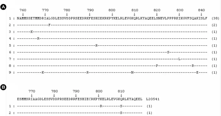
A comparison of the amino acid sequences from position 758 to 842 of the VP1/2A region of HAV strains are pre- sented in Fig. 2. Amino acid substitutions were found 12 and 2 different sites in genotype IIIA and IA, respectively (Table 2). Within genotype IIIA, the amino acid sequence was conserved with a few exceptions, while greater differences Figure 1. Neighbor-joining phylogenetic tree nucleotide sequences in the VP1/P2A region of HAV isolates in Korea. The tree includes reference strains L20541, L07676, L20553, L07722 and L07717 in genotype IA, L07703 in genotype IB, L07729 in genotype IIB, L07725, L0766 and AJ2994678 in genotype IIIA, L205328 in genotype IIIB, L077328 in genotype IV, L07731 in genotype VI. The horizontal bar at the bottom represents a genetic distance of 0.02.
were evident in the amino acid sequences between genotype IIIA and IA of HAV. Among the genotype IIIA strains, 38
isolates showed 100% identity in terms of consensus amino acid sequences with the reference strain AJ299467, a representative HAV genotype IIIA strain. The amino acid change of L772F was found in two IIIA strains. Amino acid changes of E764K, M767R, R769K, K789R, H793R, K809N, S820P, P825S, I830L, Q836R, and K838E were found in each of the different genotype IIIA strains (Table 2). These changes in amino acid sequence of genotype IIIA have never been reported in Koreans. Two HAV genotype IA strains were compared to reference strain L20541. The amino acid change of K801R was found in one strain, and that of Q810S was found in both strains (Table 2).
DISCUSSION
More recently, HAV infection has increased in Korean peoples. Vaccination of HAV is recommended to persons who don't have anti-HAV IgG. In the present study, we examined HAV genotypes in the HAV patients who showed positive reaction for anti-HAV IgM. Compared to other RNA viruses, the amino acid and nucleotide sequences of Table 2. Amino acid changes of Korean HAV strains in VP1/2A
region of HAV from 2007 to 2008 Amino acid
position Genotype Prototype Substitution Number 764 3A E K 1 767 3A M R 1 769 3A R K 1 772 3A L F 2 789 3A K R 1 793 3A H R 1 809 3A K N 1 820 3A S P 1 825 3A P S 1 830 3A I L 1 836 3A Q R 1 838 3A K E 1 801 1A K R 1 810 1A Q S 2 A
B
Figure 2. Comparison of the predicted amino acid sequences of the VP1/VP2 junction region of HAV. The consensus amino acid sequences of AJ299467 for HAV genotype IIIA and L20541 for HAV genotype IA are shown on the top line of (A) and (B), respectively.
"-" indicates conserved amino acid; differences are shown by the appropriate single letter amino acid code. The numbers above the consensus amino acid sequence indicate the predicted amino acid number from the start of HAV full amino acid. The numbers of the right ends indicate the number of HAV strains we found in the present study.
HAV are highly conserved (10). In recent years, studies on the nucleic acid heterogeneity of HAV isolates have allowed the characterization of strains and groupings into different genotypes and sub-genotypes. Studies of genotypes and changes in the nucleotide and amino acid sequences of HAV may provide valuable information with regards to the epidemiological aspects of a particular region (7).
Among several genome regions of HAV, including the C terminus of the VP3 protein (22), the N terminus of the VP1 protein (12), the junction region of VP1/2A proteins (23, 24) and the entire VP1 protein (25) have been used for HAV genotyping. The VP1/2A junction region selected in our study has been most frequently used for HAV genotype analysis in Korea (17~19). Among HAV isolates from Korean patients with acute hepatitis A from 1994 to 1998, all 18 isolates were clustered within genotype IA, irrespec- tive of the patients' geographical location (26). Moreover outbreak cases of HAV caused by HAV genotype IA have been reported in Korea (16, 17). Yun et al. (19) reported the prevalence of IA and IIIA to be 83.3% and 16.7%, respectively; these data showed that the prevalent HAV genotype was IA, however the newly identified IIIA strain was co-circulating with the genotype IA strain during 2005~2006. They suggested that new IIIA strains might have been imported from high-endemic countries into Korea.
In the present study, a phylogenetic analysis of the sequences obtained from distinct HAV isolates revealed that most of the strains were genotype IIIA (95.9%); IA was found in only 4.1% of the specimens (Fig. 1). Executing a comparison of the partial nucleotide sequences of the HAV genome is a useful approach to defining the differences among HAV isolates (27). The molecular characters and amino acid sequences homologies of HAV isolates at the VP1/2A region of HAV genome were compared to reference strains, AJ299467 for genotype IIIA and L20541 for geno- type IA, which were used in a previous study (19). HAV strains detected in this study showed over 96.0% identity compared to reference strains. Most genotype IIIA isolates showed the same amino acid sequences compared to reference strain; other isolates were conserved, with one or
two amino acid changes (Fig. 2). This is the first report of amino acid changes of genotype IIIA in the VP1/2A region of HAV in Korean population. In two genotype IA strains, one strain was changed from K to R in position 801, while the other strain was changed from Q to S in position 810 (Table 2). The amino acid change of K801R of genotype 1A strain is the first reported in this study, whereas Q810S was frequently found in a previous study (19). The present study indicates that HAV genotype in Korea seemed to be changed from genotype IA to IIIA. These changes suggest that there is a need for continual surveillance that focuses on strain variation, and that it is important for under- standing the epidemiology and development of a strategy for disease control and prevention.
REFERENCES
1) De Paula VS, Niel C, Teves SC, Villa LM, Virgolino H, Gaspar AM. Molecular epidemiology of hepatitis A virus in Brazilian Amazon. J Gastroenterol Hepatol 2006;21:1435-8.
2)Gust ID. Epidemiological patterns of hepatitis A in different parts of the world. Vaccine 1992;10:S56-8.
3)Barzaga BN. Hepatitis A shifting epidemiology in South-East Asia and China. Vaccine 2000;18:S61-4.
4) Fix AD, Martin OS, Gallicchio L, Vial PA, Lagos R.
Age-specific prevalence of antibodies to hepatitis A in Santiago, Chile: risk factors and shift in age of infection among children and young adults. Am J Trop Med Hyg 2002;66:628-32.
5)Sohn YM, Rho HO, Park MS, Park JH, Choi BY, Ki MR, Jang WI. The changing epidemiology of hepatitis A in children and the consideration of active immuni- zation in Korea. Yonsei Med J 2000;41:34-9.
6)Costa-Mattioli M, Ferre V, Monpoeho S, Garcia L, Colina R, Billaudel S, Vega I, Perez-Bercoff R, Cristina J. Genetic variability of hepatitis A virus in South America reveals heterogeneity and co-circulation during epidemic outbreaks. J Gen Virol 2001;82:2647-52.
7)Robertson BH, Jansen RW, Khanna B, Totsuka A, Nainan OV, Siegl G, Widell A, Margolis HS, Isomura S, Ito K. Genetic relatedness of hepatitis A virus strains
recovered from different geographical regions. J Gen Virol 1992;73:1365-77.
8) Fujiwara K, Yokosuka O, Imazeki F, Saisho H, Saotome N, Suzuki K, Okita K, Tanaka E, Omata M. Analysis of the genotype-determining region of hepatitis A viral RNA in relation to disease severities. Hepatol Res 2003;
25:124-34.
9) De Paula VS, Baptista ML, Lampe E, Niel C, Gaspar AM. Characterization of hepatitis A virus isolates from subgenotypes IA and IB in Rio de Janeiro, Brazil. J Med Virol 2002;66:22-7.
10) Chironna M, Grottola A, Lanave C, Villa E, Barbuti S, Quarto M. Genetic analysis of HAV strains recovered from patients with acute hepatitis from Southern Italy.
J Med Virol 2003;70:343-9.
11) Villar LM, Morais LM, Aloise R, Melo MM, Calado IA, Lampe E, Gaspar AM. Co-circulation of genotypes IA and IB of hepatitis A virus in Northeast Brazil. Braz J Med Biol Res 2006;39:873-81.
12) Robertson BH, Khanna B, Nainan OV, Margolis HS.
Epidemiologic patterns of wild-type hepatitis A virus determined by genetic variation. J Infect Dis 1991;163:
286-92.
13) Stene-Johansen K, Skaug K, Blystad H, Grinde B. A unique hepatitis A virus strain caused an epidemic in Norway associated with intravenous drug abuse. The Hepatitis A Study Group. Scand J Infect Dis 1998;30:
35-8.
14) Heitmann A, Laue T, Schottstedt V, Dotzauer A, Pichl L. Occurrence of hepatitis A virus genotype III in Germany requires the adaptation of commercially available diagnostic test systems. Transfusion 2005;45:
1097-105.
15) Davidkin I, Zheleznova N, Jokinen S, Gorchakova O, Broman M, Mukomolov S. Molecular epidemiology of hepatitis A in St. Petersburg, Russia, 1997~2003. J Med Virol 2007;79:657-62.
16)Kim JS, Kim SH. Molecular epidemiology of an outbreak of hepatitis A in Korea. Korean J Clin Pathol 2001;21:114-8.
17) Park JY, Lee JB, Jeong SY, Lee SH, Lee MA, Choi HJ.
Molecular characterization of an acute hepatitis A outbreak among healthcare workers at a Korean hospital.
J Hosp Infect 2007;67:175-81.
18) Park SH, Byun KS, Song JW, Kim JH, Song KJ, Baek LJ, Kwon OS, Yeon JE, Kim JS, Pak YT, Lee CH.
Molecular epidemiology of Korean strains of hepatitis A virus. Korean J Hepatol 2000;6:276-86.
19) Yun H, Kim S, Lee H, Byun KS, Kwon SY, Yim HJ, Lim YS, Jeong SH, Jee Y. Genetic analysis of HAV strains isolated from patients with acute hepatitis in Korea, 2005~2006. J Med Virol 2008;80:777-84.
20) Takahashi H, Yotsuyanagi H, Yasuda K, Koibuchi T, Suzuki M, Kato T, Nakamura T, Iwamoto A, Nishioka K, Iino S, Koike K, Itoh F. Molecular epidemiology of hepatitis A virus in metropolitan areas in Japan. J Gastroenterol 2006;41:981-6.
21) Thompson JD, Gibson TJ, Plewniak F, Jeanmougin F, Higgings DG. The CLUSTAL_X windows interface:
flexible strategies for multiple sequence alignment aided by quality analysis tools. Nucleic Acids Res 1997;25:
4876-82.
22)Jansen RW, Siegl G, Lemon SM. Molecular epide- miology of human hepatitis A virus defined by an antigen-capture polymerase chain reaction method.
Proc Natl Acad Sci USA 1990;87:2867-71.
23) Pina S, Buti M, Jardi R, Clemente-Casares P, Jofre J, Girones R. Genetic analysis of hepatitis A virus strains recovered from the environment and from patients with acute hepatitis. J Gen Virol 2001;82:2955-63.
24) Wattanasri N, Ruchusatsawat K, Wattanasri S. Phylo- genetic analysis of hepatitis A virus in Thailand. J Med Virol 2005;75:1-7.
25) Costa-Mattioli M, Cristina J, Romero H, Perez-Bercof R, Casane D, Colina R, Garcia L, Vega I, Glikman G, Romanowsky V, Castello A, Nicand E, Gassin M, Billaudel S, Ferre V. Molecular evolution of hepatitis A virus: a new classification based on the complete VP1 protein. J Virol 2002;76:9516-25.
26) Byun KS, Kim JH, Song KJ, Baek LJ, Song JW, Park SH, Kwon OS, Yeon JE, Kim JS, Bak YT, Lee CH.
Molecular epidemiology of hepatitis A virus in Korea.
J Gastroenterol Hepatol 2001;16:519-24.
27) Brown EA, Jansen RW, Lemon SM. Characterization of a simian hepatitis A virus (HAV): Antigenic and genetic comparison with human HAV. J Virol 1989;63:
4932-7.