Shear bond strength of resin cement to an acid etched and a laser irradiated ceramic surface
Pinar Kursoglu1*, DDS, PhD, Pelin Fatma Karagoz Motro1, DDS, MSc, Haktan Yurdaguven2, DDS, PhD
1Department of Prosthodontics, Faculty of Dentistry, Yeditepe University, Istanbul, Turkey
2Department of Restorative Dentistry, Faculty of Dentistry, Yeditepe University, Istanbul, Turkey
PURPOSE. To evaluate the effects of hydrofluoric acid etching and Er,Cr:YSGG laser irradiation on the shear bond strength of resin cement to lithium disilicate ceramic. MATERIALS AND METHODS. Fifty-five ceramic blocks (5 mm × 5 mm × 2 mm) were fabricated and embedded in acrylic resin. Their surfaces were finished with 1000-grit silicon carbide paper. The blocks were assigned to five groups: 1) 9.5% hydrofluoric-acid etching for 60 s; 2-4), 1.5-, 2.5-, and 6-W Er,Cr:YSGG laser applications for 60 seconds, respectively; and 5) no treatment (control). One specimen from each group was examined using scanning electron microscopy. Ceramic primer (Rely X ceramic primer) and adhesive (Adper Single Bond) were applied to the ceramic surfaces, followed by resin cement to bond the composite cylinders, and light curing. Bonded specimens were stored in distilled water at 37℃ for 24 hours. Shear bond strengths were determined by a universal testing machine at 1 mm/min crosshead speed. Data were analyzed using Kruskal–Wallis and Mann–Whitney U-tests (α=0.05). RESULTS.
Adhesion was significantly stronger in Group 2 (3.88 ± 1.94 MPa) and Group 3 (3.65 ± 1.87 MPa) than in Control group (1.95 ± 1.06 MPa), in which bonding values were lowest (P<.01). No significant difference was observed between Group 4 (3.59 ± 1.19 MPa) and Control group. Shear bond strength was highest in Group 1 (8.42 ± 1.86 MPa; P<.01). CONCLUSION. Er,Cr:YSGG laser irradiation at 1.5 and 2.5 W increased shear bond strengths between ceramic and resin cement compared with untreated ceramic surfaces. Irradiation at 6 W may not be an efficient ceramic surface treatment technique. [J Adv Prosthodont 2013;5:98-103]
KEY WORDS: Ceramic; Resin cement; Er,Cr: YSGG Laser; Shear bond strength
INTRODUCTION
Interest in the use of ceramic laminate veneers, inlays, and onlays has grown considerably in recent years due to their excellent esthetic properties.1,2 However, ceramic restora- tions are very brittle and have limited flexural strength;
thus, adhesive cementation increases the risks of fracture3-6 and debonding.7,8 Successful treatment depends in part on
the strength of the bond between the composite resin and the ceramic restoration.9 In addition, the repair of ceramic restoration fractures caused by inadequate design or occlu- sal forces requires surface preparation.10-12 The ceramic sur- face must be roughened to achieve successful chemical bonding and micromechanical interlocking with the resin cement.13-20
Various techniques can be used to optimize bond strength at the ceramic–cement interface. Common treat- ment options include abrasion with a diamond rotary cut- ting instrument18,19 or aluminum oxide particles,21,22 as well as acid etching.17,23,24 Etching with a hydrofluoric (HF) acid solution can achieve proper ceramic surface texture13-17 by dissolving the glass matrix to expose the crystalline phase.25,26 However, this method is hazardous for both the operator and patient. The operator must be protected from skin and eye damage, and any remaining HF acid must be removed completely with an ultrasonic cleaner before cementation. Given these limitations, HF acid etching can- not be used in some cases, such as ceramic restoration
Corresponding author:
Pinar Kursoglu
Department of Prosthodontics, Faculty of Dentistry, Yeditepe University, Bagdat Street. No:238 Goztepe, 34728 Istanbul, Turkey
Tel. 902163636044, 905322862350: e-mail, [email protected] Received October 2, 2012 / Last Revision February 14, 2013 / Accepted April 25, 2013
© 2013 The Korean Academy of Prosthodontics
This is an Open Access article distributed under the terms of the Creative Commons Attribution Non-Commercial License (http://creativecommons.
org/licenses/by-nc/3.0) which permits unrestricted non-commercial use, distribution, and reproduction in any medium, provided the original work is properly cited.
repair;27 and so more biofriendly techniques are needed.
Relatively safe and easy methods of laser irradiation for surface modification have been proposed.28-33 Studies have examined the use of several laser types, including neodymi- um: yttrium aluminum garnet (Nd:YAG), erbium (Er):YAG,34 erbium, chromium: yttrium, scandium, gallium, garnet (Er,Cr:YSGG),35 and carbon dioxide36lasers. The use of pulsed erbium lasers, such as Er:YAG and Er,Cr:YSGG lasers, has been considered for surface treatment. Previous reports have shown that the Er,Cr:YSGG laser is capable of producing surface roughness comparable to that pro- duced by acid etching of enamel and dentin surfaces.37,38 Many studies have evaluated the effects of different surface treatments,12,15,39-41 including laser treatments,29,34,42-44 on the shear bond strength of resin cement to ceramic restora- tions. Shear bond strengths in Nd:YAG and Er:YAG laser- irradiated ceramic surfaces have also been investigated,44 but not in those treated with a Er,Cr:YSGG laser. In addi- tion, no consensus on the appropriate power settings for particular laser types has been reached.29,34,43
Given the lack of information regarding the effects of different Er,Cr:YSGG laser power settings on ceramic sur- face roughness, we aimed to compare the effects of differ- ent ceramic surface modification techniques (HF acid etch- ing and Er,Cr:YSGG laser irradiation) and laser power set- tings on the shear bond strength of resin cement to lithium disilicate ceramic.
MATERIALS AND METHODS
Fifty-five lithium disilicate ceramic blocks (5 mm × 5 mm
× 2 mm; IPS Empress 2, Ivoclar Vivadent, Schaan, Liech- tenstein) were fabricated using the hot pressing technique according to the manufacturer’s instructions and embedded in acrylic resin. All specimen surfaces were wet ground with 300-1000 grit silicon carbide paper (English Abrasives &
Chemicals Ltd., London, UK) on a grinding device (Phoenix Beta, Buechler, IL, USA) and ultrasonically cleaned (CD-4800 Digital Ultrasonic Cleaner; Jeken, Dongguan, China) in distilled water for 180 seconds to remove contaminants before surface treatment. They were then dried with oil-free air and assigned to five groups (n=11).
Specimens in Group 1 were subjected to 9.5% HF acid etching (buffered porcelain etch; Ultradent Products, South Jordan, UT, USA) for 60 seconds, rinsing for 5 minutes to remove residual acid, ultrasonic cleaning for 180 seconds, and then air drying. The ceramic surfaces of specimens in groups 2-4 were subjected to Er,Cr:YSGG laser irradiation (Millenium; Biolase Technology, Inc., San Clemente, CA, USA) with a 2.78 µm wavelength, pulsed laser-powered hydrokinetics, and energy parameters of 300 mJ at 1.5, 2.5, and 6 W, respectively. The air and vapor were adjusted to 50% of the laser unit. The optical fiber of the laser (400 μm diameter, 4 mm length) was aligned perpendicular to each specimen at a distance of 1 mm and moved manually in a sweeping fashion over the entire area during a 60 sec- onds exposure period. The laser-irradiated specimens were
then cleaned ultrasonically for 180 seconds and air dried.
Specimens in Group 5 received no surface treatment and served as controls. One specimen from each group was selected for scanning electron microscopic (SEM) evaluation.
After surface treatment, a ceramic primer (Rely X; Lot
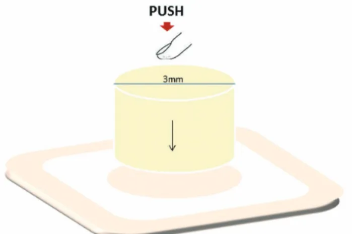
#2721; 3M ESPE, St. Paul, MN, USA) was applied to the ceramic surfaces allowed to evaporate for 3 minutes and air-dried for 30 seconds. Then, an adhesive (Adper Single Bond; Lot #1122; 3M ESPE, St. Paul, MN, USA) was applied to the ceramic surfaces and air dried to achieve thinning without light curing, according to the manufactur- er’s instructions. To standardize the areas subjected to resin cement bonding, a perforated sticker (3 mm diameter) was placed in the center of each specimen (Fig. 1). Cylindrical composite resin blocks (3 mm diameter, 4 mm length;
Filtek Z250, Lot #6020A2; 3M ESPE, St. Paul, MN, USA) were created using a transparent rubber ring mold and then bonded to the ceramic surfaces under consistent pressure by the same operator with a resin luting cement (Rely X ARC, Lot #CPCY; 3M ESPE, St. Paul, MN, USA). Care was taken to position the resin blocks on the intact ceramic surfaces, and excess resin cement was removed with an explorer before light curing for 40 seconds (Elipar Freelight 2; 3M ESPE, St. Paul, MN, USA). The stickers were then r e m ove d c a r e f u l l y f r o m t h e c e r a m i c s u r f a c e s.
Polymerization was performed for 20 seconds and distru- bed equally around the circumference of each composite resin cylinder. The bonded specimens were stored for 24 hours at 37℃ in distilled water in an incubator (UM 400;
Memmert GmbH, Schwabach, Germany). Shear bond strength was then tested using a universal testing machine (Model 3345; Instron, Norwood, MA, USA) at 1 mm/min crosshead speed. One specimen from each group was examined by SEM (JSM-6335; JEOL, Tokyo, Japan) to assess the surface texture.
The data were analyzed by Kruskal-Wallis and Mann- Whitney U-tests using the SPSS software (version 15.0 for Windows; SPSS Inc., Chicago, IL, USA). P values <.05 were considered to indicate statistical significance.
Fig. 1. Demostration of the composite cylinder bonding to the ceramic.
RESULTS
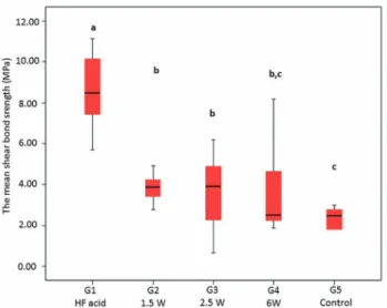
Bond strength was significantly higher in Group 1 (HF acid etching; 8.42 ± 1.86 MPa) than in other groups (P<.01).
Adhesion was significantly stronger in Group 2 (3.88 ± 1.94 MPa) and 3 (3.65 ± 1.87 MPa), which were laser-irradi- ated, than in Group 5 (1.95 ± 1.06 MPa; P<.01). There was no significant difference between Group 4 (3.59 ± 1.19 MPa), which was subjected to laser irradiation, and the con- trol group (P>.05; Fig. 2).
SEM evaluation of the Er,Cr:YSGG laser-treated lithi- um disilicate ceramic surfaces revealed that surface struc- ture depended on the surface treatment method and laser parameter. Lithium disilicate crystals were visible on the
specimen from Group 1 (Fig. 3). More surface irregularities were observed on the specimen from Group 2 than on the control specimen (Fig. 4). In turn, more surface irregulari- ties were observed in Group 3 than in Group 2, despite the absence of a significant difference in shear bond strength values (Fig. 5). The specimen from Group 4 (6 W laser irra- diation) also exhibited increased surface irregularity, as well as severely affected lithium disilicate crystals and over destruction of the surface (Fig. 6). The specimen from Group 5 had a typical untreated ceramic surface (Fig. 7).
Laser irradiation increased shear bond strengths com- pared with the control group. Higher laser power settings reduced bonding strengths between all-ceramic restorations and resin cement.
Fig. 2. The means and the standard deviations of the shear bond strength. Same lowercase letters indicate groups that were not statistically different (P>.05).
Fig. 3. Scanning electron microscopic image of hydrofluoric acid etched surface (×1,000 magnification).
Fig. 4. Scanning electron microscopic image of 1.5 W laser irradiated surface (×1,000 magnification).
Fig. 5. Scanning electron microscopic image of 2.5 W laser irradiated surface (×1,000 magnification).
DISCUSSION
In the current study, bond strength between resin cement and ceramic restorations was evaluated by applying differ- ent surface treatment methods. Modification of the lithium disilicate ceramic surfaces affected shear bond strength.
Dental materials are influenced by laser ablation rates.
The effectiveness and safety of laser treatment, and thus bond strength, are related to the use of adequate parame- ters, such as the duration, frequency, and power of irradita- tion.42
Er:YAG, Nd:YAG, and Er,Cr:YSGG lasers have been used to condition the surfaces of dental materials.34,35 Because no previous study has evaluated ceramic-surface conditioning using a Er,Cr:YSGG laser and because the wavelengths of Er:YAG (2.94 µm) and Er,Cr:YSGG (2.78 µm) lasers are similar, the results of the present study are compared to those using the Er:YAG laser.
Many in vitro studies have assessed shear bond strengths after surface treatment of ceramic restorations using differ- ent laser power settings. One study44 used a 3 W power set- ting for Er:YAG laser irradiation, while another29 assessed surfaces treated with 6, 12, and 18 W settings. Shiu et al.43 evaluated Er:YAG laser treatment of feldspathic ceramic using 500 mJ pulses at a frequency of 4 Hz, and da Silva Ferreira et al.34 applied Al2O3 using the same parameters.
These studies illustrate the lack of consensus on appropri- ate power settings for laser irradiation.
Akyil et al.44 compared the shear bond strengths of feld- spathic ceramics repaired with composite resin after six sur- face treatments, and found lower bond strengths in speci- mens treated with 3 W Er:YAG laser irradiation than in control specimens that received no surface treatment.
In the present study, all laser treatments were performed using 300 mJ pulses, but the power settings differed; 1.5
and 2.5 W laser irradiation enhanced shear bond strengths compared with the control group, but 6 W irradiation did not produce a significant difference. These findings differ from those previous study,44 perhaps due to the use of dif- ferent ceramic materials, initial surface treatments (grinding in the present study), laser types, and/or ablation rates.
Gökçe et al.29 subjected Empress 2 ceramic specimens to various surface treatments: 9.5% HF acid etching for 30 seconds and Er:YAG laser treatment at 20 Hz with 300, 600, and 900 mJ pulses. Bond strength values were highest in the 300 mJ laser treatment group. Based on this finding, a 300 mJ 20 Hz (6 W) treatment was included in the pres- ent study. In the same study, bond strength was found to decrease with increasing energy per pulse. The authors attributed the low bond strengths in the 600 and 900 mJ groups to the inadequacy of microdepths created by the Er:YAG laser, and assumed that they might be due to overdestruction of the crystal and/or matrix phases or cre- ation of a heat-damaged layer.29 In the present study, SEM analysis revealed overdestruction and weakening of the sur- face, rather than heat damage, in the specimen from Group 4 (6 W laser irradiation). No heat damage could be attribut- ed to the hydrokinetic system of the Er,Cr:YSGG laser; the absorption of laser energy by water microdroplets is believed to be partially responsible for its hard tissue-cut- ting effects.45,46
In the present study, testing of the same ceramic materi- al and power setting (6 W) used by Gökçe et al.29 yielded different results, perhaps due to differences in the laser type and timing of irradiation. Lower power settings resulted in significantly higher bond strengths than the control group and with the 6 W power setting. These higher bond strengths were associated with the creation of surface irreg- ularities, whereas overdestruction of the surface was observed in the 6 W group. Increased output power created Fig. 6. Scanning electron microscopic image of 6 W
laser irradiated surface (×1,000 magnification). Fig. 7. Scanning electron microscopic image of no treated surface (×1,000 magnification).
rough surfaces. Increase of shear bond strength was expected as a result of increased surface roughness.
However, bonding ability decreased on the contrary.
Therefore, it is supposed that despite an increase in pene- tration of the cement, bonding between the ceramic and cement decreases because of the overdestructive effect of the high power laser. As a result, it is speculated that high power laser application may decrease bonding ability of the ceramic material. In addition, HF acid etching was more effective than all laser treatments. These findings suggest that shear bond strength decreased with increasing output power; various energy settings should be evaluated in future studies.
A previous study comparing the effects of 10 surface treatments (Er:YAG laser irradiation and HF acid etching) on bonding strength between feldspathic ceramic and resin cement found that bond strength values did not differ sig- nificantly between laser-treated groups and the control group.43 In contrast, 1.5 and 2.5 W laser treatments resulted in bond strengths that differed significantly from those of the control group in the present study, although 6 W laser treatment did not. This difference may be related to the use of different laser and ceramic types and power settings.
HF acid etching was found to result in superior bond strengths in the present study (8.42 MPa); it achieves proper ceramic surface texture by dissolving the glass matrix to expose the crystalline phase and several other studies have also demonstrated that HF acid etching is the most effec- tive surface treatment.22,24 Akyil et al.44 found the bond strength of HF acid etching higher than the present study, the difference can be attributed to the different etching time. However, HF acid etching cannot be used intraoral- ly;47 thus, laser irradiation at low-power settings may be used to obtain the highest possible shear bond strength val- ues.Only one type of laser with three power settings and one type of ceramic material were used in this study.
Various laser treatments and parameters are known to affect ceramic materials differently.29 Thus, evaluation of other ceramic and laser types and energy parameters may yield different results; therefore, further studies are required.
CONCLUSION
The application of 1.5 and 2.5 W laser irradiation increased shear bond strengths between lithium disilicate ceramic and resin cement compared with untreated surfaces. The use of an Er,Cr:YSGG laser at a 6 W power setting may not be an efficient ceramic surface treatment technique. HF acid etch- ing increased the shear bond strength between resin cement and ceramic surfaces more effectively than any laser treat- ment.
REFERENCES
1. McLaughlin G. Porcelain veneers. Dent Clin North Am 1998;
42:653-6.
2. Blatz MB. Long-term clinical success of all-ceramic posterior restorations. Quintessence Int 2002;33:415-26.
3. Jensen ME, Sheth JJ, Tolliver D. Etched-porcelain resin- bonded full-veneer crowns: in vitro fracture resistance.
Compendium 1989;10:336-8, 340-1, 344-7.
4. Bergman MA. The clinical performance of ceramic inlays: a review. Aust Dent J 1999;44:157-68.
5. Hayashi M, Wilson NH, Yeung CA, Worthington HV.
Systematic review of ceramic inlays. Clin Oral Investig 2003;
7:8-19.
6. Krämer N, Frankenberger R. Clinical performance of bond- ed leucite-reinforced glass ceramic inlays and onlays after eight years. Dent Mater 2005;21:262-71.
7. Sjögren G, Molin M, van Dijken JW. A 5-year clinical evalua- tion of ceramic inlays (Cerec) cemented with a dual-cured or chemically cured resin composite luting agent. Acta Odontol Scand 1998;56:263-7.
8. Braga RR, Ballester RY, Daronch M. Influence of time and adhesive system on the extrusion shear strength between feldspathic porcelain and bovine dentin. Dent Mater 2000;16:
303-10.
9. Davidson CL. Luting cement, the stronghold or the weak Link in ceramic restoration? Adv Eng Mater 2001;3:763-7.
10. Kelly JR, Giordano R, Pober R, Cima MJ. Fracture surface analysis of dental ceramics: clinically failed restorations. Int J Prosthodont 1990;3:430-40.
11. Thompson JY, Anusavice KJ, Naman A, Morris HF. Fracture surface characterization of clinically failed all-ceramic crowns. J Dent Res 1994;73:1824-32.
12. Haselton DR, Diaz-Arnold AM, Dunne JT Jr. Shear bond strengths of 2 intraoral porcelain repair systems to porcelain or metal substrates. J Prosthet Dent 2001;86:526-31.
13. Chen JH, Matsumura H, Atsuta M. Effect of etchant, etching period, and silane priming on bond strength to porcelain of composite resin. Oper Dent 1998;23:250-7.
14. Chen JH, Matsumura H, Atsuta M. Effect of different etch- ing periods on the bond strength of a composite resin to a machinable porcelain. J Dent 1998;26:53-8.
15. Sorensen JA, Engelman MJ, Torres TJ, Avera SP. Shear bond strength of composite resin to porcelain. Int J Prosthodont 1991;4:17-23.
16. Wolf DM, Powers JM, O’Keefe KL. Bond strength of com- posite to porcelain treated with new porcelain repair agents.
Dent Mater 1992;8:158-61.
17. Bailey LF, Bennett RJ. DICOR surface treatments for en- hanced bonding. J Dent Res 1988;67:925-31.
18. Ferrando JM, Graser GN, Tallents RH, Jarvis RH. Tensile strength and microleakage of porcelain repair materials. J Prosthet Dent 1983;50:44-50.
19. Jochen DG, Caputo AA. Composite resin repair of porcelain denture teeth. J Prosthet Dent 1977;38:673-9.
20. Semmelman JO, Kulp PR. Silane bonding porcelain teeth to acrylic. J Am Dent Assoc 1968;76:69-73.
21. Lacy AM, LaLuz J, Watanabe LG, Dellinges M. Effect of por- celain surface treatment on the bond to composite. J Prosthet Dent 1988;60:288-91.
22. Calamia JR. Etched porcelain veneers: the current state of the art. Quintessence Int 1985;16:5-12.
23. Spohr AM, Sobrinho LC, Consani S, Sinhoreti MA, Knowles JC. Influence of surface conditions and silane agent on the bond of resin to IPS Empress 2 ceramic. Int J Prosthodont 2003;16:277-82.
24. Stangel I, Nathanson D, Hsu CS. Shear strength of the com- posite bond to etched porcelain. J Dent Res 1987;66:1460-5.
25. Matinlinna JP, Vallittu PK. Bonding of resin composites to etchable ceramic surfaces - an insight review of the chemical aspects on surface conditioning. J Oral Rehabil 2007;34:622- 30.
26. Ozcan M, Alkumru HN, Gemalmaz D. The effect of surface treatment on the shear bond strength of luting cement to a glass-infiltrated alumina ceramic. Int J Prosthodont 2001;14:
335-9.
27. Bertolini JC. Hydrofluoric acid: a review of toxicity. J Emerg Med 1992;10:163-8.
28. Ersu B, Yuzugullu B, Ruya Yazici A, Canay S. Surface rough- ness and bond strengths of glass-infiltrated alumina-ceramics prepared using various surface treatments. J Dent 2009;37:
848-56.
29. Gökçe B, Ozpinar B, Dündar M, Cömlekoglu E, Sen BH, Güngör MA. Bond strengths of all-ceramics: acid vs laser etching. Oper Dent 2007;32:173-8.
30. Akova T, Yoldas O, Toroglu MS, Uysal H. Porcelain surface treatment by laser for bracket-porcelain bonding. Am J Orthod Dentofacial Orthop 2005;128:630-7.
31. Spohr AM, Borges GA, Júnior LH, Mota EG, Oshima HM.
Surface modification of In-Ceram Zirconia ceramic by Nd:YAG laser, Rocatec system, or aluminum oxide sandblast- ing and its bond strength to a resin cement. Photomed Laser Surg 2008;26:203-8.
32. Cavalcanti AN, Pilecki P, Foxton RM, Watson TF, Oliveira MT, Gianinni M, Marchi GM. Evaluation of the surface roughness and morphologic features of Y-TZP ceramics af- ter different surface treatments. Photomed Laser Surg 2009;
27:473-9.
33. Jacobsen NL, Mitchell DL, Johnson DL, Holt RA. Lased and sandblasted denture base surface preparations affecting resil- ient liner bonding. J Prosthet Dent 1997;78:153-8.
34. da Silva Ferreira S, Hanashiro FS, de Souza-Zaroni WC, Turbino ML, Youssef MN. Influence of aluminum oxide sandblasting associated with Nd:YAG or Er:YAG lasers on shear bond strength of a feldspathic ceramic to resin ce- ments. Photomed Laser Surg 2010;28:471-5.
35. Kimyai S, Mohammadi N, Navimipour EJ, Rikhtegaran S.
Comparison of the effect of three mechanical surface treat- ments on the repair bond strength of a laboratory compos- ite. Photomed Laser Surg 2010;28:S25-30.
36. Chen JR, Oka K, Kawano T, Goto T, Ichikawa T. Carbon di- oxide laser application enhances the effect of silane primer on the shear bond strength between porcelain and composite resin. Dent Mater J 2010;29:731-7.
37. Hossain M, Nakamura Y, Yamada Y, Suzuki N, Murakami Y, Matsumoto K. Analysis of surface roughness of enamel and dentin after Er,Cr:YSGG laser irradiation. J Clin Laser Med
Surg 2001;19:297-303.
38. Usumez A, Aykent F. Bond strengths of porcelain laminate veneers to tooth surfaces prepared with acid and Er,Cr:
YSGG laser etching. J Prosthet Dent 2003;90:24-30.
39. Güler AU, Yilmaz F, Ural C, Güler E. Evaluation of 24-hour shear bond strength of resin composite to porcelain accord- ing to surface treatment. Int J Prosthodont 2005;18:156-60.
40. Fabianelli A, Pollington S, Papacchini F, Goracci C, Cantoro A, Ferrari M, van Noort R. The effect of different surface treatments on bond strength between leucite reinforced feld- spathic ceramic and composite resin. J Dent 2010;38:39-43.
41. Aida M, Hayakawa T, Mizukawa K. Adhesion of composite to porcelain with various surface conditions. J Prosthet Dent 1995;73:464-70.
42. Gökçe B. Effects of Er:YAG laser irradiation on dental hard tissues and all-ceramic materials: SEM Evaluation. In:
Viacheslav Kazmiruk, editor. Scanning Electron Microscopy.
New York; In tech; 2012. p. 179-212.
43. Shiu P, De Souza-Zaroni WC, Eduardo Cde P, Youssef MN.
Effect of feldspathic ceramic surface treatments on bond strength to resin cement. Photomed Laser Surg 2007;25:291- 6.
44. Akyil MS, Yilmaz A, Karaalioğlu OF, Duymuş ZY. Shear bond strength of repair composite resin to an acid-etched and a laser-irradiated feldspathic ceramic surface. Photomed Laser Surg 2010;28:539-45.
45. Eversole LR, Rizoiu I, Kimmel AI. Pulpal response to cavity preparation by an erbium, chromium:YSGG laser-powered hydrokinetic system. J Am Dent Assoc 1997;128:1099-106.
46. Kursoglu P, Yurdaguven H, Kazazoglu E, Çalýkkocaoglu S, Gursoy T. Effect of Er,Cr:YSGG laser on ceramic surface.
Balk J Stomatol 2006;10:103-9.
47. Ozcan M, Allahbeickaraghi A, Dündar M. Possible hazardous effects of hydrofluoric acid and recommendations for treat- ment approach: a review. Clin Oral Investig 2012;16:15-23.