Research Article Open Access
Effects of Inspiratory Muscle Training on Diaphragm Thickness, Pulmonary Function, and Chest Expansion in Chronic Stroke Patients
Ju-Hyeon Jung, PT, Nan-Soo Kim, PT, PhD
1†7)Department of Physical Therapy, Graduate School, Catholic University of Pusan,
1
Department of Physical Therapy, Catholic University of Pusan
흡기근 저항훈련이 만성 뇌졸중 환자의 횡격막 두께와 폐기능에 미치는 효과
정주현⋅김난수
1†부산가톨릭대학교 일반대학원 물리치료학과,
1부산가톨릭대학교 물리치료학과
Received: October 8, 2012 / Revised: November 2, 2012 / Accepted: November 5, 2012
ⓒ 2013 Journal of the Korean Society of Physical Medicine
| Abstract |
연구목적: 본 연구는 만성 뇌졸중 환자를 대상으로 흡 기근 저항훈련이 횡격막 두께와 폐기능 및 흉곽 확장에 미치는 효과를 알아보고자 수행하였다.
연구방법: 연구 대상자는 총 29명(남자 17명, 여자 12 명)으로 흡기근 저항훈련군(15명)과 대조군(14명)으로 분류하였다. 모든 대상자는 6개월 이상된 만성 뇌졸중 환 자로 일반적인 신경발달치료를 받고 있으며, 같은 기간 동안 흡기근 저항훈련군에 역치부하 흡기근육 단련기 (threshold IMT device)를 제공하고 주 3회 ×1회 20분씩 6 주간 시행하였다. 마비측과 비마비측 횡격막 두께측정 을 위해 초음파의 7.5MHz linear probe를 사용하여 최대 흡기시(Tdi.con)와 휴식시(Tdi.rel)의 두께를 측정하고 수 축률(TR)을 계산하였다. 또한 폐 활량계를 사용하여 노 력성 폐활량을 측정하였으며, 줄자를 사용하여 흉곽 확
†Corresponding Author : [email protected]
This is an Open Access article distributed under the terms of the Creative Commons Attribution Non-Commercial License (http://creativecommons.org/licenses/by-nc/3.0) which permits unrestricted non-commercial use, distribution, and reproduction in any medium, provided the original work is properly cited.
장을 측정하였다.
연구결과: 6주간 중재 후 흡기근 저항훈련군에서 최 대흡기시 횡격막 두께(Tdi.con)와 수축률(TR)은 유의한 증가를 보였다(p<.05). 1초간 노력성 호기량 (FEV
1)과 최 대 호기 속도(PEF)도 유의한 증가를 보였으나(p<.05), 노 력성 폐활량(FVC)과 1초간 노력성 호기량의 노력성 폐 활량에 대한 비(FEV
1/FVC), 흉곽 확장은 유의한 증가는 보이지 않았다(p>.05).
결론: 본 연구는 만성뇌졸중 환자를 대상으로 흡기근 저항훈련의 적용이 횡격막의 수축력과 폐기능 및 흉곽 확장력을 향상시켜 호흡근의 협응력을 증가시키고, 비 활동성으로 인해 감소된 운동내성을 증가하게 함으로써
향후 재활에서 만성 뇌졸중 환자에게 2차적인 기능향상
에 도움을 줄수 있을 것으로 보여진다.
주제어: 흡기근 저항훈련, 뇌졸중, 횡격막 두께, 폐기
능, 흉곽확장
Ⅰ. Introduction
A stroke is a major cause of social and physical dysfunctions, and it is reported that around 25 to 30 percent of stroke patients suffer from severe movement disorders (Duncan et al., 2002). Stroke patients have a poor cardiopulmonary function, given that a prolonged period of hospitalization and a bed-ridden living pattern deteriorates oxygen-transport ability and lowers their frequency of physical activity (Kashihara et al., 1994). In addition, the damage caused by a stroke to the motor cortex and the pyramidal tract leads to hemiplegia. As well, postural and muscle tonus and abnormal voluntary movement cause motor-control disorders and trunk-muscle synergy. This further damages coordination of respiratory-muscle contraction and deteriorates exercise capacity (De Almeida et al. 2011).
Stroke-induced hemiplegia injures the respiratory muscles of the paretic side and chest-wall mechanics (Annoni et al., 1990; Lanini et al., 2003). Previous studies reported that stroke patients suffered from a restrictive lung disease (Fugl-Meyer et al., 1983; Kim et al., 2011). Some 40 percent of stroke patients with restrictive ventilator impairment showed a decline in diaphragm displacement.
Their forced vital capacity (FVC), forced expiratory volume in the first second (FEV1), and peak expiratory flow (PEF) were about half the level of healthy adults. As well, changes in oxygen saturation and respiratory pattern were observed in stroke patients’arteries (Khedr et al., 2000). For this reason, stroke patients are more likely to feel fatigue in the inspiratory muscle and are at a greater risk of pulmonary complications (Larson et al., 1988; Loveridge et al., 1992).
Meanwhile, previous studies showed that a variety of exercises, such as muscular-strength exercises, independent pulmonary-muscle training, and aerobic exercise, effectively enhanced pulmonary function of stroke patients who were experiencing a movement disorder, deteriorated aerobic-exercise capacity, and cardiopulmonary function. Thus, to help stroke patients perform daily activities independently, it
is important to enhance their pulmonary functions (Lee, 2008).
Inspiratory muscle training (IMT) is one such exercise:
it applies a load to the diaphragm and to the accessory inspiratory muscles to improve the strength and endurance of these muscles (Moodie et al., 2011). IMT is known to enhance ventilator capacity of patients with inspiratory- muscle impairment regardless of whether the disorder is neurological or non-neurological (Petrovic et al., 2009).
IMT is conducted in the same way to train skeletal muscles, based on the principles of overload, specificity, and reversibility.
Studies that compared healthy adults and those with a chronic obstructive pulmonary disease revealed that IMT enhanced the strength and endurance of inspiratory muscles (Gosselink et al., 2011; McConnell and Romer, 2004).
Similarly, for patients with multiple sclerosis or Parkinson’s disease, it was shown that IMT effectively improved respiratory function, as well as the strength and endurance of the inspiratory muscles (Fry et al., 2007; Inzelberg et al. 2005). Only a few studies have been undertaken concerning IMT’s impact on the pulmonary function of chronic stroke patients with restrictive respiratory patterns.
Therefore, this study applies IMT to chronic stroke patients over a period of six weeks and observes changes in their diaphragm thickness, pulmonary function, and chest expansion.
Ⅱ. Methods
1. Participants
The experiment subjects were people who had been
diagnosed with a stroke through computed tomography,
had partial impairment in the cerebral hemisphere, and had
a secondary injury of hemiplegia. The subjects had been
with effects of a stroke for longer than six months and
were undergoing general physical therapy, including
neurological treatment, though without special treatment to enhance pulmonary function. Those who had an innate thorax deformity, were incapable of performing the respiratory-system mechanics due to a rib fracture, or had a disease related to lungs, kidneys, the endocrine system, orthopedics, or rheumatology were excluded from the experiment. The patients also could not have undergone abdominal surgery in the past We used a randomized controlled design in which the assessor was blind to the group allocation of the subject. Blinding the patients was not possible because of the nature of the treatment. In the end, 33 stroke patients participated in the experiment. They had a full understanding of its objective and consented to participation. However, There were 3 dropouts, 2 from the experimental group (lack of time) and 2 from the control group (high blood pressure). They were divided into an intervention group (n=15) and a control group (n=14) There were (De Almeida et al., 2011).
2. Measurement Protocol
The measurements were done in the following order:
lung function, diaphragm thickness, and chest expansion.
This was in consideration of the fact that an increase in lung volume affects diaphragm thickness (Ueki et al., 1995).
1) Lung-function measurements
The subjects’ FVC was measured with a spirometer (CHESTGRAPH HI 101, Chest M.I. Inc, Japan). A full explanation was given in advance, along with a demonstration. Lung function was examined three times, and the results with formability and probability were chosen for the analysis. The subjects kept an upright sitting position, and the measurement values included FVC, FEV
1, theFEV
1/FVCratio,andPEF.
2) Assessment of diaphragm thickness
An ultrasonography using a B-mode M12L high-frequency linear transducer (5.0-14.0MHz) (Logiq 7, GE, USA) was
used to measure diaphragm thickness. To assess diaphragm function, probes were arranged in a row on the Tdi zone, and an observation was made regarding the brightness of the echogenic focus instead of displacement of the diaphragm dom. Because a probe in the upper abdomen may interfere with an abdominal movement (Cohen et al., 1994). high-resolution ultrasound was used to measure Tdi.
All the subjects kept a standing position. Their mid axillary line, between the 8
thand 9
thcostae, was marked, and then the subjects changed to an upright sitting position. The linear transducer (5.0-14.0MHz) was projected onto the chest wall at a perpendicular angle, and the intercostal spaces were read on a flat image. Diaphragm thickness indicates a distance between two bright parallel lines seen in the center of the pleura and the peritoneum, and it was measured three times to calculate a mean value (Enright et al., 2006).
To assess functional residual capacity (FRC), diaphragm thickness was measured first. The subject changed to an upright sitting position and wore a spirometer nose clip and mouth piece in order that the diaphragm thickness at functional residual capacity (Tdi.rel) could be measured (ICC .86, 95%CI .81 to .91). The subjects were asked to keep the maximum inspiratory mouth pressure (PImax) possible for two seconds so that diaphragm thickness at total lung capacity (Tdi.cont) could be measured (ICC .94, 95%CI .92 to .96). A formula proposed by Ueki et al.
was used to calculate a standardized value of the diaphragm thickness ratio (TR) regardless of the changing lung volume (Fig. 1).
TR = Diaphragm thickness during MIP maneuver of FRC Mean thickness while relaxing at FRC Fig. 1. Formula for the calculation of the diaphragm
thickening ration (TR)
3) Chest expansion
To measure chest expansion when breathing, a subject’s
chest wall was measured in an upright sitting position with a tapeline. The chest wall was exposed, and the circumference was measured in a released state, maximal inspiration, and maximal inspiration. The tapeline crossed over the areas that connect the xiphoid process and the corpus sterni. The extent of chest expansion was calculated by determining differences in circumferences of the chest wall in a state of maximal expiration and maximal inspiration (Kim et al., 2011).
3. Inspiratory Muscle Training Protocols Patients in the intervention group had a Threshold IMT device (Threshold Inspiratory Muscle Trainer, Respironics Health Scan, Inc., USA) that is easily adaptable and achieves a PImax of 41cm H
2O or higher in an accurate and safe manner (Fregonezi et al., 2009). Prior to intervention, the researcher explained the training method to the subjects, and the subjects practiced the method to acquire an adequate level of skill. A six-week training session followed(Training was performed 3 times weekly on nonconsecutive days). The IMT program was designed based on the previously published protocol, and a progressive load was applied (Appendix) (Enright et al., 2006; Fry et al., 2007).
The IMT session consisted of a five-minute warm-up period, a ten-minute overload period, and a five-minute cool-down period (Brannon et al., 1998; Skinner, 1993).
To measure the degrees of consciousness concerning the subjects’ fatigue, the Borg Rating of Perceived Exertion (RPE) was used (Borg, 1982). The subjects recorded a log documenting their exercise adherence, the Borg RPE, and daily symptoms. Patients who developed lightheadedness were removed from the training and resumed the training after the symptom was gone. When a subject complained about lightheadedness, pressure resistance was reduced by 2cm H
2O.
A 60-second break was given after the first IMT set, and the break became shorter by ten seconds each time.
Two 60-second breaks were given during the warm-up and the cool-down periods, and the break for the overload period gradually decreased (Enright et al., 2006).
4. Data Analysis
The collected data were analyzed using PASW (Predictive Analytics Software) Statistics for Windows (version 18.0) at a significance level (α) of 0.05. The study conducted a paired t-test to examine the effect of the training intervention and conducted an independent t-test to observe differences between the two groups.
Ⅲ. Results
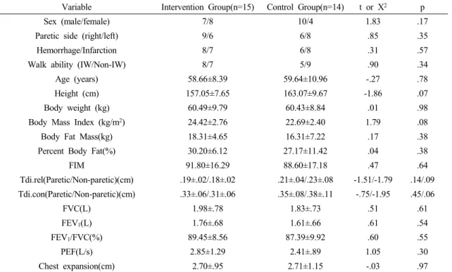
1. Study-Group Characteristics: Baseline Analysis The intervention group had seven males and eight females; among them, nine had right hemiplegia and six had left hemiplegia. The control group had nine males and six females; among them, six had right hemiplegia and nine had left hemiplegia. General characteristics of the two groups were examined in advance, and no significant difference was found in terms of age, height, body weight, diaphragm thickness, lung function, and chest expansion (Table 1).
2. Changes in diaphragm thickness and thickening ratio
Table 2 shows how the patients’diaphragm thickness
changed after six weeks of IMT. In terms of Tdi.rel, neither
group showed significant changes for paretic or non-paretic
side. In the intervention group, Tdi.con and TR significantly
increased after the training. More specifically, Tdi.cont
significantly increased on both paretic and non-paretic sides
(p=.001). A comparison between the two groups showed
that the increase in Tdi.cont in the intervention group was
significantly larger than in the control group for both paretic
and non-paretic sides (p=.001). Also, in the intervention
Pretest Posttest
t p Post-Pre
t p
Group Paretic side M±SD M±SD M±SD
Tdi.rel (cm)
Intervention(n=15)
Paretic .19±.02 .20±.02 1.32 .20 .00±.01
1.20 .24
Control(n=14) .21±.04 .20±.02 -.71 .48 -.00±.03
Intervention(n=15)
Non-paretic .18±.02 .19±.02 1.37 .19 .00±.02
1.75 .09
Control(n=14) .23±.08 .20±.02 -1.41 .18 -.02±.07
Tdi.cont (cm)
Intervention(n=15)
Paretic .33±.06 .46±.10 5.12 .00 .13±.99
3.88 .00
Control(n=14) .35±.83 .35±.07 .01 .99 .00±.08
Intervention(n=15)
Non-paretic .31±.06 .43±.08 5.04 .00 .11±.09
3.52 .00
Control(n=14) .38±.11 .36±.09 -.59 .56 -.01±.11
TR
Intervention(n=15)
Paretic 1.72±.23 2.32±.44 5.85 .00 .60±.39
4.25 .00
Control(n=14) 1.65±.23 1.71±.27 .73 .47 .05±.27
Intervention(n=15)
Non-paretic 1.67±.26 2.18±.27 7.01 .00 .51±.28
3.73 .00
Control(n=14) 1.69±.29 1.80±.38 1.42 .17 .11±.29
Tdi.rel: diaphragm thickness at functional residual capacity, Tdi.cont: diaphragm thickness at total lung capacity.
TR: thickening raito
Table 2. Comparison of diaphragm thickness between Intervention group and Control group
(n=29) Variable Intervention Group(n=15) Control Group(n=14) t or X2 p
Sex (male/female) 7/8 10/4 1.83 .17
Paretic side (right/left) 9/6 6/8 .85 .35
Hemorrhage/Infarction 8/7 6/8 .31 .57
Walk ability (IW/Non-IW) 8/7 5/9 .90 .34
Age (years) 58.66±8.39 59.64±10.96 -.27 .78
Height (cm) 157.05±7.65 163.07±9.67 -1.86 .07
Body weight (kg) 60.49±9.79 60.43±8.84 .01 .98
Body Mass Index (kg/m2) 24.42±2.76 22.69±2.40 1.79 .08
Body Fat Mass(kg) 18.31±4.65 16.31±7.22 .17 .38
Percent Body Fat(%) 30.20±6.12 27.17±11.42 .04 .38
FIM 91.80±16.29 88.60±17.18 .47 .64
Tdi.rel(Paretic/Non-paretic)(cm) .19±.02/.18±.02 .21±.04/.23±.08 -1.51/-1.79 .14/.09 Tdi.con(Paretic/Non-paretic)(cm) .33±.06/.31±.06 .35±.08/.38±.11 -.75/-1.95 .45/.06
FVC(L) 1.98±.78 1.83±.73 .51 .61
FEV1(L) 1.76±.68 1.61±.66 .61 .54
FEV1/FVC(%) 89.45±8.56 87.39±9.92 .60 .55
PEF(L/s) 2.85±1.29 2.41±.89 1.05 .30
Chest expansion(cm) 2.70±.95 2.71±1.15 -.03 .97
IW=Independent walking; Modified motor assessment scale(MMAS)≥4, Non-IW=Non-independent walking group; MMAS<4, Tdi.rel=diaphragm thickness at functional residual capacity, Tdi.con=diaphragm thickness at total lung capacity, FVC=forced vital capacity, FEV1=forced expiratory volume at one second, PEF=peak expiratory flow.
Table 1. Baseline characteristics of study participants
(n=29)
Pretest Posttest
t p Post-Pre
t p
Group M±SD M±SD M±SD
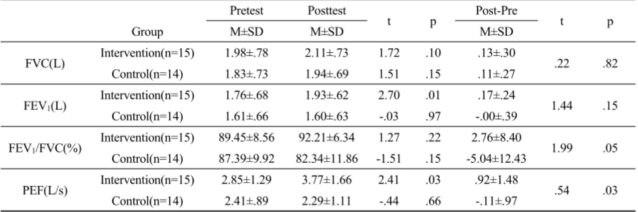
FVC(L) Intervention(n=15) 1.98±.78 2.11±.73 1.72 .10 .13±.30
.22 .82
Control(n=14) 1.83±.73 1.94±.69 1.51 .15 .11±.27 FEV1(L) Intervention(n=15) 1.76±.68 1.93±.62 2.70 .01 .17±.24
1.44 .15 Control(n=14) 1.61±.66 1.60±.63 -.03 .97 -.00±.39
FEV1/FVC(%) Intervention(n=15) 89.45±8.56 92.21±6.34 1.27 .22 2.76±8.40
1.99 .05 Control(n=14) 87.39±9.92 82.34±11.86 -1.51 .15 -5.04±12.43
PEF(L/s) Intervention(n=15) 2.85±1.29 3.77±1.66 2.41 .03 .92±1.48
.54 .03
Control(n=14) 2.41±.89 2.29±1.11 -.44 .66 -.11±.97 FVC=forced vital capacity, FEV1=forced expiratory volume at one second, PEF=peak expiratory flow.
Table 3. Comparison of pulmonary function between Intervention group and Control group
(n=29)
Pretest Posttest
t p Post-Pre
t p
Group M±SD M±SD M±SD
Chest expansion (cm)
Intervention(n=15) 2.70±.95 2.96±.78 1.41 .17 .26±.72
1.15 .25 Control(n=14) 2.71±1.15 2.60±.92 -.40 .69 -.10±1.00
Table 4. Comparison of chest expansion between Intervention group and Control group
(n=29)
group, TR significantly increased after the training (p=.001) for both paretic and non-paretic sides. Again, the increase in TR was significantly greater than for the control group (p=.001). Meanwhile, in the control group, neither Tdi.con nor TR showed a significant change after the training.
3. Changes in lung function
The subjects’ lung functions went through some changes following the six weeks of IMT (Table 3). In the intervention group, neither the FEV
1/FVC ratio nor FVC increased significantly, and no significant difference was found between the two groups. In the intervention group, FEV
1increased significantly(p=.001), but a comparison between the two groups did not find a significant difference.
In the intervention group, PEF also rose significantly after the training (p=.003). A comparison between the two groups showed that the increase in PEF in the intervention group was significantly larger than in the control group. In the
control group, no significant change was observed after the training in terms of the FEV
1/FVC ratio, FVC, FEV
1, and PEF (Table 4).
4. Changes in chest expansion
Lastly, the patients’ chest expansion changed after the six weeks of IMT, as shown in Table 4. In both groups, the degree of chest expansion did not increase significantly after the training, nor was there a significant difference between the two groups in terms of an increase in chest expansion.
Ⅳ. Discussion
Stroke patients often experience a deteriorated pulmonary
function. This easily causes fatigue for patients who need
intensive rehabilitation training and who thus perform
aerobic exercises that demand muscle endurance. The deteriorated pulmonary function also limits activities of daily living, leads to a lower success rate in the rehabilitation process, and obstructs full recovery of bodily functions (Estenne et al., 1993). Recognizing that IMT could benefit patients with a cardiac disease or COPD (Chronic Obstructive Pulmonary Disease), this study established a hypothesis that IMT could improve pulmonary function in chronic stroke patients and the researchers conducted an experiment accordingly.
The inspiratory muscles, including the diaphragm, are categorized as skeletal muscles from a morphological and functional viewpoint, and they show the same response as other locomotor muscles when an adequate physiological load is applied (Kraemer et al., 2002). Thus, when pressure resistance is applied to the diaphragm during weight training, its thickness changes (McCool et al., 1997).
Enright examined the changes in diaphragm thickness when healthy adults went through IMT. After eight weeks of training, Tdi.rel did not increase significantly, but Tdi.cont and TR did, which is consistent with the results of this study (Enright et al., 2006), even though the previous study’s subjects were not stroke patients. The study results suggest that IMT applies a load to the inspiratory muscles, increases diaphragm thickness, and enhances pulmonary mechanics. This study showed that continued application of IMT also affects diaphragm thickness in stroke patients, and its impact on stroke patients is consistent with the overload principle (Kraemer et al., 1998).
Sutbeyaz et al., in their recent study of 45 subacute stroke patients, applied IMT to fifteen patients, six times a week for six weeks, and examined their pulmonary functions using both a breathing-retraining group (IMT group) and a control group (Sutbeyaz et al., 2010). The results showed that FEV1andFVC increased significantly in the IMT group, but PEF did not. As well, Fry et al.
examined 46 multiple sclerosis patients and applied a ten-week IMT session to twenty patients in an experimental
group that used an inspiratory-muscle threshold trainer.
Following the training, FEV1andFVC significantly increased in the experimental group, as did FEV (by 25 to 75 percent).
However, the FEV1/FVC ratio did not increase significantly (Fry et al., 2007). The outcomes of this study showed results similar to those of previous studies in terms of changes in FEV1 and PEF. Regarding FVC, its average value increased but not at a statistically significant level.
Also, regarding the FEV1/FVC ratio, the average value increased in the IMT group and decreased in the control group, and yet the difference was not found to be statistically significant. Previous studies identified the most effective length of an IMT program to be six to eight weeks (Teixeira-Salmela et al., 2005). Presumably, FVC and the FEV1/FVC ratio in this study would have shown significant increases if the IMT had lasted longer than six weeks.
It is noteworthy that in Sutbeyaz’s study, the pre-test baseline values of the participants were 30 percent to 40 percent higher than the pulmonary-function baseline values of this study. This is presumably because as a stroke develops into a chronic stage, muscle efficiency declines due to an instable chest wall and an inactive lifestyle. Also, Sutbeyaz et al. (2010) reported that stroke patients often fail to keep the minimum fitness level required to maintain independent living and fall into a low aerobic fitness. This leads to poor cardiorespiratory fitness and increases both their risk of cardiovascular disease and their mortality rate (Sutbeyaz et al., 2010). These suggest that as a stroke becomes a chronic condition, a patient’s lifestyle becomes more inactive, and pulmonary function continues to decline.
Thus, IMT could be an effective intervention method for improving the pulmonary function of chronic stroke patients.
Fry argued that IMT improves deep breathing ability
and expiratory volume, which clinically confirms the
positive indirect effect of IMT (Fry et al., 2007). In this
study, inspiratory training led to a significant increase in
PEF, indicating better expiratory pulmonary function.
Concerning indirect effects of IMT, Klefbeck and Hamrah Nedjad (2003) reported that a ten-week IMT period improved the physical activity of patients who had developed a debilitating physical condition due to long hours spent on a bed and in a wheelchair. He reported that IMT also improved fatigue symptom and increased body temperature (Klefbeck and Hamrah Nedjad, 2003). These findings further support the idea that IMT is a good intervention method that not only trains the inspiratory muscles but also provides several indirect effects, such as improved expiratory pulmonary function.
Patients in the IMT group showed an increase in chest expansion following the training. IMT strengthens the inspiratory muscles and increases maximum chest-wall expansion, thus improving the chest expansion of stroke patients (Minoguchi et al., 2002). According to previous studies, healthy adults between 55 and 64 years of age recorded chest-expansion values between 4.0cm and 5.5cm (Moll and Wright, 1972). However, the subjects of this study had quite a limited chest-expansion value prior to the training (2.70±0.9cm). Presumably, this is because of the reduction in thoracic mobility and chest-muscle activity as a stroke develops into a chronic stage. As well, a study that examined myasthenia gravis patients reported that their chest expansion increased significantly after IMT (Fregonezi et al., 2005). Similarly, Minoguchi examined COPD patients and observed chest expansion in the xiphoid area (Minoguchi et al., 2002).
In this study, chest expansion increased in the intervention group, but it actually decreased in the control group. Although the inter-group difference was not statistically significant, it conformed to the results of previous studies.
It is surmised that the difference was statistically insignificant because, as shown in Minoguchi’s research, IMT intervention affects the lower chest wall more than it does the axilla or xiphoid area. IMT ameliorates chest-wall stiffness while increasing maximum chest-wall expansion (Minoguchi et al., 2002). In addition, it was observed that IMT increased
maximal inspiratory pressure (MIP), with an increase in chest expansion being a secondary effect (Sahin et al.,2004).
We admit some limitations of the study. The researchers conducted a six-week experiment, and due to the short period of experiment, it was difficult to examine the long-term effects of IMT. Also, the subjects were not randomly selected. It is yet to be confirmed how long the effect of IMT lasts for chronic stroke patients. For these reasons, it will be necessary to conduct follow-up research that includes a more comprehensive analysis of the impact of IMT intervention on chronic stroke patients.
Ⅴ. Conclusion
This study examined how inspiratory muscle training (IMT) affected the diaphragm thickness, pulmonary function, and chest expansion of chronic stroke patients.
The results showed that stroke patients had experienced a decline in exercise capacity and deteriorated coordination of the respiratory muscle due to a damaged motor cortex.
As a stroke develops into a chronic stage, patients’
pulmonary function keeps declining due to an inactive lifestyle. The study applied IMT to stroke patients for six weeks, and the results showed that their diaphragm thickness, thickness ratio (TR), and pulmonary function increased. These findings suggest that IMT could be an adequate intervention method for enhancing the diaphragm mobility and pulmonary function of chronic stroke patients.
Reference
Annoni JM, Ackermann D, Kesselring J. Respiratory function in chronic hemiplegia. Int Disabil Stud. 1990;12(2):
78-80.
Brannon FJ, Foley MW, Starr JA et al. Cardiopulmonary rehabilitation basic theory and application. 3nd Ed.
Philadelphia: F.A. Davis Co. 1998.
Borg GA. Psychophysical bases of perceived exertion. Med Sci Sports Exerc. 1982;14(5):377-81.
Cohen E, Mier A, Heywood P et al. Excursion-volume relation of the right hemidiaphragm measured by ultrasonography and respiratory airflow measurements. Thorax.
1994;49(9):885-9.
De Almeida IC, Clementino AC, Rocha EH et al. Effects of hemiplegy on pulmonary function and diaphragmatic dome displacement. Respir Physiol Neurobiol.
2011;178(2):196-201.
Duncan PW, Homer RD, Reker DM et al. Adherence to post acute rehabilitation guidelines is associated with functional recovery in stroke. Stroke. 2002;33(1):
169-77.
Enright SJ, Unnithan VB, Heward C et al. Effect of high-intensity inspiratory muscle training on lung volumes, diaphragm thickness, and exercise capacity in subjects who are healthy. Phys Ther. 2006;86(3):345-54.
Estenne M, Gevenois PA, Kinnear W et al. Lung volume restriction in patients with chronic respiratory muscle weakness: the role of microatelectasis. Thorax.
1993;48(7):698-701.
Fregonezi GA, Resqueti VR, Guell R et al. Effects of 8-week, interval-based inspiratory muscle training and breathing retraining in patients with generalized myasthenia gravis. Chest. 2005;128(3):1524-30.
Fregonezi GA, Azevedo IG, Araujo TL et al. Adaptation of the threshold IMT with double spring load allows higher inspiratory pressure for muscle training. Clin Physiol Funct Imaging. 2009;29(6):462-4.
Fry DK, Pfalzer LA, Chokshi AR et al. Randomized control trial of effects of a 10-week inspiratory muscle training program on measures of pulmonary function in persons with multiple sclerosis. J Neurol Phys Ther.
2007;31(4):162-72.
Fugl-Meyer AR, Linderholm H, Wilson AF. Restrictive ventilatory dysfunction in stroke: its relation to locomotor function. Scand J Rehabil Med Suppl.
1983;9:118-24.
Gosselink R, De Vos J, van den Heuvel SP et al. Impact of inspiratory muscle training in patients with COPD:
what is the evidence? Eur Respir J. 2011;37(2):416-25.
Inzelberg R, Peleg N, Nisipeanu P et al. Inspiratory muscle training and the perception of dyspnea in Parkinson’s disease. Can J Neurol Sci. 2005;32(2):213-7.
Lee JH The effect of pulmonary function in the stroke patients after feedback breathing exercise. Daegu university graduate school of rehabilitation sciences. Master‘s thesis. 2008.
Kashihara H, Haruna Y, Suzuki Y et al. Effects of mild supine exercise during 20 days bed rest on maximal oxygen uptake rate in young humans. Acta Physiol Scand Suppl. 1994;616:19-26.
Khedr EM, El Shinawy O, Khedr T et al. Assessment of corticodiaphragmatic pathway and pulmonary function in acute ischemic stroke patients. Eur J Neurol.
2000;7(5):509-16.
Kim K, Fell DW, Lee JH. Feedback respiratory training to enhance chest expansion and pulmonary function in chronic stroke: a double-blind, randomized controlled study. Journal of Physical Therapy Science. 2011;
23(1):75-9.
Klefbeck B, Hamrah Nedjad J. Effect of inspiratory muscle training in patients with multiple sclerosis. Arch Phys Med Rehabil. 2003;84(7):994-9.
Kraemer WJ, Duncan ND, Volek JS. Resistance training and elite athletes: adaptations and program considerations.
J Orthop Sports Phys Ther. 1998;28(2):110-9.
Kraemer WJ, Adams K, Cafarelli E et al. American college of sports medicine position stand. progression models in resistance training for healthy adults. Med Sci Sports Exerc. 2002;34(2):364-80.
Lanini B, Bianchi R, Romagnoli I et al. Chest wall kinematics in patients with hemiplegia. Am J Respir Crit Care Med. 2003;168(1):109-13.
Larson JL, Kim MJ, Sharp JT et al. Inspiratory muscle training
with a pressure threshold breathing device in patients
with chronic obstructive pulmonary disease. Am Rev Respir Dis. 1988;138(3):689-96.
Loveridge B, Sanii R, Dubo HI. Breathing pattern adjustments during the first year following cervical spinal cord injury. Paraplegia. 1992;30(7):479-88.
McConnell AK, Romer LM. Respiratory muscle trainnig in healthy humans: resolving the controversy. Int J Sports Med. 2004;25(4):284-93.
McCool FD, Conomos P, Benditt JO et al. Maximal inspiratory pressures and dimensions of the diaphragm. Am J Respir Crit Care Med. 1997;155(4):1329-34.
Minoguchi H, Shibuya M, Miyagawa T et al. Cross-over comparison between respiratory muscle stretch gymnastics and inspiratory muscle training. Intern Med. 2002;41(10):805-12.
Moll JM, Wright V. An objective clinical study of chest expansion. Ann Rheum Dis. 1972;31(1):1-8.
Moodie L, Reeve J, Elkins M. Inspiratory muscle training increases inspiratory muscle strength in patients weaning from mechanical ventilation: a systematic review. J Physiother. 2011;57(4):213-21.
Petrovic M, Lahrmann H, Pohl W et al. Idiopathic diaphragmatic
paralysis-satisfactory improvement of inspiratory muscle function by inspiratory muscle training. Respir Physiol Neurobiol. 2009;165(2-3):266-7.
Sahin G, Calikoğlu M, Ozge C et al. Respiratory muscle strength but not BASFI score relates to diminished chest expansion in ankylosing spondylitis. Clin Rheumatol.
2004;23(3):199-202.
Skinner JS. Exercise testing and exercise prescription for special cases. 2nd Ed. Philadelelephia. Lea & Febiger. 1993.
Sutbeyaz ST, Koseoglu F, Inan L et al. Respiratory muscle training improves cardiopulmonary function and exercise tolerance in subjects with subacute stroke:
a randomized controlled trial. Clin Rehabil. 2010;
24(3):240-50.
Teixeira-Salmela LF, Parreira VF, Britto RR et al. Respiratory pressures and thoracoabdominal motion in community-dwelling chronic stroke survivors. Arch Phys Med Rehabil. 2005;86(10):1974-8.
Ueki J, De Bruin PF, Pride NB. In vivo assessment of diaphragm contraction by ultrasound in normal subjects. Thorax.
1995;50(11):1157-61.
Appendix
Frequency: IMT performed daily for 6 weeks.
Overload: Repetitions and Sets: Ten sets of 15 repetitions*
Resistance: Initial training load was submaximal, was based on 30% of the patient’s PImax(H2Ocm).
Progression: 30% of the patient’s PImax was given during the warm-up and the cool-down periods, and the resistance for the overload period gradually increased.
Subject’s Baseline MIP Pressure: <41 cm H2O
Borg RPE <13 13 to 15 >15 >17
Pressure resistance (cm H2O) Increased by 2 Increased by 1 Maintained at same level Reduced by 2 Subject’s Baseline MIP Pressure: >41 cm H2O
Borg RPE <13 13 to 15 >15 >17
Pressure resistance (cm H2O) Increased by 4 Increased by 2 Maintained at same level Reduced by 2 If subjects developed symptoms (ie, dizziness, lighteadedness, or shortness of breath) while performing IMT, the
resistance, the resistance was adjusted as follows until no symptoms persisted.
*If a subject achieved the maximum IMT Trainer pressure resistance of 41 cm H2O and resistance could no longer be increased, a fourth set of exercises was added along with an incereased number of repetitions up to a maximum of 15 repetitions.
Abbreviations: IMT, inspiratory muscle strength training; MIP, maximal inspiratory pressure; RPE, rating of perceived exertion
Six-Week Inspiratory Muscle Training Protocol