• 검색 결과가 없습니다.

The Effect of Barbaloin on LPS-stimulated Inflammatory Reaction in Mice Peritoneal Macrophages

N/A
N/A
Protected

Academic year: 2021

Share "The Effect of Barbaloin on LPS-stimulated Inflammatory Reaction in Mice Peritoneal Macrophages"

Copied!
7
0
0

로드 중.... (전체 텍스트 보기)

전체 글

(1)

Introduction

Inflammation is a protective mechanism of blood vessel tissues. In inflammatory conditions, pathogens, cell injury and infection cause impairment (Yagi et al., 2009). Generally, the activated immune cells, such as macrophages and lymphocytes, accompany inflammation (Limtrakul et al., 2015). Particularly, macrophages play significant roles in the inflammation reaction and immune responses are included in variety of disease processes (Qian et al., 2015). Activated macrophages, stimulated by lipopolysaccharide (LPS), produce pro-inflammatory cytokines that activate other macrophages and recruit immune cells (Medzhitov et al., 1997.). Macrophages are distributed in all tissues of the body and eliminate unnecessary elements of human body such as aging cells and cancer cells as well as bacteria and viruses. In addition, macrophages play an important mediator role in maximizing secondary immune response by releasing antigen and various cytokines (NamKoong et al., 2012).

The nitric oxide (NO), produced by inducible nitric oxide

synthase (iNOS), is secreted in activated macrophages. Also, inflammatory cytokines such as tumor necrosis factor (TNF)- α, interleukin (IL)-6 and IL-1βare produced in activated macrophages. Inflammatory mediators and cytokines are essential to repair tissue injury for host condition (Chen et al., 2014). Inhibition of the production of inflammatory cytokines and mediators serves as a key mechanism to control inflammatory reactions. Various anti-inflammatory factors against NO, IL-6, TNF- α and prostaglandin E2 have already entered clinical trials as treatment for inflammatory diseases (Reinhart et al., 2001). Therefore, the drugs that regulate the expression of inflammatory mediators have potential interest as therapeutics for the treatment of inflammatory diseases (Park et al., 2011).

Inflammation induces the phosphorylation of p38 mitogen- activated protein kinase (MAPKs), c-Jun NH2-terminal kinase (JNK) and extracellular signal-related kinase (ERK)-1/2, which induce the transcription of inflammatory genes such as nuclear factor-kappa B (NF-κB) (Ajizian et al., 1999). In unstimulated cell conditions, NF-κB in bound to the inhibitory protein I kappa B (IκB) in cytosol. In response to LPS- stimulation, I κB is rapidly phosphorylated by IκB kinase

The Effect of Barbaloin on LPS-stimulated Inflammatory Reaction in Mice Peritoneal Macrophages

Yong-Deok Jeon

1

and Jong-Hyun Lee

2

*

1

Department of Oriental Medicine Resources, Chonbuk National University, Iksan 54596, Korea

2

College of Pharmacy, Dongduk Women’s University, Seoul 02748, Korea

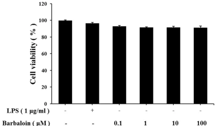
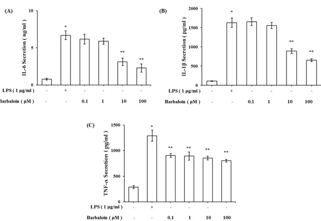
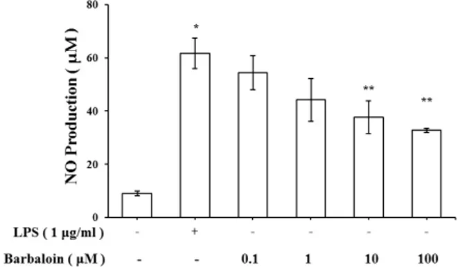
Abstract - Barbaloin is a major component of Aloe vera, which has been used for a laxative. Also, barbaloin is C-glucoside of aloe emodin anthrone which is founded in Aloe vera. Barbaloin has varieties of pharmacological activity such as inhibitory effects on inflammation, histamine release, cancer and microbial infection. But the effect of barbaloin on lipopolysaccharide (LPS)-stimulated macrophages has not been understood. In this study, we evaluated the effects of barbaloin against LPS-stimulated production of nitric oxide (NO), inflammatory cytokines and MAPKs activation in macrophage. We treated barbaloin (0.1 , 1 , 10 , 100 µM) in LPS-stimulated mice peritoneal macrophage. Our results showed that barbaloin significantly inhibited production of NO and cytokines of tumor necrosis factor (TNF)-α, interleukin (IL)-6, interleukin (IL)-1β in LPS-stimulated peritoneal macrophage. Moreover, barbaloin inhibited the phosphorylation of ERK and JNK in a dose dependent manner. These results indicated that barbaloin could be useful for inflammatory diseases.

Key words - Barbaloin, Cytokine, Inflammation, Macrophage, Nitric oxide

*Corresponding author. E-mail : [email protected] Tel. +82-2-940-4485

ⓒ 2017 by The Plant Resources Society of Korea

Original Research Article

(2)

(IKK) and degraded. Consequently, the free NF-κB dimers translocate to the nucleus regulating the transcription of target genes, which include cyclooxygenage-2 (COX-2) and iNOS (Li et al., 2002). Therefore, regulation of MAPKs and transcription factor pathway is a potential therapeutic strategy for inflammatory diseases.

Barbaloin is C-glucoside of aloe emodin anthrone which is founded in Aloe vera, called as healing plant (Tina et al., 2003). Barbaloin can be recrystallized for increase purity (Hattori et al., 1988). Barbaloin is known as having various pharmacological activities such as inhibition of histamine release, anti-cancer and anti-microbial. Also, barbaloin is known as having anti-inflammatory effect by regulating COX-2 pathway (Park et al., 2009). However, it has not been reported that barbaloin has anti-inflammatory effects on LPS-stimulated mice peritoneal macrophages by suppressing secretion of inflammatory cytokines.

In this study, we investigated the anti-inflammatory effects and mechanisms of barbaloin in LPS-stimulated mice peritoneal macrophages. The findings suggest that barbaloin suppresses inflammatory mediators and may be potentially useful in the treatment of inflammatory diseases.

Materials and Methods

Reagents

LPS were purchased from Sigma Aldrich (St. Louis, MO, USA). Anti-mouse TNF- α, biotinylated anti-mouse TNF-α, recombinant mouse TNF-α, anti-mouse IL-6, biotinylated anti-mouse IL-6, and recombinant mouse IL-6 were purchased from BD Pharmingen (San Diego, CA, USA). The specific Anti-phospho-ERK, -p38 and -JNK were purchased from Cell Signalling (Massachusetts, MA, USA). Anti-ERK, -JNK, -p38 and –COX-2 antibodies were purchased from Santa Cruz Biotechnology (Woodland, CA, USA). Anti-GAPDH antibody and β-actin were purchased from Thermo scientifc (Pittsburgh, PA, USA). Barbaloin was obtained from the Wako Pure Chemical Industry Co (Osaka, Japan) and purified by column chromatography on silica gel, followed by re- crystallization from EtOH.

Animals

Male C57BL/6 (6 weeks old) mice were purchased from SAMTAKO (Osan, Korea), and the animals were maintained in the college of Oriental Herb Science Medicine, Chonbuk National University. The mice were housed five to six per cage in a laminar air-flow room maintained at a temperature of 25-27℃ throughout the study. The research was conducted in accordance with the internationally accepted principles for laboratory animal use and care as found in Jeon Buk University guidelines.

Isolation of peritoneal microphages

All experiment protocols (CBNU2016-0016) were approved by the Committee on the Care of Laboratory Animal Resources, Chonbuk National University and were conducted in accordance with the Guide for the Care and Use of Laboratory Animals. Mice were i.p. injected with 3 mL of thiogllicholate (TG), and TG-elicited macrophages were harvested after 3days. By using 7 mL of Dulbecco’s Modified Eagles Medium (DMEM), peritoneal lavage was performed.

Then, the cells were distributed in DMEM, which was supplemented with 10% heat-inactivated Fetal bovine serum (FBS), in 24-well cell culture plates (3×10

5

cells/well) incubated for 24 h at 37℃ in an atmosphere of 5% CO

2

.

MTT assay

Cell viability was determined using MTT assay. Briefly, 500 ㎕ of peritoneal macrophage cells suspension (3×10

5

cells) was cultured in 24-well plates for 24h after treatment with various concentrations of barbaloin. 50 ㎕ of MTT solution (5 ㎎/㎖) was added and then cells were incubated for 4 h at 37 ℃. After washing the supernatant out, the insoluble formazan product was dissolved in DMSO. Then, optical density of 96-well culture plates was measured using enzyme-linked immunosorbent assay (ELISA) reader at 540 ㎚. The optical density of formazan formed in untreated control cells was taken as 100% of viability.

Enzyme-linked immunosorbent assay (ELISA)

Cytokine assay was performed by a modified enzyme-

linked immunosorbent assay (ELISA). In briefly, the wells of

96-well plates were pre-coated with mouse monoclonal Abs

(3)

specific for TNF-α, IL-6 and IL-1β overnight at 4℃. The pre-coated plates were washed with phosphate-buffered saline (PBS) containing 0.05% Tween 20 prior to subsequent steps in the assay. After washing with PBST, samples and standards were incubated for 2 hours at room temperature.

Recombinant IL-6, TNF-α and IL-1β were used as standards.

Serial dilutions from 10 ng/ ㎖ solution were used to establish the standard curve. The assay plates were sequentially exposed to biotinylated mouse IL-6, TNF-α and IL-1β secondary Abs, avidin peroxidase, and 2,20-azino-bis(3- ethylbenzthiazoline-6-sulfonic acid) (ABTS) substrate solution containing 30% distilled water. The absorbance values of the plate were measured at 405 ㎚.

Nitrite Assay

To measure the nitrite content, 100 ㎕ aliquots were removed from the conditioned medium and incubated with an equal volume of Griess reagent (1% sulfanilamide/0.1%

N-(1-naphthyl)-ethylenediamine dihydrochloride/2.5% H

3

PO

4

) for 10 minutes at room temperature. The absorbance was measured by a microplate reader at a wavelength of 540 ㎚.

The NO

2-

level was determined using sodium nitrite as a standard. The value was recorded in each experiment and subtracted from that obtained for the medium containing the peritoneal macrophages.

Western blot analysis

A total of 3 × 10

5

mouse macrophages per well were grown in 6 ㎝ dish and treated with barbaloin at different concentrations (1 μM, 10 μM, 100 uM) after 1 hour later, stimulated with LPS (1 ㎍/㎖) for 30 min. Then, cells were harvested on ice and washed once with ice-cold PBS. Lysis buffer with phos- phatase and protease inhibitors (iNtRON Biotech, Republic of Korea) was added to lyse the cells. After incubating 30 min, cell extracts were centrifuged at 14,463×g in a refrigerated centrifuge (5418R, Eppendorf, Germany) at 4°C for 10 min to collect cell total proteins, the amount of which was quantified using a BCA protein assay kit (Sigma. USA).

SDS-PAGE (10%) was used to separate proteins, which were electro-transferred to PVDF membranes (Roche, UK).

Membranes were blocked with 5 % (wt/vol) dried skimmed milk for 1 h, and incubated with various specific primary

antibodies, namely, anti-P38, anti-P-P38, anti-ERK, anti-P- ERK, anti-JNK, anti-P-JNK, to probe corresponding target proteins. Bound antibodies were detected using peroxidase conjugated secondary antibodies, and the amount of bound antibody was assessed by enhanced chemiluminescence (ECL). Relative levels of target proteins were obtained based on the optical density of electrophoresis bands with GAPDH serving as an internal control. The protein-antibody complexes were visualized by ECL Western blotting luminol reagent (Santa Cruz Biotech, USA) and detected by LAS-4000 image reader (Fugi film).

Statistical Analysis

All results are presented as the mean ± S.E.M. Results were analyzed using Graph Pad Prism version 5.0 program (Graph Pad Software, Inc, La Jolla, CA, USA). One-way analysis of variance with Tukey post hoc test was used to determine statistically significant differences. P <0.05 was considered significant.

Results

Effect of barbaloin on cell viability in mice peritoneal macrophages

The effect of barbaloin on the cell viability was examined using MTT assay. The cells were treated with various concentrations (0.1 μM – 100 μM) of barbaloin. And then the

Fig. 1. Effect of barbaloin on cell viability in mice peritoneal

macrophages. Cells (5× 10

5

cell/well) were pre-treated with

various concentration of barbaloin (0.1 – 100 μM) for 4 hours

and then stimulated with LPS (1 ㎍/㎖) for 24 hour. Cell

viability was measured by MTT assay.

(4)

absorbance of formazan crystal was measured at 540 ㎚.

Barbaloin did not show a cytotoxic effect up to 100 μM (Fig. 1).

Effect of barbaloin on secretion of inflammatory cytokines in LPS-stimulated mice peritoneal macrophages

To investigate the anti-inflammatory effect of Barbaloin, the production of IL-6, IL-1β and TNF-α were measured by ELISA. The cells were pre-treated with various concentrations of barbaloin (100 nM – 100 μM). After 4 hours later, 1 μM of LPS were treated for 24 hours. The stimulation of LPS significantly increased the secretion of IL-6, IL-1β and TNF- α. However, the treatment of barbaloin suppressed the secretion of IL-6, IL-1 β and TNF-α in a dose dependent manner in LPS-stimulated peritoneal macrophages (Fig. 2).

Effect of barbaloin on NO production in LPS-stimulated mice peritoneal macrophages

To investigate the regulatory effect of barbaloin on NO production, Griess assay was performed. The cells were pre-treated with various concentrations of barbaloin (0.1 μM – 100 μM). After 4 hours later, 1 μM of LPS were treated for 48 hours. NO was increased by LPS treatment in mice peritoneal macrophages. However, barbaloin decreased LPS-stimulated NO production in a dose dependent manner (Fig. 3).

Effect of barbaloin on phosphorylation of MAPKs in LPS-stimulated mice peritoneal macrophages

The western blot analysis was performed to investigate the effect of barbaloin on MAPK phosphorylation. Phosphorylation of MAPK pathway plays a role in inflammatory reactions.

Among the MAPKs, barbaloin inhibited the phosphorylation

Fig. 2. Effect of barbaloin on secretion of inflammatory cytokines in LPS-stimulated mice peritoneal macrophages. Cells (5× 10

5

cell/well) were pre-treated with various concentration of barbaloin (0.1 – 100 μM) for 4 hours and then stimulated with LPS (1 ㎍/㎖) for 24 hour. The secretion of cytokines was measured by ELISA. (A) IL-6, (B) IL-1 β, (C) TNF-α. Values represent mean ± S.E.M..

Data were analyzed by Tukey post hoc test (*P < 0.05 versus non-treatment and **P < 0.05 versus LPS alone).

(5)

of ERK and JNK, but not p38 in LPS-stimulated peritoneal macrophages (Fig. 4).

Discussion

Barbaloin is the main constituent of Aloe vera which has been used for traditional medicine. Aloe vera has been reported that has beneficial effects for health such as anti-inflammation, anti-cancer and disinfection (Barbara et al., 2014). However, the exact effects and mechanism of barbaloin have not been understood well. Barbaloin is a C-glycoside that can be hydrolyzed in the gut. Barbaloin and aloe-emodin were known to have possibility to remedy chronic inflammatory diseases (Park et al., 2009). In this study, the anti-inflammatory effect of barbaloin was tested in mice peritoneal macrophages.

The macrophage was used to evaluate the anti-inflammatory effect. Macrophage is known to have crucial role in both non-specific and acquired immune responses. LPS-stimulated macrophages lead to activate phospholipase A2, which produces lipid metabolites of arachidonic acid, and the production of NO, and the secretion of inflammatory cytokines such as IL-6, IL-1β and TNF-α (Kim et al., 2014).

Inflammatory cytokines such as IL-6, IL-1β and TNF-α are important role in inflammatory diseases. IL-6, IL-1 β and TNF- α are involved in the inflammatory responses, which are associated with autoimmune diseases such as inflammatory bowel disease, asthma, and psoriasis (Rincon et al., 2012). In this study, we demonstrated that the treatment of barbaloin suppressed the secretion of IL-6, IL-1 β and TNF-α in LPS-stimulated mice peritoneal macrophages (Fig. 2).

NO has been known as an important regulatory molecule in various physiological functions including inflammation, vasodilation, and cellular communication (MacMicking et al., 1997). The overproduction of NO has been proven to be harmful and resulted in septic shock and auto immune diseases (Kim et al., 2015). The present result showed that barbaloin attenuated inflammation by reducing NO production in LPS-stimulated macrophages.

The phosphorylation of MAPKs has been known to regulate inflammatory mediators in LPS-stimulated macrophages.

Activation of p38 regulates TNF-α and iNOS production through NF- κB pathway (Kaminska, 2005). The activation of JNK regulates various inflammatory mediators such as the phosphorylation of c-Jun and activation of AP-1 (Karin et al., Fig. 3. Effect of barbaloin on NO production in LPS-stimulated

mice peritoneal macrophages. Cells (5× 10

5

cell/well) were pre-treated with various concentration of barbaloin (0.1 – 100 μM) for 4 hours and then stimulated with LPS (1 ㎍/㎖) for 48 hour. The production of nitric oxide was measured by Griess assay. Values represent mean ± S.E.M.. Data were analyzed by Tukey post hoc test (*P < 0.05 versus non-treatment and **P <

0.05 versus LPS alone).

Fig. 4. Effect of barbaloin on phosphorylation of MAPKs in

LPS-stimulated mice peritoneal macrophages. Cells (5× 10

6

cell/well) were pre-treated with various concentration of

barbaloin (1 – 100 μM) for 1 hours and then stimulated with

LPS (1 ㎍/㎖) for 30 minutes. The phosphorylation of MAPKs

was measured by western blot analysis.

(6)

1997). In addition, ERK is involved in iNOS expression and in the production of cytokines in monocytes and macrophages (Chang et al., 2003). In this study, the treatment of Barbaloin attenuated LPS-induced phosphorylation of ERK and JNK, but not that of p38 (Fig. 4). This result showed that barbaloin inhibits the expression of inflammatory mediators by blocking the phosphorylation of ERK and JNK in LPS-stimulated peritoneal macrophages.

This study demonstrated anti-inflammatory effect of barbaloin through down-regulation of phosphorylation of MAPKs (ERK and JNK) and suppression of inflammatory cytokine secretion. Taken together, these results suggest that barbaloin is an effective medicine for anti-inflammatory agent.

References

Ajizian, S.J., B.K. English and E.A. Meals. 1999. Specific inhibitors of p38 and extracellular signal regulated kinase mitogen-activated protein kinase pathway block inducible nitric oxide synthase and tumor necrosis factor accumulation in murine macrophages stimulated with lipopolysaccharide and interferon-gamma. J. Infect. Dis. 7(14):939 –944.

Barbara, B.J, M. Niemcewicz, J. Kocik, L. Jung, E. Skopinska- rozewska and P. Skopinski. 2014. Oral administration of Aloe vera gel, antimicrobial and anti-inflammatory herbal remedy, stimulates cell-mediated immunity and antibody production in a mouse model. Cent. Eur. J. Immunol.

39(2):125-126.

Chang, F., L.S. Steelman, J.T. Lee, J.G. Shelton, P.M. Navolani, W.L. Blalock, R.A. Franklin and J.A. McCubrey. 2003.

Signal transduction mediated by the Ras/Raf/MEK/ERK pathway from cytokine receptors to transcription factors:

Potential targeting for therapeutic intervention. Leukemia 17(7):1263-1293.

Chen, Y.Y., M.J. Hsu, C.Y. Hsieh, L.W. Lee, Z.C. Chen and J.R. Sheu. 2014. Andrographolide inhibits nuclear factor-kB activation through JNK-Akt-p65 signaling cascade in tumor necrosis factor-α-stimulated vascular smooth muscle cells.

ScientificWorld Journal. Doi:10.1155/2014.130381.

Hattori, M., T. Kanda, Y.Z. Shu, T. Akao, K. Kobashi and T.

Namba. 1988. Metabolism of barbaloin by intestinal bacteria.

Chem. Pharm. Bull. 36(11):4462-4466.

Kaminska, B. 2005. MAPK signaling pathways as molecular targets for anti-inflammatory therapy from molecular mecha- nisms to therapeutic benefits. Biochim. Biophys. Acta.

1754(1-2):253-262.

Karin, M.Z, G. Liu and E. Zandi. 1997. AP-1 function and egulation. Curr. Opin. Cell Biol. 9(2):240-246.

Kim, S.J., W.S. Chung, S.G. Kim and J.Y. Um. 2011. Anti- inflammatory effect of Oldenlandia diffusa and its constituent, hentriacontane, through suppression of caspase-1 activation in mouse peritoneal macrophages. Phytother Res. 25(10):

1537-1546

Kim, S.J., H.J. Shin, B.J. Lee, D.S. Kim, J.H. Lee, M.Y. Jeong, H.L. Kim, J. Park, H. Lim, S.H. Kim, S.H. Hong, M.W.

Hwang and J.Y. Um. 2014. The antiinflammatory mechanism of igongsan in mouse peritoneal macrophages via suppression of NF-kB/Caspase-1 activation. Phytother Res. 28(5):736-744.

Limtrakul, P., S. Yodkeeree, P. Pitchakarn and W. Punfa. 2015.

Suppression of inflammatory responses by black rice extract in RAW 264.7 macrophage cells via downregulation of NF-kB and AP-1 signaling pathways. Asian Pac J Cancer Prev. 16(10):4277-4283.

Li, Q. and I.M. Verma.2002. NF-kappaB regulation in the immune system. Nat. Rev. Immunol. 2(10):725 –734.

MacMicking. J., Q.W. Xie and C. Nathan. 1997. Nitric Oxide and macrophage function. Annu. Rev. Immunol. 15:323-330.

Medzhitov, R. and C.A. Janeway Jr. 1997. Innate immunity:

The virtues of a nonclonal system of recognition. Cell 91(3):295 –298.

Namkoong, S., Y.J. Kim, T. Kim and E.H. Sohn. 2012. Study on the immunomodulatory effects of ellagic acid and their mechanisms related to toll-like receptor 4 in macrophages.

Korean J. Plant Res. 25(5):561-567.

Park, Y.D., C.H. Ji, D.S. Choi, M.W. Byun and J.Y. Jeong.

2011. Biological evaluation of isoegomaketone isolated from Perilla frutescens and its synthetic derivatives as anti- inflammatory agents. Arch. Pharm. Res. 34(8):1277-1282.

Park, M.Y., H.J. Kwon and M.K. Sung. 2009. Evauation of aloin and aloe-emodin as anti-inflammatory agents in aloe by using murine macrophages. Biosci. Biotechnol. Biochem.

73(4):828-832.

Qian, Z., Z. Wu, L. Huang, H. Qiu, L. Wang, L. Li, L. Yao, K.

Kang, J. Qu, Y. Wu, J. Luo, J.J. Liu, Y. Yang, W. Yang and

D. Gou. 2015. Mulberry fruit prevents LPS-induced NF- κ

B/pERK/MAPK signals in macrophages and suppresses

acute colitis and colorectal tumorigenesis in mice. Sci Rep.

(7)

1-12.

Reinhart, K. and W. Karzai. 2001. Anti-tumor necrosis factor therapy in sepsis: Update on clinical trials and lessons learned. Crit. Care. Med. 29 (Suppl. 7):S121-S125.

Rincon, M., C.G. Irvin. 2012. Role of IL-6 in asthma and other inflammatory pulmonary diseases. Int. J. biol. Sci. 8(9):

1281-1290.

Tina, J., J. Liu, J. Zhang, Z. Hu and X. Chen. 2003. Fluorescence studies on the interactions of barbaloin with bovine serum albumin. Chem. Pharm. Bull. 51(5):589-582.

Yagi, A., S. Hegazy, A. Kabbash and E.A. Wahab. 2009.

Possible hypoglycemic effect of Aloe vera L. high molecular weight fractions on type 2 diabetic patients. Saudi Pharm J.

17(3):209-215.

(Received 29 April 2017 ; Revised 9 June 2017 ; Accepted 14 June 2017)

수치

Fig. 1. Effect of barbaloin on cell viability in mice peritoneal  macrophages. Cells (5× 10 5  cell/well) were pre-treated with  various concentration of barbaloin (0.1  – 100 μM) for 4 hours  and then stimulated with LPS (1  ㎍/㎖) for 24 hour
Fig. 2. Effect of barbaloin on secretion of inflammatory cytokines in LPS-stimulated mice peritoneal macrophages
Fig. 4. Effect of barbaloin on phosphorylation of MAPKs in  LPS-stimulated mice peritoneal macrophages

참조

관련 문서

Anti-inflammatory Activity of Beta vulgaris Extract Using Biorenovation in LPS-stimulated RAW 264.7 Cells.. Hyehyun Hong, Taejin Park, Min-Sung Kang, and

Anti-inflammatory Effects of Hemistepta lyrata Bunge in LPS-stimulated RAW 264.7 Cells through Regulation of MAPK..

The treatment with LOF extracts significantly decreased the generation of NO and pro-inflammatory cytokines(TNF-α, IL-6) in LPS-stimulated macrophage cells. Furthermore

These results demonstrated that the production and expression of inflammation mediators, such as NO and PGE2, as well as pro-inflammatory cytokines, are strongly inhibited by

Lipopolysaccharide-induced mouse peritoneal cells showed that cordycepin treatment increased the expression of the inflammatory cytokines interleukin (IL)-1β, IL-12, and tumor

The inhibitory effects of BENDU381 on expression of pro-inflammatory mediators such as COX-2, iNOS, inflammatory cytokines in LPS- stimulated RAW264.7 cells may be mediated by

LPS increased nitric oxide (NO) production, pro-inflammatory cytokines (TNF-α, IFN-γ, IL-1β, and IL-6), and inflammation related genes (iNOS and COX-2) in mice splenocytes.. LPS+CD

PGE 2 production was significantly inhibited in LPS-stimulated RAW 264.7 macrophages treated with 100 and 200 μg/mL of AOF hot aque- ous extract compared to that in the