Immediate Effects of Low-Dye Taping on the Ankle Motion and Ground Reaction Forces in the Pronated Rear-Foot During Gait
Sung-shin Kim1, PhD, PT, Jae-yeop Chung2, PhD, OT
1Dept. of Physical Therapy, The Graduate School, Hallym University
2Dept. of Occupational Therapy, Kyongbuk Science College
Abstract
Background: Increased foot pronation causes biomedchanical changes at the lower limbs, which may result in musculoskeletal injuries at the proximal joints. Pronation rear-foot leads to plantar fasciitis, Achilles tendonitis, and posterior tibial tendonitis pathologically. According to the recent meta-analysis, They showed that therapeutic adhesive taping is more effective than foot orthoses and motion control footwear, low-Dye (LD) taping has become the most popular method used by physiotherapists.
Objects: The purpose of this study was to determine the immediate effects of LD taping results in different ankle motion and ground reaction force (GRF) as before and after applied LD taping on pronated rear-foot during gait.
Methods: Twenty-four participants were recruited for this study. The gait data were recorded using an 8-camera motion capture system and two force platforms. At first, the experiments were carried out that participants walked barefoot without LD taping. And then they walked both feet was applied LD taping.
Results: The ankle inversion minimum was significantly greater after LD taping than before LD taping (p=.04); however, in the GRF, there were no significant differences in the inversion maximum or total motion of the stance phase (p=.33, p=.07), or in the vertical (p=.33), posterior (p=.22), and lateral (p=.14) peak forces.
Conclusion: The application of taping to pronation rear-foot assists in increased ankle inversion.
Key Words: Ground reaction force; Inversion angle; Low-dye taping; Pronated rear-foot.
Introduction
Excessive foot pronation is defined as the flat- tening of the foot, or a reduction in the medical lon- gitudinal arch (Franco, 1987; Scranton et al, 1982).
Although pronated rear-foot during gait is a natural dynamic movement in a standing phase, excessive pronated rear-foot collapses the medical arch, and affects the kinematics of the lower extremities (e.g.
hip, knee, and ankle joints). Pronated rear-foot leads to calcaneal eversion, while the talus reacts to ad- duction and plantar flexion. As a result, the move- ment of the talus reveals two outcomes. The first is that a pronation or drop occurs in the navicular bone, and the second is that the tibia undergoes me-
dial or internal rotation.
Medial tibial rotation leads to increasing stress on the musculoskeletal structures, including the tibialis anterior muscle and anterior compartment, which are related to the tibia and foot (Landorf et al, 2005).
Also excessive pronation causes pain in the lower extremities and heel, and result in medial tibial stress syndrome. Further this pathological pronated rear-foot causes plantar fasciitis, Achilles tendonitis, and posterior tibial tendonitis (Graham et al, 2011;
Raissi et al, 2009; Whitaker et al, 2003).
Recently, non-steroidal anti-inflammatory medi- cation, stretching, and temporary and custom ortho- ses have been used to cure the diseases related to excessive foot pronation (Yoho et al, 2012). However,
Corresponding author: Jae-yeop Chung [email protected]
recent research conducted via a meta-analysis showed that therapeutic adhesive taping is more effective than foot orthoses and motion control footwear, and that low-Dye (LD) taping is the most popular meth- od used by physiotherapists and podiatrists (Cheung et al, 2011).
LD taping is made up of an anchor strip and transverse strip. The anchor strip is attached to the first metatarsal head, passing behind the heel from the fifth metatarsal head, while the transverse strip is attached to the anchor strip, from the lateral to the medial direction, on the bottom of the foot. This taping suppresses pronation by causing the subtalar joint to produce a supination force (Ator et al, 1991;
Childs et al, 1996; Dye, 2007). The foot arch is ele- vated, while the subtalar joint is fixed, which re- duces some of the tension on the plantar aponeu- rosis, resulting in instant effects for the patients with pronated rear-foot (Hyland et al, 2006: van de Water and Speksnijder, 2010).
In the previous research on LD taping, the analy- ses were conducted on the height of the longitudinal arch, electromyography of the lower extremities, foot pressure, and the changes in the kinematic elements of the ankle and knee joints when walking (Franettovich et al, 2010; Franettovich et al, 2012;
O’Sullivan et al, 2008; Radford et al, 2006; Vicenzino et al, 2005). The results showed that the application of tape raises the longitudinal arch of the foot, changing the posture, and stimulating changes in the lever arms of the foot related muscles (tibialis poste- rior, tibialis anterior, and peroneus longus) and cuta- neous receptors. This reduces the demand on these muscles while walking, thus diminishing muscle fa- tigue (Franettovich et al, 2008). Diminishing pronation when walking was reported for LD taping, but it had little statistical effect on the inversion of the foot during the stance phase. Although the LD taping had the advantage of increasing the stability of the pro- nated rear-foot, its effect on the actual movements while walking was minimal (Radford et al, 2006).
The ground reaction force (GRF), a relevant gait
parameter, is known to yield a lower peak force in the vertical GRF values than with a normal arch during the propulsion period of the stance phase; and the pronated rear-foot subjects’ peak vertical GRF values were particularly low during the early and late stance phases (Chang et al, 2014; Kerr et al, 2014).
The subjects with pronated rear-foot had difficulties with forming normal arches, and the collapse of the arch by the weight while walking made it hard to demonstrate proper repulsive power. This also requires more muscle activity, and is known to increase the foot’s structural stress (Landorf et al, 2005). In addi- tion, in some of the antecedent research, the subjects with pronated rear-foot showed higher GRF values than the normal subjects in various athletic tasks.
Because of this repetitive impact, the researchers con- cluded that there is a correlation between flat feet and overuse injuries (Queen et al, 2009).
Up to the present, the research studying the ef- fects of the application of LD taping to a pronated rear-foot during the stance phase, has multiple inter- pretations, and theses studying the change in the GRF value are inadequate. Another study showed that the orientation of the subtalar joint axis links rear-foot eversion with lower limb internal rotation (Nigg et al, 1993; Souza et al, 2009; Souza et al, 2010). Therefore, the purpose of this study was to investigate the immediate effect of LD taping, by analyzing the changes in the range of motion of the ankle and the GRF when LD taping was applied to the pronated rear-foot.
Methods
Subjects
The sample size was determined to be the number of participants necessary to reach a statistical power of 80%, with a significance level of .05, considering an expected moderate effect size (d=.6) (Resende et al, 2015). Total sample size calculated 24 by G-pow- er software ver. 3.1 (Franz Faul, University of Kiel,
A B
Figure 1. (A) Conventional gait model consisting of shank segments, showing anatomically placed skin markers on the femur lateral/medial epicondyles (RFLE and RFME), shank (RSK), fibular apex of the lateral malleolus (RFAL), and tibial apex of the medial malleolus (RTAM). (B) Anatomically placed skin markers on the foot, including the posterior surface of the calcaneus (CA), head of the 2nd metatarsus (SMH), and head of the 5th metatarsus (VMH).
Kiel, Germany). All participants were voluntarily re- cruited for this study, and were without neuromuscular disease or any biomechanical abnormalities that af- fected gait ability. An initial screening session de- termined if the subjects had excessive pronation, using the navicular drop test, which is a commonly used method for measuring excessive pronation in healthy individuals, having good intra-rater reliability (Vinicombe et al, 2001). Excessive pronation was defined as a na- vicular drop of >10 ㎜ (O’Sullivan et al, 2008). Moreover, tape allergy testing was performed at the initial screening, for which a piece of zinc oxide tape was applied to the right ankle, and left in situ for at least 24 hours.
Finally, this study was conducted 24 participants
Procedures
Initially, the heights and masses of the participants were obtained. Then, the gait data were recorded at 100
㎐ using an 8-camera motion capture system (Oqus, Qualisys, Göthenburg, Sweden) and two force platforms (Type 9260AA, Kistler, Winterthur, Switzerland). The force platforms registered the GRF data at a fre- quency of 1000 ㎐, which was subsequently re- sampled to 100 ㎐ to match the motion capture data.
Spherical markers (15 ㎜ diameter) were fixed to the skin according to a conventional gait model (Davis et al, 1991) (Figure 1), and all of the foot markers remained on the skin for both the standing calibra- tion trials and the dynamic trials. The shank was defined and tracked using an existing shank model and marker set (Manal et al, 2000), and the shank segment was defined by four segment definition mark- ers: two proximal markers (medial and lateral femo- ral epicondyles), and two distal markers (the medial and lateral malleoli). Instead of a rigid cluster, we attached only one shank marker, which used to define the coronal plane of the tibia (ankle flexion/extension axis location and orientation). Anatomical markers were used to determine the coordinates of the shank and foot using the data obtained with the participants in a standing position on a force platform. The experi- ments were carried out that participants walked bare-
foot without LD taping. And then they walked both feet was applied LD taping. Although we collected data from both feet, the results presented are only from the data of the right lower limb. According to the methods described by Dye (2007), a standard LD taping technique using rigid 3.8 ㎝ wide zinc oxide tape was used, similar to other trials (Lange et al, 2004; Russo and Chipchase, 2001). To enhance the consistency, the same investigator applied all taping, following a standardized protocol (Figure 2). Each participant first performed a practice walk, then at least three walking trials for each condition, on a 9.6 m walkway. The participants performed the walking task at a 1.76 ㎧ walking velocity, 75 ㎝ step length, 10 ㎝ step width, and 100 step/min cadence, accord- ing to auditory rhythmic stimulation, in order to re- duce variables in the individual differences.
Data reduction
Synchronized raw signals of ankle motion and GRF were processed using Visual 3D (C-motion Inc., Germantown, MD, USA). The raw data of ankle motion and GRF were filtered using a low-pass fourth order Butterworth filter, with cut-off frequen- cies set at 6 ㎐ (Winter, 2009) and 18 ㎐ (Resende et al, 2015). The heel contact and toe-off were de-
Variables n (%) Mean SDa
Gender Male 6 (25.0)
Female 18 (75.0)
Age (year) 21.0 1.3
Body weight (㎏) 53.6 4.9
Height (㎝) 163.4 7.5
astandard deviation.
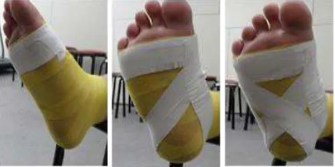
Table 1. General characteristics of subjects (N=24) Figure 2. Low-Dye taping for excessively
pronated rear-foot.
termined automatically in Visual 3D using the verti- cal GRF with a threshold of 20 N. In addition, the ankle inversion-eversion, with respect to the shank, was calculated, and the kinematic data were calcu- lated based on the Cardan flexion/extension, adduc- tion/abduction, and internal/external rotational se- quences (Kadaba et al, 1990). The peak GRF data were normalized to the body weight (㎏), with re- spect to the percent body weight, and the gait meas- urements were normalized to 101 data points, one for each percentage of the stance phase.
Statistical analysis
The data of ankle motion and GRF were averaged across three trials for each participant, while the an- kle motion included the (1) inversion minimum, (2) inversion maximum, and (3) total motion. The peak vertical GRF and shear force (anterio-posterior, me- dio-lateral) were identified during the stance phase of gait. Paired t-tests were used to identify the imme- diate effect of the applied LD taping. A significance level of p<.05 was set. Finally, the effect sizes were com- puted to infer the importance of the mean differences:
small (.2), medium (.5), and large (.8) (Cohen, 2013).
Results
Geranal characteristics of subjects Twenty-four pronated rear-foot participants (6 males and 18 females), with an average age, mass,
and height of 21.0 years (standard deviation 1.3), 53.6
㎏ (standard deviation 4.9), and 163.4 ㎝ (standard deviation 7.5), respectively, were recruited for this study (Table 1).
Ankle motion
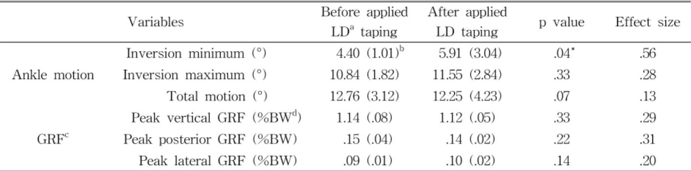
The ankle inversion minimum increased significantly from 4.40° before applied LD taping to 5.91° after ap- plied LD taping and there was statistically medium level in ES (p=.04; Cohen’s d=.56). However, no sig- nificant differences were found in the inversion max- imum or total motion of the stance phase (p=.33;
Cohen’s d=.28, p=.07; Cohen’s d=.13) (Table 2).
Ground reaction force
There were no significant differences in the verti- cal (p=.33), posterior (p=.22), and lateral (p=.14) peak forces. But ES were statistically small level in verti- cal, posterior and lateral peak forces (Cohen’s d=.29, Cohen’s d=.31, Cohen’s d=.20) (Table 2).
Discussion
This study intended to determine the differences in the ankle motion and GRF in pronated rear-foot dur- ing gaits, and to clarify the effects of LD taping.
The results of the analysis of the ankle motion in a stance phase, showed that there was a difference in the angle of inversion as before/after applied LD taping. This is generally consistent with the ad- vanced research results, which showed less inversion in the flat foot gait when compared to the normal foot
Variables Before applied LDa taping
After applied
LD taping p value Effect size Ankle motion
Inversion minimum (°) 4.40 (1.01)b 5.91 (3.04) .04* .56
Inversion maximum (°) 10.84 (1.82) 11.55 (2.84) .33 .28
Total motion (°) 12.76 (3.12) 12.25 (4.23) .07 .13
GRFc
Peak vertical GRF (%BWd) 1.14 (.08) 1.12 (.05) .33 .29
Peak posterior GRF (%BW) .15 (.04) .14 (.02) .22 .31
Peak lateral GRF (%BW) .09 (.01) .10 (.02) .14 .20
alow-Dye, bmean (standard deviation), cground reaction force, dbody weight, *p<.05.
Table 2. Comparison of ankle angles and GRF as before/after applied LD taping (N=24)
gait (Twomey et al, 2010). The applied tape is believed to limit the eversion phenomenon of the pronated rear-foot, when holding up the body weight by sup- porting the arch structures of pronated rear-foot. While the body is engaged in a dynamic activity, such as walking or running, the maintenance of the inversion of the pronated rear-foot acts to control the load, distribute pressure, and prevent damage (Hargrave et al, 2003).
The inversion of the subtalar joint opens the tar- sometatarsal joints and lowers the inner longitudinal foot arch, providing flexibility in the foot and ab- sorbing the pressure when the body weight is loaded. However, via the abnormal function of the subtalar joint and inner longitudinal foot arch, the normal functioning of the foot is damaged, showing excessive eversion, which leads to various damage in the lower limb (Khamis and Yizhar, 2007). The de- crease in the ankle joint inversion is thought to eventually form a foot arch in a flat foot with a lowered arch, decreasing the eversion phenomenon, and playing a positive role in absorbing the impact of the foot. A greater degree of pronation is clin- ically thought to put greater stress and strain upon the plantar fascia.
In this study, LD taped foot showed less foot ever- sion during stance, and it could be prevent the in- ternal rotaion on the tibia and foot. Therefore, LD taping would be considered effective therapeutic meth- od in clinic. and also in this study, LD taping was increased 1.51° in minimum inversion ankle motion.
The results of the analysis of GRF in this study, showed that there was a difference in the vertical,
posterior, and lateral peak forces as before and after applied LD taping. But according to one study, plan- tar fascitis’s patients appeared a diminished peak vertical GRF during propulsion in comparison to healthy individuals (Chang et al, 2014).
The GRF measured on plywood appeared to be lower in pronated rear-foot when compared to nor- mal feet. This is contrary to the results obtained by Twomey and McIntosh (2012), which reported that pronated rear-foot caused a higher GRF when com- pared to normal feet at the instant of initial contact, when the heel touched the ground. However, this study seems to have failed to accurately distinguish the upright type and flaccid type of the flat feet when classifying the subjects. Although the upright type of pronated rear-foot is said to have less im- pact absorbance at initial contact, due to the absence of arches, the flaccid type seems to absorb more GRF than normal feet when the body weight is loaded, since it exhibits lower arches with the load via the relaxation of the surrounding tissues, or weakening of the muscles despite normal foot arches.
It appears that further study on the differences in the GRF between the flaccid type and upright type of flat foot is still needed.
Our findings the application of LD taping on pro- nated rear-foot was not increased GRF but the in- version of ankle motion. These are similar to findings of Yoho et al (2012), who identified the immediate ef- fect of the LD taping on ankle motion biomechanically.
These findings may suggest important treatment on pronated rear-foot for clinical practice and also LD
taping has benefit more comfortable and easy than other brace device which is rigid forms of immobilization.
The limitations of this study include the fact that the age range was limited to the early 20s, and that the classification of pronated rear-foot failed to properly separate the flaccid and upright types. Furthermore, in order to maximize the effects of the variable on taping, the subjects’ walking speed, stride length, and cadence were limited, preventing the gait from falling into a general walking pattern, and creating an envi- ronment that failed to fit the individual characteristics.
Conclusion
The purpose of this study was to determine wheth- er LD taping has different ankle motion and GRF on pronated rear-foot during gait. Overall, there were significant differences in the ankle inversion mini- mum in the stance phase, but not in the inversion maximum and the total inversion-eversion motion.
These data suggest that those with LD taping re- duce ankle angle of foot pronation. Therefore, the application of taping to pronated rear-foot assists in increased ankle inversion, and is expected to be used as a supplementary means of pronated rear-foot treat- ment in the future.
References
Ator R, Gunn K, McPoil TG, et al. The effect of ad- hesive strapping on medial longitudinal arch sup- port before and after exercise. J Orthop Sports Phys Ther. 1991;14(1):18-23.
Chang R, Rodrigues PA, Van Emmerik RE, et al.
Multi-segment foot kinematics and ground re- action forces during gait of individuals with plantar fasciitis. J Biomech. 2014;47(11):2571-2577.
http://dx.doi.org/10.1016/j.jbiomech.2014.06.003 Cheung RT, Chung RC, Ng GY. Efficacies of differ-
ent external controls for excessive foot prona-
tion: A meta-analysis. Br J Sports Med. 2011;45(9):
743-751. http://dx.doi.org/10.1136/bjsm.2010.079780 Childs RA, Olson BA, McPoil TG, et al. The effect of three treatment techniques in reducing meta- tarsal head pressures during walking. Lower Extremity. 1996;3:25-29.
Cohen J. Statistical Power Analysis for the Behavioral Sciences. 2nd ed. London, Routledge, 2013:24-27.
Davis RB, Õunpuu S, Tyburski D, et al. A gait anal- ysis data collection and reduction technique. Hum Mov Sci. 1991;10(5):575-587.
Dye RW. A strapping. 1939. J Am Podiatr Med Assoc.
2007;97(4):282-284.
Franco AH. Pes cavus and pes planus. Analyses and treatment. Phys Ther. 1987;67(5):688-694.
Franettovich M, Chapman A, Blanch P, et al. Continual use of augmented low-Dye taping increases arch height in standing but does not influence neuro- motor control of gait. Gait Posture. 2010;31(2):247-250.
http://dx.doi.org/10.1016/j.gaitpost.2009.10.015 Franettovich M, Chapman A, Vicenzino B. Tape that
increases medial longitudinal arch height also re- duces leg muscle activity: A preliminary study. Med Sci Sports Exerc. 2008;40(4):593-600. http://dx.doi.org/
10.1249/MSS.0b013e318162134f
Franettovich MM, Murley GS, David BS, et al. A comparison of augmented low-Dye taping and ankle bracing on lower limb muscle activity during walking in adults with flat-arched foot posture. J Sci Med Sport. 2012;15(1):8-13. http://dx.doi.org/
10.1016/j.jsams.2011.05.009
Graham ME, Jawrani NT, Goel VK. Evaluating plan- tar fascia strain in hyperpronating cadaveric feet following an extra-osseous talotarsal stabiliza- tion procedure. J Foot Ankle Res. 2011;50(6):
682-686. http://dx.doi.org/10.1053/j.jfas.2011.07.005 Hargrave MD, Carcia CR, Gansneder BM, et al.
Subtalar pronation does not influence impact forces or rate of loading during a single-leg landing. J Athl Train. 2003;38(1):18-23.
Hyland MR, Webber-Gaffney A, Cohen L, et al.
Randomized controlled trial of calcaneal taping,
sham taping, and plantar fascia stretching for the short-term management of plantar heel pain.
J Orthop Sports Phys Ther. 2006;36(6):364-371.
Kadaba MP, Ramakrishnan HK, Wootten ME.
Measurement of lower extremity kinematics during level walking. J Orthop Res. 1990;8(3):
383-392.
Kerr CM, Stebbins J, Theologis T, et al. Normalized ground reaction force peaks are reduced in symp- tomatic flatfoot. Gait Posture. 2014;39(Suppl 1):S71-S72.
Khamis S, Yizhar Z. Effect of feet hyperpronation on pelvic alignment in a standing position. Gait Posture.
2007;25(1):127-134.
Landorf KB, Radford JA, Keenan A, et al. Effectiveness of low-Dye taping for the short-term management of plantar fasciitis. J Am Podiatr Med Assoc. 2005;
95(6):525-530.
Lange B, Chipchase L, Evans A. The effect of low-Dye taping on plantar pressures, during gait, in subjects with navicular drop exceeding 10 ㎜. J Orthop Sports Phys Ther. 2004;34(4):201-209.
Manal K, McClay I, Stanhope S, et al. Comparison of surface mounted markers and attachment meth- ods in estimating tibial rotations during walking:
An in vivo study. Gait Posture. 2000;11(1):38-45.
Nigg BM, Cole GK, Nachbauer W. Effect of arch height of the foot on angular motion of the low- er extremities in running. J Biomech. 1993;26(8):
909-916.
O’Sullivan K, Kennedy N, O’Neill E, et al. The effect of low-Dye taping on rearfoot motion and plantar pressure during the stance phase of gait. BMC Musculoskelet Disord. 2008;9:111. http://dx.doi.org/
10.1186/1471-2474-9-111
Queen RM, Mall NA, Nunley JA, et al. Differences in plantar loading between flat and normal feet dur- ing different athletic tasks. Gait Posture. 2009;29(4):
582-586. http://dx.doi.org/10.1016/j.gaitpost.2008.12.010 Radford JA, Burns J, Buchbinder R, et al. The effect of
low-Dye taping on kinematic, kinetic, and elec- tromyographic variables: A systematic review. J Orthop Sports Phys Ther. 2006;36(4):232-241.
Raissi GR, Cherati AD, Mansoori KD, et al. The rela- tionship between lower extremity alignment and medial tibial stress syndrome among non-pro- fessional athletes. Sports Med Arthrosc Rehabil Ther Technol. 2009;1(1):11.
Resende RA, Deluzio KJ, Kirkwood RN, et al. Increased unilateral foot pronation affects lower limbs and pelvic biomechanics during walking. Gait Posture.
2015:41(2);395-401. http://dx.doi.org/10.1016/j.gaitpost.
2014.10.025
Russo SJ, Chipchase LS. The effect of low-Dye taping on peak plantar pressures of normal feet during gait. Aust J Physiother. 2001;47(4):239-244.
Scranton PE Jr, Pedegana LR, Whitesel JP. Gait analysis. Alterations in support phase forces us- ing supportive devices. Am J Sports Med. 1982;
10(1):6-11.
Souza TR, Pinto RZ, Trede RG, et al. Late rearfoot eversion and lower-limb internal rotation caused by changes in the interaction between forefoot and support surface. J Am Podiatr Med Assoc 2009;99(6):503-511.
Souza TR, Pinto RZ, Trede RG, et al. Temporal cou- plings between rearfoot-shank complex and hip joint during walking. Clin Biomech (Bristol, Avon). 2010;
25(7):745-748. http://dx.doi.org/10.1016/j.clinbiomech.
2010.04.012
Twomey DM, McIntosh AS. The effects of low arched feet on lower limb gait kinematics in children.
Foot (Edinb). 2012;22(2):60-65. http://dx.doi.org/
10.1016/j.foot.2011.11.005
Twomey D, McIntosh AS, Simon J, et al. Kinematic differences between normal and low arched feet in children using the heidelberg foot measurement method. Gait Posture. 2010;32(1):1-5. http://dx.doi.org/
10.1016/j.gaitpost.2010.01.021
van de Water AT, Speksnijder CM. Efficacy of tap- ing for the treatment of plantar fasciosis: A sys- tematic review of controlled trials. J Am Podiatr Med Assoc. 2010;100(1):41-51.
Vicenzino B, Franettovich M, McPoil T, et al. Initial effects of anti-pronation tape on the medial lon-
This article was received November 1, 2015, was reviewed November 1, 2015, and was accepted December 1, 2015.
gitudinal arch during walking and running. Br J Sports Med. 2005;39(12):939-943.
Vinicombe A, Raspovic A, Menz HB. Reliability of navicular displacement measurement as a clinical indicator of foot posture. J Am Podiatr Med Assoc.
2001;91(5):262-268.
Whitaker JM, Augustus K, Ishii S. Effect of the low-Dye strap on pronation-sensitive mechanical attributes of the foot. J Am Podiatr Med Assoc.
2003;93(2):118-123.
Winter DA. Biomechanics and Motor Control of
Human Movement. 4th ed. New Jersey, John Wiley & Sons, 2009:33-38.
Yoho R, Rivera JJ, Renschler R, et al. A biomechanical analysis of the effects of low-Dye taping on arch deformation during gait. Foot (Edinb). 2012;22(4):
283-286. http://dx.doi.org/10.1016/j.foot.2012.08.006