48
Introduction
Anterior cruciate ligament (ACL) injury is a well-rec- ognized risk factor for developing knee osteoarthritis (OA) and degenerative radiographic changes are noted in 16~90% of the subjects 5~15 years after such an injury10,15,17,22). Degenerative changes in the knee after ACL injury, as well as after ACL reconstruction, are common in the long term7,16,25). The purpose of ACL reconstruction is not only to restore the stability and function of the knee, but also to prevent degenerative changes that can occur later on.
The most commonly used grafts for ACL reconstruction
are bone-patellar tendon-bone (BPTB) and hamstring tendon (HT) autografts. The use of BPTB and HT auto- grafts for ACL reconstructions is well established with good, reproducible results1-3,10). The medium- and long- term results are more or less similar for both tech- niques9,18,26). Several meta-analyses have shown only minor differences between the 2 methods6,8,31). However, there are very few reported studies in the medical litera- ture on the long term results after ACL surgeries, and especially, there are few studies that have compared the incidence and the risk factors for developing osteoarthritis after ACL reconstruction with using BPTB or HT auto- grafts.
The aim of this study was to evaluate the radiographic prevalence of OA and the clinical outcome in the long term after ACL reconstruction with using either a BPTB or HT autograft. The secondary aim was to evaluate the influ- ence of various factors on the prevalence of OA.
전방 십자 인대 재건술 후 골관절염의 발생 빈도 및 위험 인자들에 대한 비교 - 자가 슬개건과 자가 슬괴건을 이용한 방법 -
송은규∙선종근∙김형순
1∙강경도∙변재욱
화순전남대학교병원 관절센터, 서남대학교 남광병원 정형외과1
목적: 자가 골-슬개건-골과 자가 슬괵건을 이용한 전방 십자 인대 재건술 후, 골관절염의 발생률과 위험인자를 비교하고자 하였다.
대상 및 방법: 8년 이상 추시가 가능하였던, 자가 슬개건 53예 및 자가 슬괵건 40예를 이용한 전방 십대 인대 재건술을 시행한 총 93예를 대상으로 하였다. Kellgren and Lawrence의 분류에 따라 방사선상의 골관절염의 변화를 관찰하였고, 슬관절의 임 상적 기능 (Lysholm 슬관절 점수, Tegner 운동 지수), 신체검사상의 전방 이완 정도 (Lachman 검사, Pivot-Shift 검사) 및 Telos�기구를 이용한 전방 이완 정도를 비교 평가하였다.
결과: 자가 골-슬개건-골을 이용한 대상 중 24예 (45.3%)에서, 자가 슬괵건을 이용한 대상 중 14예 (35.0%)에서 방사선 검 사상 골관절염 변화가 확인되었다. 동반된 연골판 손상 (골-슬개건-골 군 p<0.001, 슬괵건군 p=0.091), 수상에서 재건술까 지 12개월 이상 경과 (골-슬개건-골 군 p=0.037, 슬괵건 군 p=0.021), 재건술 당시 연령 25세 이상 (골-슬개건-골 군 p=0.003, 슬괵건 군 p=0.048) 등이 골관절염의 유의한 위험 인자로 나타났다. 하지만, 골관절염의 발생과 임상적 결과, 방사 선적 안정성과의 연관성은 없었다.
결론: 자가 골-슬개건-골 및 자가 슬괵건을 이용한 전방 십자 인대 재건술 후 평균 10년 추시상 두 군 모두에서 임상적으로 양호한 결과를 얻었으나, 상당한 비율에서 방사선적 골관절염 변화가 관찰되었다. 또한 동반된 연골판 손상, 수상 후 재건술까 지의 지연된 시간, 25세 이상의 연령 등의 다양한 인자가 이에 관련됨을 알 수 있었다.
색인 단어: 전방 십자 인대, 골-슬개건-골, 슬괵건, 관절염, 재건술
통신저자: 강 경 도
전남 화순군 화순읍 일심리 160 화순전남대학교병원 관절센터
TEL: 061) 379-7676∙FAX: 061) 379-7681 E-mail: [email protected]
Materials and Methods
Between December 1990 and December 1996, 165 consecutive patients underwent arthroscope-assisted ACL reconstruction using a BPTB or HT autograft in our institution. All of the patients were considered to have a unilateral ACL injury that was clinically verified by a his- tory of trauma, a positive Lachman test and/or a positive pivot-shift test. The exclusion criteria were associated previous knee ligament reconstruction or known con- tralateral knee ligament injury, and radiographic OA changes of the knee in any compartment more than grade I according to the rating systems of Kellgren and Lawrence’s classification at the time of the index surgery. Among these patients, 72 patients were exclud- ed because 31 patients were lost to follow-up because of address changes or occupation changes, 20 patients had a contralateral knee ligament injury or previous ligament reconstruction, 14 patients had later received another form of ligamentous surgery other than ACL due to the trauma and 7 patients had various degrees of radiographic OA changes more than grade I according to Kellgren and Lawrence’s classification at the index procedure.
Therefore, the 93 remaining patients composed the study group for this retrospective, minimum 8-year follow-up
study (82 males and 11 females).
Among them, there was no one who had obvious carti- lage injury. And 53 underwent reconstruction with using ipsilateral BPTB autografts and 40 with ipsilateral quadruple hamstring tendon autografts, alternatively.
Fortunately, there has been no graft failure among the patients during the follow-up period. The median age at surgery was 27 years (range: 16.5 to 59.9 years), and the reconstructions were performed at a median of 7.3 months (range: 0.2 to 292 months) after the injury. ACL reconstructions were done in 23 patients (25%) in the acute period within 12 weeks after injury. Regarding the chronicity of injury, there was no difference between both groups. The operative records were retrospectively reviewed to document meniscal injury (Table 1). Among 64 meniscal injured patients, 31 patients had undergone partial menisectomy for medial meniscus injury, 20 patients, partial menisectomy for lateral meniscus injury, 2 patients, subtotal menisectomy for lateral meniscus injury, and 1 patient, total menisectomy for lateral menis- cus injury. And 7 patients of remaining 10 patients had received meniscal sutures for lateral meniscus injury, 3 patients, meniscal sutures for medial meniscus injury.
Table 1. Meniscal injuries treated during the index operation.
BPTB group (N=53) HT group (N=40) p-value
Meniscus injuries combined with 33 (62%) 31 (77.5%) >0.05
the index ACL injury
Medial meniscus injury 26 (49%) 25 (62.5%) >0.05
Lateral meniscus injury 16 (30%) 12 (30%) >0.05
Medial and lateral meniscus injury 09 (17%) 06 (15%) >0.05
BPTB: Bone-patellar tendon-bone HT: Hamstring tendon
ACL: Anterior cruciate ligament NS: Non-specific
Table 2. Grades of radiographic osteoarthritic changes by the Kellgren and Lawrence’s classification. (p=0.757).
Normal or Grade I Grade II Grade III* Total
BPTB group (N) 29 17 7 53
HT group (N) 26 10 4 40
Total (N(%)) 56 (60.2%) 26 (28.0) 11(11.8%) 93 (100%)
BPTB: Bone-patellar tendon-bone HT: Hamstring tendon
* There was no grade IV osteoarthritis by the Kellgren and Lawrence’s classification in our patients.
1. Radiographic assessment
The radiographic assessment of the patient groups involved 2 views, namely, the standing anteroposterior view, the lateral view with 20 to 30 degrees of flexion of the knee at the last follow-up. We could obtain the radi- ographic images at all included patients. At the final fol- low-up, the patients with more than grade II radiographic OA changes who showed clear osteophytes were defined as having osteoarthritis according to the rating systems of Kellgren and Lawrence’s classification13). To ensure the reproducibility of radiographic OA changes, two indepen- dent radiologists not involved in the surgical procedures independently reviewed preoperative and last follow-up radiographs.
2. Clinical assessments
Clinical evaluations of the knee function and laxity were performed preoperatively and at the last follow-up by the two senior authors of this study. The preoperative and postoperative Lysholm Knee Scores and the Tegner activity scores were obtained by means of a self-admin- istered questionnaire and the scores were evaluated at the time of follow up to access the functional capability of the knee29). All the functional assessments were per- formed at a minimum 8-year-follow up. Assessment of knee stability was undertaken using the Lachman, anterior drawer and pivot shift tests. Instrumented laxity testing was determined using the Telos� (Telos stress device;
Austin & Associate, Inc., Polston, US) (20 lbs) by mea- suring the side-to-side differences in anterior displace- ment at 20 degrees of knee flexion5). The range of motion was measured using a goniometer. In addition, we evalu- ated the relationships between the patient’s age at the time of reconstruction, the time from injury to recon- struction and any accompanying meniscal injury with the presence of osteoarthritis at the final follow-up.
3. Graft harvest site morbidity
At the last follow up, the patients were asked to note any kneeling pain, paresthesia or numbness at the auto- graft harvest site, crepitus and anterior knee pain.
Kneeling pain was reported if it was present after the patients kneeled on a carpeted floor.
4. Statistical analysis
Descriptive statistics (arithmetic means, SDs, and ranges) were calculated using standard formulas. The results such as range of motion, the Lysholm score age, time to surgery and the follow-up period were compared between the BPTB and HT autograft groups by use of independent t test. Pearson’s chi-square test, Fisher’s exact test and odds ratios were used for analyzing the ordered categorical variables such as the OA grade, the Tegner activity score, Lachman test. Inter-rater agree- ment for radiographic osteoarthritic changes was esti- mated using Cohen’s κstatistic. The nomenclature for describing the level of reliability associated with a specific value of κ, as presented by Landis and Koch, was follows:
less than 0.00 poor, 0.00 to 0.20, slight, 0.21 to 0.40, moderate, 0.61 to 0.80, substantial, 0.81 to 1.00, almost perfect. Tested comparisons with p <0.05 were consid- ered to be significantly different, and all aspects of the statistical analysis were reviewed independently by a statistician.
Results
There were no statistically significant differences between the preoperative data of the two groups with respect to gender, age, the time from injury to operation, the knee laxity tests, range of motion and meniscus injury. In the BPTB group, the mean preoperative Lysholm knee scores was slightly lower than that in the HT group (57 vs. 66, respectively, p=0.001) and the fol- low up duration was longer than in the HT group (10.7 vs.
9.0, respectively, p=0.00). Preoperatively, the mean range of motion was -1.42�to 135.5�in the BPTB group and it was -0.38�to 134.3�in the HT group (p>0.05).
1. Radiographic assessment
Standard weight-bearing radiography showed no sig- nificant differences between the BPTB group and the HT group with respect to radiographic OA changes at final follow-up period. Overall, radiographic OA changes was identified in 38 patients (40.8%) among 93 patients (24 patients (45.3%) in the BPTB group and 14 patients (35%) in the HT group, p=0.318) according to the Kellgren and Lawrence’s classification system. Inter- rater agreement for radiographic osteoarthritic changes
was almost perfect (Cohen’s κ, 0.83).With regard to the severity and the sites of osteoarthritis at the final follow- up, as determined by Kellgren and Lawrence’s classifi- cation, there were no significant differences between the two groups (Table 2, 3). Patients with meniscal injuries during the index operation had significantly more radi- ographic OA changes than the patients without such injuries, according to the Kellgren and Lawrence’s clas- sification system (p<0.001, odds ratio= 7.083) (Table 4).
Patients with an age of more than 25 years at the time of
reconstruction which were mean 33.2 years (range, 25.1 to 59.9) had significantly more radiographic OA changes than the patients below 25 years of age at the time of reconstruction (p<0.001, odds ratio= 5.217) (Table 5).
The patients with more than 12 months from injury to the time of reconstruction had significantly more radiographic OA changes than patients with less than 12 months from the time of injury until reconstruction (p=0.001, odds ratio= 4.105) (Table 6). However, there was no signifi- cant difference in the incidence of radiographic OA
Table 3. Radiographic osteoarthritic changes according to the compartment of the knee.
Medial Lateral Patello-femoral
Total*
compartment compartment compartment
BPTB group (N) 23 9 8 24
HT group (N) 11 6 2 14
Total (N(%)) 34 (89.5%) 15 (39.5%) 10 (26.3%) 38
p value 0.133 0.771 0.340 0.318
BPTB: Bone-patellar tendon-bone HT: Hamstring tendon
*There were 6 OA knees involving two compartments (BPTP group, 4; HT group, 2) and 7 knees involving all compartments (BPTB group, 5; HT group, 2).
Table 4. Radiographic OA changes according to meniscal injuries during the index operation.
Meniscal injury BPTB group (N) HT group (N) Total (N(%))
Non-OA group No 17 08 25 (45.5%)
Yes 12 18 30 (54.5%)
OA group No 03 01 04 (10.5%)
Yes 21 13 34 (89.5%)
p value 0.001 0.091 <0.001
Odds ratio 9.917 5.778 7.083
OA: Osteoarthritis
BPTB: Bone-patellar tendon-bone HT: Hamstring tendon
Table 5. Radiographic OA changes according to an age at the time of reconstruction.
Age at index operation BPTB group (N) HT group (N) Total (N(%))
Non-OA group < 25 years old 18 14 32 (58.2%)
≥ 25 years old 11 12 23 (41.8%)
OA group < 25 years old 05 03 08 (21.1%)
≥ 25 years old 19 11 30 (78.9%)
P value 0.003 0.048 <0.001
Odds ratio 6.218 4.278 5.217
OA: Osteoarthritis
BPTB: Bone-patellar tendon-bone HT: Hamstring tendon
changes between two groups according to 3 months from injury to the time of reconstruction.
2. Clinical assessment
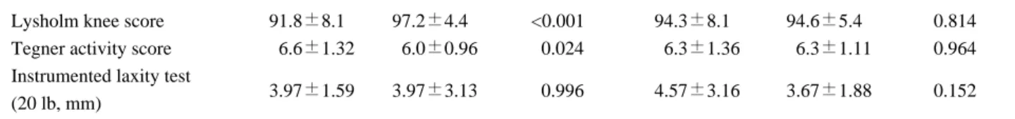
At the last follow-up, the Lysholm knee and the Tegner activity score were significantly improved in both groups. There was no difference between the patients with OA and those without OA with respect to the Lysholm knee score (p=0.814) and the Tegner activity score (p=0.964) (Table 7). Clinical laxity assessment according to the Lachman test and the pivot shift test is summarized in Table 8. There was no difference between the patients with and without radiographic OA changes with respect to the result of Lachman test (p=0.345) and the pivot-shift test (p=0.425). At the last follow up, both groups were improved in terms of the Lysholm knee score, the Tegner activity score, the Lachman test, the pivot-shift test and the side-to-side difference of the anterior laxity on the Telos� stress radiography (p<0.001).
3. Graft harvest site morbidity
were four patients (7.5%) with kneeling pain in the BPTB group, but no patients in the HT group complained of this. In both groups, there was no difference in the incidence of symptoms arising from their graft harvest site such as paresthesia or numbness. Incision site paresthesia or numbness was reported by 28.3% of the patients in the BPTB group and by 27.5% of the patients in the HT group. Crepitus was reported by 6 patients (11%) in the BPTB group and by 5 patients (13%) in the HT group.
Discussion
After ACL injury, 60% to 90% of individuals develop radiographic evidence of knee OA within 10 to 20 years of their injury17,22). Reconstruction of a ruptured ACL of the knee of an active patient is widely recommended to prevent knee instability, recurrent injury and further intraarticular injury4,28). However, long-term studies have shown that ACL reconstruction fails to slow the progres-
Table 6. Radiographic OA changes according to a time from injury to reconstruction.
Time from injury
BPTB group (N) HT group (N) Total (N(%)) to reconstruction
Non-OA group < 12 months 18 18 36 (65.5%)
≥ 12 months 11 08 19 (44.5%)
OA group < 12 months 08 04 12 (31.6%)
≥ 12 months 16 10 26 (68.4%)
p value 0.037 0.021 0.001
Odds ratio 3.273 5.625 4.105
OA: Osteoarthritis
BPTB: Bone-patellar tendon-bone HT: Hamstring tendon
Table 7. Postoperative clinical assessment of the patients
By operation methods By radiographic OA
BPTB group HT group p value OA group Non-OA group p value Lysholm knee score 91.8±8.10 97.2±4.40 <0.001 94.3±8.10 94.6±5.40 0.814 Tegner activity score 06.6±1.32 06.0±0.96 00.024 06.3±1.36 06.3±1.11 0.964 Instrumented laxity test
3.97±1.59 3.97±3.13 00.996 4.57±3.16 3.67±1.88 0.152
(20 lb, mm) OA: Osteoarthritis
BPTB: Bone-patellar tendon-bone HT: Hamstring tendon
sion of knee arthritis that may be caused by altered joint mechanics, concomitant meniscal and cartilage injuries prior to or after ACL reconstruction, changes in the physiologic environment or even a genetic predisposi- tion30). Most studies have concentrated on the radiological changes following ACL rupture and reconstruction. The long-term outcome in patients who are symptomatically stable following an ACL rupture is uncertain, although in a small cohort of elite athletes, all of them had degenerative changes 35 years after injury and eight out of 19 (42%) had undergone total knee replacement20). At 20 years fol- low-up, the reported risk of developing osteoarthritis is lower after ACL reconstruction (14%~26% with a normal medial meniscus and 37% with meniscectomy) than that for untreated ruptures (60~100%)17). According to our literature review, we think that ACL reconstruction can reduce the risk of OA.
The reported incidences of OA after ACL reconstruc- tion have varied according to the type of graft that is used, the follow up period and other related factors. The frequency and causes of OA currently remain controver- sial. In this study, the average follow-up after ACL reconstruction with using a patellar or hamstring tendon autograft was 10 years, and degenerative osteoarthritis developed in 38 of 93 patients (40.8%), and especially in the medial compartment. Some authors have reported that OA developed significantly more often in the BPTB group than that in the HT group12,24,27). Yet in this study, radi- ographic OA changes developed more frequently in the BPTB group (45%) than that in the HT group (35%), but this was not significant, which was consistent with other studies14,18,23). The frequency of OA according to the graft type currently remains controversial.
In terms of the risk factors for osteoarthritis, the fol-
lowing have all been suggested7,15,17,21)
: the patient’s age at the time of reconstruction, accompanying meniscal injury, the time from injury to reconstruction, anterior instability and persistent anterior subluxation. When the ACL had been reconstructed, the loss of the medial meniscus still shows an increased risk of OA. In our study, 64 patients (69%) had a combined meniscal injury. Among them, 34 patients (53%) had radiographic OA changes at the final follow up. In the other hand, among the 29 patients with- out meniscal injury, only 4 patients (13.8%) showed radiographic OA changes at the final follow up. Meniscal injury is an important factor related with OA after ACL reconstruction. Previous studies also have shown the impact of not only ACL rupture, but also damaged menisci on the development of OA7,11,19). It is plausible that abnormal kinematics and damaged menisci are additional risk factors for the development of OA. The time from injury to reconstruction also influences the long term results. In the present study, this averaged 27 months, but Jomha et al11). reported an average interval of 5.6 years. In their study, of the 53 cases that underwent early ACL reconstruction within 12 weeks of injury, degenera- tive changes were detected in 27% at 7-year postopera- tively, and of the cases that underwent construction at more than 12 weeks after injury, degenerative changes were detected in 54%. Thus, it was suggested that reconstruction during the early period helps prevent the development of degenerative osteoarthritis. In our study, for the 45 patients who underwent reconstruction over 12 months after injury, radiographic OA changes developed in 26 patients (58%) and for the 48 patients with less than 12 months of time from injury to reconstruction, radiographic OA changes developed in 12 patients (25%).
There was a significant difference in the incidence of
Table 8. Clinical laxity assessment according to the Lachman test and the pivot shift test
Lachman test Pivot shift test
Grade Preoperative Last follow-up Preoperative Last follow-up
(0/+/++/+++) (0/+/++/+++) (0/+/++/+++) (0/+)
BPTB group (N) -/17/30/6 44/7/2/- 7/28/13/5 46/7
(%) (0/32.1/56.6/11.3%) (83/13.2/3.8/0%) (13/52.9/24.6/9.5%) (86.8/13.2%)
HT group (N) -/14/15/11 32/7/1/- 7/23/4/6 34/6
(%) (0/35/37.5/27.5%) (80/17.5/2.5/0%) (17.5/57.5/10/15%) (85/15%)
p value 0.080 0.811 0.308 0.210
BPTB: Bone-patellar tendon-bone HT: Hamstring tendon
radiographic OA changes according to the time to recon- struction. Therefore, in our assessment, more delayed operation than 12 months from injury is strongly related with incidence of radiographic OA changes. Moreover, the grades of OA were related with a more delayed time to reconstruction (p=0.001). These findings indicate that a longer interval from injury to reconstruction increased the risk of developing osteoarthritis as well as its grade. With regard to the patients’ages and the development of degenerative osteoarthritis, it has been reported that the frequency of developing osteoarthritis is significantly higher for those patients over 25 years of age at the time of injury7,11). In the present study, 30 of the patients (56%) who were over 25 years of age at the time of surgery developed osteoarthritis, which was a signifi- cantly higher rate than that for the patients (8 patients (20%)) who were 25 years or younger.
The relation between the development of osteoarthritis after ACL reconstruction and anterior instability is still controversial. A number of authors have proposed that anterior instability induces degenerative osteoarthritis in the knee joint. However, it has recently been reported that anterior instability is not related to developing osteoarthritis after reconstruction, since degenerative changes were detected in the patients who were without anterior instability during their long-term follow-up7). In our study, the overall mean laxity of knee checked by Telos device appears to be well above 3 mm side-to- side difference and the OA group is 4.6 mm. Although we did not identify a statistically significant difference, this may be clinically significant. Also, our testing was per- formed at only 20 lbs of force. A larger force would have likely resulted in increase side-to-side differences.
There is also a concern that the Telos data did not corre- late with the clinical Lachman’s test data. After ACL reconstruction, the Lysholm knee scores and Lachman and pivot-shift test results were as good as those reported by others researchers. In our study, there was no significant difference between the groups with respect to the Lachman or pivot-shift tests at the final follow-up.
The mean value of the objective anterior laxity measure- ments revealed no significant differences between the hamstring tendon graft group and the patellar tendon graft group. However, the pivot shift clinical examination may also underestimate true rotational instability. The combi- nation of these factors may have played an important role in the development of radiographic OA changes, which
further investigation will be needed.
The present study has some limitations. First, the study involved a retrospective review of a single institutional database. It is likely that very large multi-institutional databases would provide additional significant information.
Second limitation in the design of our study is that there were meniscal injuries in addition to the ACL tear. It is difficult to obtain patients with solitary ACL tears and who are without any other intra-articular lesions.
In conclusion, radiographic osteoarthritic changes were identified in 40.8% of patients, although there was an improvement in the clinical results at an average follow- up of 10 years after anterior cruciate ligament recon- struction with using a patellar or hamstring tendon auto- graft. The frequency of radiographic degenerative changes of the knee was high in patients with accompa- nying meniscal injury, for patients with a protracted time from injury to reconstruction and for patients who were more than 25 years old at the time of reconstruction.
REFERENCES
01. Brandsson S, Faxen E, Eriksson BI, Sward L, Lundin O, Karlsson J: Reconstruction of the anterior cruciate ligament: Comparison of outside-in and all-inside tech- niques. Br J Sports Med, 33: 42-45, 1999.
02. Brown CH, Steiner ME, Carson EW: The use of ham- string tendons for anterior cruciate ligament reconstruc- tion. Technique and results. Clin Sports Med, 12: 723-756, 1993.
03. Buss DD, Warren RF, Wickiewicz TL, Galinat BJ, Panariello R: Arthroscopically assisted reconstruction of the anterior cruciate ligament with use of autogenous patellar-ligament grafts. Results after twenty-four to forty- two months. J Bone Joint Surg Am, 75: 1346-1655, 1993.
04. Daniel DM, Stone ML, Dobson BE: Fate of the ACL- injured patient: A prospective outcome study. Am J Sports Med, 22: 632-644, 1994.
05. Franklin JL, Rosenberg TD, Paulos LE, France EP, City SL: Radiographic assessment of instability of the knee due to rupture of the anterior cruciate ligament. J Bone Joint Surg Am, 73: 365-372, 1991.
06. Freedman KB, D’’Amato MJ, Nedeff DD, Kaz A, Bach BR Jr: Arthroscopic anterior cruciate ligament recon- struction: A metaanalysis comparing patellar tendon and hamstring tendon autografts. Am J Sports Med, 31: 2-11, 2003.
07. Gillquist J, Messner K: Anterior cruciate ligament reconstruction and the long-term incidence of gonarthro- sis. Sports Med, 27: 143-156, 1999.
08. Goldblatt JP, Fitzsimmons SE, Balk E, Richmond JC:
Reconstruction of the anterior cruciate ligament: Meta- analysis of patellar tendon versus hamstring tendon auto- graft. Arthroscopy, 21: 791-803, 2005.
09. Ibrahim SA, Al-Kussary IM, Al-Misfer AR, Al-Mutairi HQ, Ghafar SA, El Noor TA: Clinical evaluation of arthroscopically assisted anterior cruciate ligament reconstruction: Patellar tendon versus gracilis and semi- tendinosus autograft. Arthroscopy, 21: 412-417, 2005.
10. Jackson DW, Jennings LD: Arthroscopically assisted reconstruction of the anterior cruciate ligament using a patella tendon bone autograft. Clin Sports Med, 7: 785- 800, 1988.
11. Jomha NM, Borton DC, Clingeleffer AJ, Pinczewski LA: Long-term osteoarthritic changes in anterior cruciate ligament reconstructed knees. Clin?Orthop, 358: 188-193, 1999.
12. Keays SL, Bullock-Saxton JE, Keays AC, Newcombe PA, Bullock MI: A 6-year follow-up of the effect of graft site on strength, stability, range of motion, function, and joint degeneration after anterior cruciate ligament recon- struction: patellar tendon versus semitendinosus and gra- cilis tendon graft. Am J Sports Med, 35: 729-739, 2007.
13. Kellgren JH, Lawrence JS: Radiological assessment of osteo-arthritis. Ann Rheum Dis, 16: 494-501, 1957.
14. Liden M, Sernert N, Rostgård-Christensen L, Kartus C, Ejerhed L: Osteoarthritic changes after anterior cruci- ate ligament reconstruction using bone-patellar tendon- bone or hamstring tendon autografts: a retrospective, 7- year radiographic and clinical follow-up study.
Arthroscopy, 24:899-908, 2008.
15. Lohmander LS, Englund PM, Dahl LL, Roos EM: The long-term consequence of anterior cruciate ligament and meniscus injuries: osteoarthritis. Am J Sports Med, 35:
1756-1769, 2007.
16. Lohmander LS, Ostengberg A, Englund M, Roos H:
High prevalence of knee osteoarthritis, pain, and function- al limitations in female soccer players twelve years after anterior cruciate ligament injury. Arthritis Rheum, 50:
3145-3152, 2004.
17. Louboutin H, Debarge R, Richou J, et al.: Osteoarthritis in patients with anterior cruciate ligament rupture: A review of risk factors. Knee, 16: 239-244, 2009.
18. Matsumoto A, Yoshiya S, Muratsu H: A comparison of
bone-patellar tendon-bone and bone-hamstring tendon- bone autografts for anterior cruciate ligament reconstruc- tion. Am J Sports Med, 34: 213-219, 2006.
19. Meunier A, Odensten M, Good L: Long-term results after primary repair or non-surgical treatment of anterior cruciate ligament rupture: A randomized study with a 15- year follow-up. Scan J Med Sci Sports, 17: 230-237, 2007.
20. Nebelung W, Wuschech H: Thirty-five years of follow-up of anterior cruciate ligament-deficient knees in high-level athletes. Arthroscopy, 31: 670-696, 2005.
21. Neuman P, Englund M, Kostogiannis I, Friden T, Roos H, Dahlberg EL: Prevalence of tibiofemoral osteoarthri- tis 15 years after nonoperative treatment of anterior cruci- ate ligament injury: a prospective cohort study. Am J Sports Med, 36: 1717-1725, 2008.
22. Neyret P, Donell ST, Dejour D, Dejour H: Partial meniscectomy and anterior cruciate ligament rupture in soccer players: A study with a minimum 20-year followup.
Am J Sports Med, 21: 455-460, 1993.
23. O’’Neill DB: Arthroscopically assisted reconstruction of the anterior cruciate ligament. A follow-up report. J Bone Joint Srug Am, 83: 1329-1332, 2001.
24. Pinczewski LA, Lyman J, Salmon LJ, Russell VJ, Roe J, Linklater J: A 10-year comparison of anterior cruciate ligament reconstructions with hamstring tendon and patel- lar tendon autograft: a controlled, prospective trial. Am J Sports Med, 35: 564-574, 2007.
25. Porat A, Roos EM, Roos H: High prevalence of osteoarthri- tis 14 years after an anterior cruciate ligament tear in male soccer players: A study of radiographic and patient relevant outcomes. Ann rheum Dis, 63: 269-273, 2004.
26. Roe J, Pinczewski LA, Russell VJ, Salmon LJ, Kawamata T, Chew M: A 7-year follow-up of patellar tendon and hamstring tendon grafts for arthroscopic ante- rior cruciate ligament reconstruction: Differences and similarities. Am J sports Med, 33: 1337-1345, 2005.
27. Sajovic M, Vengust V, Komandina R, Tavcar R, Skaza K: A prospective, randomized comparison of semitendi- nosus and gracilis tendon versus patellar tendon auto- grafts for anterior cruciate ligament reconstruction: five- year follow-up. Am J Sports Med, 34: 1933-1940, 2006.
28. Shelbourne KD, Gray T: Results of anterior cruciate lig- ament reconstruction based on meniscus and articular cartilage status at the time of surgery. Five- to fifteen-year evaluations. Am J Sports Med, 28: 446-452, 2000.
29. Tegner Y, Lysholm J: Rating systems in the evaluation of knee ligament injuries. Clin Orthop, 198: 43-49, 1985.
30. Yu J, Garrett WE: Femoral tunnel placement in anterior cruciate ligament reconstruction. Operative Techniques in Sports Medicine, 14: 45-49, 2006.
31. Yunes M, Richmond JC, Engels EA, Pinczewski LA:
Patellar versus hamstring tendons in anterior cruciate lig- ament reconstruction: A meta-analysis. Arthroscopy, 17:
248-257, 2001.
32. Wilder FV, Hall BJ, Barrett Jr Jp, Lemrow NB:
History of acute knee injury and osteoarthritis of the knee:
a prospective epidemiological assessment. The Clearwater Osteoarthritis study. Osteoarthritis Cartilage, 10: 611- 616, 2002.
Comparison of the Incidence and Risk Factors for Developing Osteoarthritis after ACL Reconstruction
- Patellar Versus a Hamstring Autograft -
Eun-Kyoo Song, M.D.Ph.D., Jong-Keun Seon, M.D., Hyung-Soon Kim, M.D.1, Kyung-Do Kang, M.D., Jae-Wook Byun, M.D.
Department of Orthopedic Surgery, Center for Joint Disease, Chonnam National University Hwasun Hospital, Jeonnam, Korea
Department of Orthopedic Surgery, Seonam University Namgwang Hospital, Gwangju, Korea1
Purpose: To compare the incidence and risk factors for osteoarthritis after anterior cruciate ligament (ACL) reconstruction between two groups using bone-patellar tendon-bone (BPTB) and hamstring tendon (HT) autograft.
Materials and Methods: 53 cases of ACL reconstruction using patellar tendon and 40 cases using hamstring tendon were followed up at least 8 years. Radiographic evaluation was done according to the Kellgren and Lawrence’s classification. Clinical functional testing (Lysholm Knee Scores, the Tegner activity scores) and laxity testing (Lachman, pivot shift tests), and the instrumented laxity test- ing with Telos�were all examined in relation to the development of osteoarthritis.
Results: Radiographic osteoarthritic changes were detected in 24 patients (45.3%) in BPTB group and 14 patients (35.0%) in HT group. Accompanying meniscal injury (BPTB p<0.001; HT p=0.091), intervals from the injury to reconstruction of > 12 months (BPTB p=0.037; HT p=0.021), and patient’
s age at reconstruction of >25 years (BPTB p=0.003; HT p=0.048) were found to be significant inde- pendent predictors of osteoarthritis. However, no statistically significant correlations were found between the development of osteoarthritis and the clinical outcome or the radiographic stability in both groups.
Conclusion: Although ACL reconstruction using BPTB or HT autograft had good clinical results at an average follow-up of 10 years, considerable incidence of radiographic osteoarthritic changes were noted. Various factors such as accompanying meniscal injury, protracted time from injury to recon- struction, more than 25 years old at the time of reconstruction were related to radiographic osteoarthritic changes.
Key Words: Anterior cruciate ligament, BPTB, HT, Osteoarthritis
Address reprint requests to Kyung-Do Kang, M.D.
Department of Orthopedic Surgery, Center for Joint Disease, Chonnam National University Hwasun Hospital 160, Ilsim-Ri, Hwasun-Eup, Hwasun-Gun, Jeonnam, Korea
TEL: 82-61-379-7676, FAX: 82-61-379-7681, E-mail: [email protected]
= ABSTRACT =