ISSN 2234-3806 • eISSN 2234-3814
540 www.annlabmed.org https://doi.org/10.3343/alm.2017.37.6.540 Ann Lab Med 2017;37:540-543
https://doi.org/10.3343/alm.2017.37.6.540
Case Report
Diagnostic Genetics
Novel 4-bp Intronic Deletion (c.1560+3_1560+6del) in LEMD3 in a Korean Patient With Osteopoikilosis
In Young Yoo, M.D.1, Ju-Sun Song, M.D.1, Chang-Seok Ki, M.D.1, Jong-Won Kim, M.D.1, Hoon-Suk Cha, M.D.2, and Yong-Ki Min, M.D.2
Department of Laboratory Medicine and Genetics1, Samsung Medical Center, Sungkyunkwan University School of Medicine, Seoul; Division of Endocrinology and Metabolism2, Department of Medicine, Samsung Medical Center, Sungkyunkwan University School of Medicine, Seoul, Korea
Osteopoikilosis is an autosomal dominant bone disorder characterized by symmetric mul- tiple osteosclerotic lesions throughout the axial and appendicular skeleton. Pathogenic variants in the LEMD3 have been identified as the cause of osteopoikilosis. LEMD3 en- codes an inner nuclear membrane protein that interacts with bone morphogenetic protein (BMP) and transforming growth factor (TGF)-β pathways. We report the case of a 19-year- old man presenting with lower back pain and sciatica. His radiograph revealed bilateral and symmetrical multiple osteosclerotic bone lesions in both scapular areas. Sanger se- quencing of LEMD3 revealed a four-base-pair deletion in intron 2 (c.1560+3_1560+6del), which was inherited from his father. We found that this four-base-pair deletion in intron 2 causes aberrant splicing and consequent deletion of exon 2. To the best of our knowl- edge, this is the first report of genetically confirmed osteopoikilosis in Korea.
Key Words: LEMD3, Osteopoikilosis, Sequencing, Deletion
Received: February 9, 2017 Revision received: April 10, 2017 Accepted: July 18, 2017
Corresponding author: Chang-Seok Ki Department of Laboratory Medicine and Genetics, Samsung Medical Center, Sungkyunkwan University School of Medicine, 81 Irwon-ro, Gangnam-gu, Seoul 06351, Korea
Tel: +82-2-3410-2709 Fax: +82-2-3410-2719 E-mail: [email protected]
Co-corresponding author: Yong-Ki Min Division of Endocrinology and Metabolism, Department of Medicine, Samsung Medical Center, Sungkyunkwan University School of Medicine, 81 Irwon-ro, Gangnam-gu, Seoul 06351, Korea
Tel: +82-2-3410-3432 Fax: +82-2-3410-3849 E-mail: [email protected]
© Korean Society for Laboratory Medicine This is an Open Access article distributed under the terms of the Creative Commons Attribution Non-Commercial License (http://creativecom- mons.org/licenses/by-nc/4.0) which permits unrestricted non-commercial use, distribution, and reproduction in any medium, provided the original work is properly cited.
INTRODUCTION
Osteopoikilosis, also known as osteopathia condensans dissemi- nata, is a rare and benign autosomal dominant disease charac- terized by symmetric but unequal distribution of osteosclerotic bone dysplasia in different parts of the skeleton. The diagnosis is often made on the basis of incidental radiologic findings with multiple small, sclerotic foci at the ends of the long bones, pel- vis, sacrum, and bones of the hands and feet. Although it is
usually asymptomatic, 15-20% of patients may have joint pain and effusion [1-4].
Although the pathogenesis of osteopoikilosis has not been fully elucidated, heterozygous loss-of-function variants in the LEM domain-containing protein 3 (LEMD3) gene have been identified as the cause of osteopoikilosis by genome-wide link- age analysis in affected families [5]. In Korea, several cases of familial osteopoikilosis have been reported; however, all of these cases were diagnosed by radiological findings and clinical mani-
1 / 1 CROSSMARK_logo_3_Test
2017-03-16 https://crossmark-cdn.crossref.org/widget/v2.0/logos/CROSSMARK_Color_square.svg
Yoo IY, et al.
LEMD3-related osteopoikilosis
https://doi.org/10.3343/alm.2017.37.6.540 www.annlabmed.org 541
festations such as pelvic pain and lower abdominal discomfort, without genetic analysis of LEMD3 [6-9]. We report a Korean
patient with osteopoikilosis carrying a novel splice site variant in the LEMD3 gene.
Fig. 2. Novel splice site variant in the LEMD3 gene. (A) Sequencing pattern of LEMD3 shows overlapping peaks due to a heterozygous variant in intron 2 (c.1560+3_1560+6del; arrow). (B) Cloning of reverse transcription (RT)-PCR products reveals two clones: a normal clone and an abnormal clone without exon 2. (C) Schematic illustration of aberrant splicing due to the heterozygous 4-bp deletion.
Exon 2
Forward Reverse
c.1560+3_1560+6del Intron 2 A
Exon 2 Exon 3
Exon 3 Exon 1
Exon 1 Normal splicing
Aberrant splicing B
Exon 2 Exon 3
Exon 1
c.1560+3_1560+6del Aberrant splicing
...CAGCA gtaa... ...ag TAGAAA... TACAA gtaagtaa ...ag GAAAG...
C
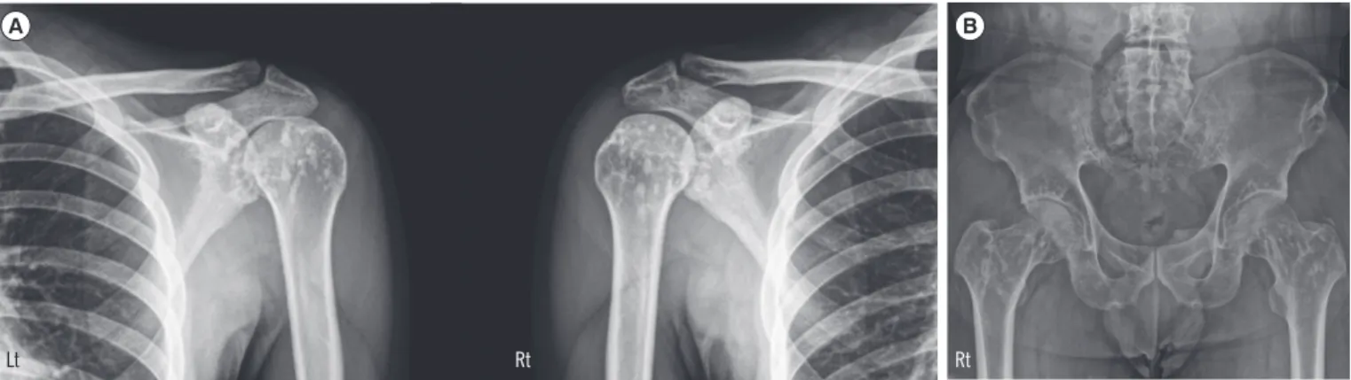
Fig. 1. Representative radiologic findings of the patients. (A) Ill-defined osteosclerotic lesions in both humoral head and scapula are ob- served in the proband. (B) Pelvis radiograph of the proband’s father shows small, roundish radio-dense lesions in both proximal femurs and the iliac bone.
Abbreviations: Lt, Left; Rt, Right.
A B
Lt Rt Rt
Yoo IY, et al.
LEMD3-related osteopoikilosis
542 www.annlabmed.org https://doi.org/10.3343/alm.2017.37.6.540
CASE REPORT
A 19-yr-old man with lower back pain and sciatica presented to our hospital to rule out bone metabolic disease. Radiographs showed multiple osteosclerotic bone lesions in both scapular ar- eas (Fig. 1A). He had a treatment history for short stature, and at the time he first visited our hospital, his height was 162 cm.
Other than short stature, there were no remarkable findings in the connective tissue or skin. The proband’s father also suffered from multiple joint pain, and the father’s radiographs showed multiple, small, roundish radio-dense lesions in both proximal femurs and the iliac bone (Fig. 1B).
After obtaining informed consents, whole blood samples were collected from the patient and his father to analyze their LEMD3 status. Genomic DNA was extracted from peripheral blood leu- kocytes, and all 13 exons were amplified by PCR using an ABI 3730xl analyzer (Applied Biosystems, Foster City, CA, USA) and the BigDye Terminator v3.1 Cycle Sequencing Kit (Applied Bio- systems) with primers designed by the authors. LEMD3 se- quences were analyzed by using Sequencher software (Gene Codes Corp., Ann Arbor, MI, USA) and compared with the refer- ence sequence (NM_014319.4).
A four-base-pair deletion (c.1560+3_1560+6del) in intron 2 of LEMD3 was identified in the patient and his father (Fig. 2A).
This variant has not been reported previously and was absent from the Single Nucleotide Polymorphism database (https://
www.ncbi.nlm.nih.gov/snp), the Exome Aggregation Consortium (ExAC) database (http://exac.broadinstitute.org/), and the Ko- rean Reference Genome database (http://152.99.75.168/
KRGDB/). Reverse-transcription PCR (RT-PCR) and amplicon cloning results confirmed aberrant splicing. As expected, one clone with a deletion in exon 2 (r.1523_1560del) was identified (Fig. 2B and C), resulting in a frameshift in the LEMD3 protein (p.Ile508Argfs*3). On the basis of these results, we regard this variant as a pathogenic variant that may cause osteopoikilosis according to the standards and guidelines of the American Col- lege of Medical Genetics and Genomics and the Association for Molecular Pathology [10].
DISCUSSION
Osteopoikilosis is an autosomal dominant bone disorder charac- terized by symmetric multiple osteosclerotic lesions throughout the axial and appendicular skeleton. Prevalence has been esti- mated to be 1/50,000 and occurs at the same rate in men and women [1]. Heterozygous loss-of-function variants in LEMD3
have been identified as the major cause of both osteopoikilosis and Buschke-Ollendorff syndrome (BOS, OMIM 166700), with or without melorheostosis [5]. BOS is a rare autosomal domi- nant disorder characterized by early-onset skin features, such as multiple cutaneous elastic hamartomas, and also osteopoiki- losis [11]. To date, many variants of LEMD3 have been found, including nonsense, frame-shift, and splice site defects resulting in exon skipping, as was found in this case [12]. However, no phenotype-specific variants have been found in the LEMD3 gene [13].
LEMD3 is an inner nuclear membrane protein that interacts with both bone morphogenetic protein (BMP) and transforming growth factor (TGF)-β signaling pathways through interactions with Smad family proteins such as Smad1 (BMP-specific) and Smad2 (TGF-β specific). When LEMD3 is deactivated, it cannot downregulate Smad1 activation resulting in the promotion of bone formation via BMP signaling [5]. Due to the antagonistic effect of LEMD3 on TGF-β signaling, increased signaling in the TGF-β pathway can cause skin lesions in patients with BOS [14]. Therefore, loss-of-function variants in LEMD3 could lead to the abnormal growth of connective and bone tissue via en- hanced BMP and TGF-β signaling [15].
In conclusion, we identified a novel splice site variant of LEMD3 causing osteopoikilosis in a Korean family. Our finding reinforces the idea that defects in the LEMD3 gene are associ- ated with osteosclerotic bone dysplasia. To the best of our knowledge, this is the first case of osteopoikilosis in Korea where diagnosis was confirmed by genetic analysis.
Authors’ Disclosures of Potential Conflicts of Interest
No potential conflicts of interest relevant to this article were re- ported.
REFERENCES
1. Korkmaz MF, Elli M, Özkan MB, Bilgici MC, Dağdemir A, Korkmaz M, et al. Osteopoikilosis: report of a familial case and review of the literature.
Rheumatol Int 2015;35:921-4.
2. Paraskevas G, Raikos A, Stavrakas M, Spanidou S, Papaziogas B. Os- teopoikilosis: a case report of a symptomatic patient. J Radiol Case Rep 2009;3:38-43.
3. Benli IT, Akalin S, Boysan E, Mumcu EF, Kiş M, Türkoğlu D. Epidemio- logical, clinical and radiological aspects of osteopoikilosis. J Bone Joint Surg Br 1992;74:504-6.
4. Debeer P, Pykels E, Lammens J, Devriendt K, Fryns JP. Melorheostosis in a family with autosomal dominant osteopoikilosis: report of a third
Yoo IY, et al.
LEMD3-related osteopoikilosis
https://doi.org/10.3343/alm.2017.37.6.540 www.annlabmed.org 543
family. Am J Med Genet A 2003;119A:188-93.
5. Hellemans J, Preobrazhenska O, Willaert A, Debeer P, Verdonk PC, Costa T, et al. Loss-of-function mutations in LEMD3 result in osteopoiki- losis, Buschke-Ollendorff syndrome and melorheostosis. Nat Genet 2004;36:1213-8.
6. Kim Y, Kim YI, Moon MK. Osteopoikilosis - Report of 6 cases. J Korean Orthop Assoc 1978;13:433-8.
7. Yune SH, Lee JK, Ahn SR, Rha SY, Park CH. A case report of familial osteopoikilosis. J Korean Orthop Assoc 1986;21:1133-6.
8. Jun S, Kim YK, Kim IJ, Nam HY, Kim BS. Innumerable small bony nod- ular sclerotic lesions with negative findings on both bone scintigraphy and F-18 FDG PET: Osteopoikilosis in a patient of breast cancer. Nucl Med Mol Imaging 2008;42:256-8.
9. Yoo JH, Park YW, Park JS, Rowe KC, Chung KJ, Kim HK, et al. Non-fa- milial osteopoikilosis around the both hip joints: a case report. J Korean Hip Soc 2010;22:86-9.
10. Richards S, Aziz N, Bale S, Bick D, Das S, Gastier-Foster J, et al. Stan- dards and guidelines for the interpretation of sequence variants: a joint consensus recommendation of the American College of Medical Genet- ics and Genomics and the Association for Molecular Pathology. Genet
Med 2015;17:405-24.
11. Reid EM, Baker BL, Stees MA, Stone SP. Buschke-Ollendorff syn- drome: a 32-month-old boy with elastomas and craniosynostosis. Pedi- atr Dermatol 2008;25:349-51.
12. Zhang Y, Castori M, Ferranti G, Paradisi M, Wordsworth BP. Novel and recurrent germline LEMD3 mutations causing Buschke-Ollendorff syn- drome and osteopoikilosis but not isolated melorheostosis. Clin Genet 2009;75:556-61.
13. Zhang Q, Mo ZH, Dong CS, Yang F, Xie YH, Jin P. Identification of a novel LEMD3 Y871X mutation in a three-generation family with osteo- poikilosis and review of the literature. J Endocrinol Invest 2016;39:679- 85.
14. Lin F, Morrison JM, Wu W, Worman HJ. MAN1, an integral protein of the inner nuclear membrane, binds Smad2 and Smad3 and antagoniz- es transforming growth factor-beta signaling. Hum Mol Genet 2005;
14:437-45.
15. Hershkovitz D, Amitai B, Sprecher E. Familial cutaneous collagenomas resulting from a novel mutation in LEMD3. Br J Dermatol 2007;156:
375-7.