ORIGINAL ARTICLE
J Korean Surg Soc 2012;82:1-7
http://dx.doi.org/10.4174/jkss.2012.82.1.1
JKSS
Journal of the Korean Surgical Society pISSN 2233-7903ㆍeISSN 2093-0488
Received July 7, 2011, Revised October 23, 2011, Accepted November 7, 2011 Correspondence to: Sung-Hyuk Choi
Department of Emergency Medicine, Korea University Guro Hospital, Korea University College of Medicine, 80 Guro 2-dong, Guro-gu, Seoul 152-703, Korea
Tel: +82-2-2626-1561, Fax: +82-2-2626-1562, E-mail: kuedchoi@korea.ac.kr
cc Journal of the Korean Surgical Society is an Open Access Journal. All articles are distributed under the terms of the Creative Commons Attribution Non-Commercial License (http://creativecommons.org/licenses/by-nc/3.0/) which permits unrestricted non-commercial use, distribution, and reproduction in any medium, provided the original work is properly cited.
Hypertonic saline downregulate the production level of lipopolysaccharide-induced migration inhibitory factor in THP-1 cells
Cheul Han, Sung-Hyuk Choi
1, Young-Hoon Yoon
1, Young-Duck Cho
1, Jung-Youn Kim
1, Yun-Sik Hong
1, Sung-Woo Lee
1, Sung-Woo Moon
1, Han-Jin Cho
1, Young-Jin Cheon
Department of Emergency Medicine, Ewha Womans University Hospital, 1Department of Emergency Medicine, The Institute for Trauma Research, Korea University College of Medicine, Seoul, Korea
Purpose: Macrophage migration inhibitory factor (MIF) may serve as a general marker for systemic inflammation in septic and nonseptic acute critical illness. Additionally, our previous experiment has demonstrated that immunosuppressant Prostaglandin E2 (PGE2) lowered MIF levels and inhibited T-cells proliferation when compared to control levels. The addition of hypertonic saline (HTS) increased MIF production as compared with PGE2-stimulated T-cells in concordance with restore PGE2-suppressed T-cells proliferation. Generally, HTS has been well known for its anti-inflammatory effect so far. Therefore, the experiments were conducted to evaluate MIF after stimulating lipopolysaccharide (LPS) either in the presence or absence of HTS in monocyte, in response to early phase injury. Methods: Human acute monocytic leukemic cell line (THP-1) cells were cultured in RPMI media, to a final concentration of 1 × 10⁶ cells/mL. The effect of HTS on LPS-induced MIF was eval- uated in monocyte with 1 μg/mL LPS. HTS at 10, 20 or 40 mmol/L above isotonicity was added. MIF concentrations of the su- pernatant were determined by enzyme-linked immunosorbent assay, and cell lysates were used for Western blots analysis to determine the MIF expression. Results: MIF concentrations in the cell supernatant increased in LPS-induced cells compared to control cells. Also, levels of MIF protein expression were higher in LPS stimulating cells. However, the addition of HTS to LPS stimulated cell restored MIF concentrations and MIF expression. Conclusion: The role of HTS in maintaining physio- logical balance in human beings, at least in part, should be mediated through the MIF pathway.
Key Words: Hypertonic saline solution, Macrophage migration-inhibitory factors, Lipopolysaccharides, Anti-inflammatory agents, Immunosuppression
INTRODUCTION
In response to tissue injury, multifactorial networks of chemical signal initiate and maintain a host response de-
signed to ‘heal’ the afflicted tissue. This involves activation and directed migration of leukocyte (neutrophils, mono- cytes and eosinophil) from the venous system to sites of damage. Inflammation is the first response of the immune
system to infection or irritation. Cytokines are regulators of host responses to immune responses, inflammation and trauma. There are two types of cytokines: pro-inflamma- tory and anti-inflammatory. Thus, inhibitors of the pro-in- flammatory cytokines have been considered as a candi- date of anti-inflammatory drugs [1].
Recently, it has become apparent that Macrophage mi- gration inhibitory factor (MIF) plays a central role in sev- eral immune responses including the modulation of sev- eral cytokines and monocyte, neutrophil and T-cell activa- tion [2]. MIF may serve as a general marker for systemic in- flammation in both septic and nonseptic acute critical ill- ness [3,4]. This cytokine appears to be a critical regulator of the inflammatory pathways, leading to systemic inflam- matory response syndrome (SIRS) and subsequent multi- ple organ dysfunction syndrome (MODS) [5,6]. Further- more, MIF was rediscovered as a neuroendocrine peptide released in a hormone-like fashion by the anterior pitui- tary gland and the adrenal cortex after exposure to endo- toxins (such as lipopolysaccharide [LPS]) of Gram-neg- ative bacteria, corticotrophin-releasing hormone, or phys- iological stress [7]. However, immune-suppressant pros- taglandin E2 (PGE2) lowered MIF levels and inhibited T-cells proliferation when compared to control levels [8,9].
Generally, hypertonic saline (HTS) has been known for its anti-inflammatory effect so far [10,11]. In addition, HTS is also known to have anti-immunoparalysis activity, dem- onstrated in our experiments by restoring PGE2-suppress- ant MIF on T-cells. Therefore we hypothesize that the ho- meostasis of humeral mediators by HTS occurs, at least in part, by an MIF mediated mechanism. Therefore, the ex- periments were conducted to evaluate MIF after stimulat- ing LPS either in the presence or absence of HTS in mono- cyte, in response to early phase injury.
METHODS
Cells culture and cells stimulation
Human acute monocytic leukemic cell line (THP-1) cells (American Type Culture Collection, Manassas, VA, USA) were maintained in RPMI-1640 (Invitrogen, Carlsbad, CA, USA) supplemented with 10% fetal bovine serum, 2 mM
glutamine, 10 mM HEPES, 100 U/mL penicillin/strep- tomycin at 37oC in 5% carbon dioxide incubator. Cells were cultured to a density of 5 × 10⁵ cells/mL. Cell viability, as determined by tyropan blue dye exclusion, was >99%.
For the protein extracts, the cells were plated at a density of 1 × 10⁶ cells/mL in 6-well flat-bottom culture plates and were stimulated with LPS (1 μg/mL) (Sigma-Aldrich Co., St. Louis, MO, USA) in the presence or absence of HTS at 10 mmol/L (HTS10), 20 mmol/L (HTS20) and 40 mmol/L (HTS40) above isotonicity, simultaneously. This resulted in sodium concentrations of 150 mmol/L, 160 mmol/L, and 180 mmol/L, which was measured by GEM Premier 3000 (Instrumentation Laboratory, Lexington, MA, USA), respectively.
3-(4,5-dimethylthiazol-2-yl)-2,5-diphenyl-tetrazolium bromide (MTT) viability assay
The tetrazolium dye, MTT, is widely used to assess the viability or the metabolic state of the cells [12]. The MTT-colorimetric monocyte mediated cytotoxicity assay, is based on the ability of living cells to reduce MTT into formazan by mitochondrial succinate dehydrogenase in viable cells. THP-1 cells were plated in 96-well flat-bottom tissue culture plates to attain a final concentration of 1 × 10⁶ cells/mL. The effect of HTS on THP-1 cell viability was evaluated in THP-1 cells with 1 μg/mL LPS, a concen- tration that induces THP-1 cell cytoxicity, in the presence or absence of HTS at 10 mmol/L, 20 mmol/L, and 40 mmol/L above isotonicity. After incubation for 24 hours at 37oC, the resultant THP-1 cell viability was determined by the MTT viability assay (ATCC, Manassas, VA, USA).
Enzyme-linked immunosorbent assasy (ELISA) for MIF
After incubation of THP-1 cells (1 × 10⁵ cells/well; 100 μL) in 96-well culture plates for 2 hours or 20 hours, MIF concentration in culture supernatants was measured by sandwich ELISA. Briefly, 2 μg/mL of monoclonal capture antibody (R&D systems Inc., Minneapolis, MN, USA) was added to a 96-well plate and incubated overnight at room temperature. After incubation, the plates were incubated in a blocking solution comprising phosphate-buffered sal- ine (PBS) containing 1% bovine serum albumin (BSA) and
0.05% Tween 20 for 2 hours at room temperature. The test samples and standard recombinant MIF (R&D systems Inc.) were added to the plates, and the plates were in- cubated for 2 hours or 20 hours at 4oC. The plates were washed four times with PBS containing Tween 20, and af- ter the addition of 200 ng/mL of biotinylated detection monoclonal antibodies (R&D systems Inc.), plates were in- cubated for 2 hours at room temperature. The plates were washed, streptavidin-alkaline-phosphatase (1:2000; Sig- ma-Aldrich Co.) was added, and the reaction was allowed to proceed for 2 hours at room temperature. The plates were washed four times, and 1 mg/mL of p-nitrophenyl- phosphate dissolved in diethanolamine (Sigma-Aldrich Co.) was added to induce color reaction, which was stop- ped by adding 50 μL of 1 N NaOH. The optical density at 405 nm was measured on an automated microplate reader (Bio-Rad Laboratories Inc., Hercules, CA, USA). A stand- ard curve was drawn by plotting optical density versus the log of the concentration of MIF.
Protein extracts
After incubation for 24 hours at 37oC, the cells were washed 2 times in cold PBS and then centrifuged for 10 minutes. Cells pellet were resuspended in 10 μL per 1 × 10⁶ cells/mL of superlysis buffer (protease Inhibitors, 1 M HEPES, 5 M NaCl, 0.5 M ethylenediaminetetraacetic acid, 1 mM NaOV4, 20% Triton X-100, 50 mM phenylmethyl- sulfonyl fluoride), incubated on ice for 7 minutes, and then centrifuged at 3,000 × g (12,000 rpm) for 15 minutes at 4oC.
The supernatant was then transferred to an eppendorf tube and used for assay. The total protein concentration was determined by the Bradford method using a commer- cially available assay kit (Thermo Fisher Scientific, Rockford, IL, USA) [13]. The prepared protein lysates were aliquoted and used for Western blot analysis.
Western blot analysis for MIF expression
By using the protein extracts, expression of MIF protein was quantified by Western blot analysis. Proteins (20 μg/
sample) were fractionated on 15% sodium dodecyl sulfate polyacrylamide gel (Bio-Rad Laboratories Inc.) and trans- ferred onto a nitrocellulose membrane. Membranes were blocked for 1h in 5% BSA (Sigma-Aldrich Co.) as the block-
ing solution and then incubated with anti-human MIF (1:250; R&D systems Inc.). After washing, membranes were incubated with 1:2000 horseradish peroxidase-la- beled goat anti-mouse antibody (R&D systems Inc.). The proteins were detected using Supersignal reagent (Pierce, Pockford, IL, USA) chemiluminescence kit.
Stastistical analysis
One-way analysis of variance was performed to eval- uate the significance of difference between the experi- mental groups. For a single comparison of two groups, Student’s t-test was used with SPSS ver. 12.0 (SPSS Inc., Chicago, IL, USA). Data are expressed as mean ± SD. A val- ue of P < 0.05 was considered significant. All experiments were performed in triplicate.
RESULTS
The effect of HTS on THP-1 cell viability
We performed the MTT test to evaluate the safety of HTS in THP-1 cells. Cells were exposed to increasing con- centrations (10 to 40 mM of culture medium) of HTS for 24 hours. No sign of any negative effect was observed after treatment with HTS. However, THP-1 cell numbers were affected after 24 hours of treatment with LPS 1 μg/mL in the presence or absence of HTS at 10 mM (81.4 ± 0.14% vs.
78.5 ± 0.14%). HTS20 (88.4 ± 0.15%) and HTS40 (87.6 ± 0.13%) were found to protect against LPS-induced cyto- toxicity in THP-1 cells (Fig. 1).
The effect of HTS on MIF concentrations in the cell supernatant
To find the relation between HTS and MIF on LPS-in- duced THP-1 cells, the MIF levels were measured in the cell supernatant. MIF levels increased by 0.69 ± 0.05 ng/mL in the supernatant of LPS-stimulated cells when compared to control levels (0.05 ± 0.01 ng/mL) at 2 hours. Also, MIF levels in LPS-stimulated cells (1.19 ± 0.65 ng/mL) were higher than control cells (0.43 ± 0.51 ng/mL) at 24 hours.
However, HTS10 restored partially and decreased the MIF level (0.49 ± 0.05 ng/mL) in LPS-stimulated THP-1 cells (P
< 0.05). LPS-stimulated THP-1 cells treated with HTS40
Fig. 1. 3-(4,5-dimethylthiazol-2-yl)- 2,5-diphenyl-tetrazolium bro- mide (MTT) assay in THP-1 cells after 24 hours treatment with lipopolysaccharide (LPS) and varying concentrations of hyper- tonic saline (HTS). Absorbance of MTT formazan was determined at 570 nm using enzyme-linked immunosorbent assay reader. *LPS induced THP-1 cell viability by 19.3% (P < 0.05). However, HTS alone has no cytotoxic effects in THP-1 cells. Besides, †HTS partially protects against LPS-induced cytotoxicity in THP-1 cells.
There was no statistical difference in protective effect by either HTS20 or HTS40.
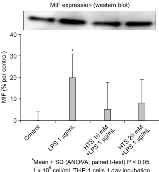
Fig. 3. Levels of Migration inhibitory factor (MIF) protein expres- sion were higher in lipopolysaccharide (LPS)-stimulating cells (25% decrease in band density) (P < 0.05). Addition of hypertonic saline (HTS) to LPS-stimulated cell decreased MIF protein expres- sion regardless of HTS concentrations.
Fig. 2. Migration inhibitory factor (MIF) levels in cell supernatant were measured. THP-1 cells in presence of lipopolysaccharide (LPS) had increase in MIF level (0.69 ng/mL ± 0.05) when compared with THP-1 cells (0.05 ng/mL ± 0.01) at 2 hours. However, hypertonic saline (HTS) 10 restored partially and decreased MIF levels (0.49 ng/mL ± 0.05) when compared with LPS-stimulated THP-1 cells (P < 0.05). LPS-stimulated THP-1 cells treated with HTS40 had lower MIF levels (0.27 ng/mL ± 0.04) than HTS10- treated cells, however, there was no statistically significant dif- ference on MIF levels according to HTS concentrations or measured times. ELISA, enzyme-linked immunosorbent assay.
had a lower MIF level (0.27 ± 0.04 ng/mL) than HTS10- treated cells. However, there was no statistically signifi- cant difference of MIF levels in the supernatant of HTS10, HTS20 or HTS40 treated cells, respectively, and in the measured times (Fig. 2).
The effect of HTS on MIF expression
To determine the effect of HTS on the MIF expression, western blot analysis was performed. Correlating with the ELISA, levels of MIF protein expression were higher in LPS stimulating cells (25% decrease in band density) (P < 0.05). The addition of HTS to LPS stimulated cell de- creased MIF protein expression regardless of HTS concen- trations (Fig. 3). However, there was no statistically sig- nificant difference of MIF levels in the supernatant of HTS10 or HTS20 treated cells.
DISCUSSION
Severe damage on the human body, incurred by trauma,
surgery, or prolonged shock may directly induce organ dysfunction. Possible pathogenesis follows: induction of ischemia, reperfusion injury, or immediate tissue destruc- tion due to trauma (first hit). The first hit may not induce a primary multi-system organ failure, but a second hit, such as infection, could further activate the immune sys- tem already primed by the first hit. The pathophysiology of sepsis and shock emphasizes the importance of humeral mediator network. Mortality and morbidity will increase with the unusual response of these mediators. Specially, in the early stage after the first hit, increased tumor necrosis factor-α and interleukin (IL) 1 cause a hyperinflammation state, leading to early MODS [1,14,15]. Later on, immuno- suppressive mediators such as PGE2, transforming growth factor-beta, IL-10 cause immuneparalysis, result- ing in late MODS [1]. The mechanism through which hy- pertonic saline solutions reduce mortality in hypotensive patients is still unclear [16]. However, recent studies have demonstrated that hypertonicity substantially alters acti- vation of inflammatory cells, consequently reducing sub- sequent organ injury from ischemia-reperfusion and de- creasing nosocomial infection [17-19].
By controlling immune and inflammatory responses, MIF is thought to play an important role in the patho- physiology of septic shock and chronic inflammatory diseases. This implies that MIF may serve as a general marker for systemic inflammation in septic and nonseptic acute critical illness [3,4]. MIF is, specifically, a cytokine se- creted by the anterior pituitary gland and immune cells in response to surgical stress, injury, and sepsis [7]. This cyto- kine appears to play a critical role in the regulation of the inflammatory pathways, contributing to SIRS and sub- sequent MODS [5,6]. MIF is also secreted by activated T-cells, and neutralizing anti-MIF antibodies inhibit T-cell proliferation and IL-2 production. T-cells also release MIF in response to glucocorticoid stimulation. MIF, in this case, acts to override glucocorticoid inhibition of T-cell pro- liferation, IL-2 and interferon γ production [8]. As for contribution of MIF to the regulation of cell survival, the inhibitory effect of MIF on apoptosis may be related to its reported stimulatory effects on cell proliferation, even though cellular apoptosis and cell proliferation are not simply the reverse of each other [6,20-22]. Our previous
experiments have shown that PGE2 inhibited T-cells pro- liferation, leading to a decrease in MIF levels when com- pared to control levels. The addition of HTS increased MIF production as compared with PGE2-stimulated T-cells in concordance with restore PGE2-suppressed T-cells pro- liferation (not shown). The role of HTS in restoring T-cell proliferation suppressed by PGE2, at least in part, is be- lieved to be mediated through an MIF pathway. Hyperos- molality may play an important role in HTS-enhancement of T-cell proliferation. Hypertonicity may contribute to in- creased IL-2 expression for T-cell proliferation [9,11,23].
LPS, a component of the cell wall of Gram-negative bac- teria, is an endotoxin that induces septic shock syndrome.
It stimulates the production of inflammatory mediators and is known to activate a number of cellular signals of hu- man monocyte cells during inflammation and infection.
Monocytes are key mediators of inflammation and widely distributed in the body [24,25]. THP-1 cells, which repre- sent an appropriate model system to study immune re- sponses, were utilized, therefore, to investigate the anti-in- flammatory effects of HTS. Our present study demon- strated that HTS is an effective inhibitor of LPS-induced MIF generation and expression in THP-1 cells. In order words, LPS increased MIF levels in THP-1 cells. However, HTS restored the MIF levels in LPS-stimulated THP-1 cells and there was no statistically significance difference of MIF levels in HTS10, HTS20 or HTS40 treated cells.
Besides, similar to ELISA, there was no statistically sig- nificance difference in MIF levels in HTS10 or HTS20 treat- ed cells. So, it is considered that HTS10 would be suffi- cient. It is presumed that HTS controls LPS-induced MIF production or secretion. Therefore, we need to study eval- uations between Toll-like receptor 4 or MIF receptors, which effect MIF production and effect, and HTS. Finally, this study indicated that HTS exhibited anti-inflammatory activities in THP-1 cells.
MIF exhibits proinflammatory activity and regulates cell proliferation and survival. In our previous and pres- ent experiment, MIF production was lower in PGE2- stimulated T-cells, and higher in LPS-stimulated THP-1 cells compared to control groups. HTS restored MIF pro- duction to control in PGE2-stimulated T-cells and LPS- stimulated THP-1 cells. We have demonstrated that HTS
acts to maintain balance between both pro- and anti-in- flammatory activities, which is critical for host defense against diseases. This implies that the role of HTS in main- taining homeostasis with humeral mediators, at least in part, should be mediated through an MIF pathway. This in vitro study, however, does not necessarily indicate re- spective in vivo effects, especially since the concentrations used are far higher than are achievable in vivo. Still, it may contribute to a better understanding of the possible bio- logical actions of HTS.
CONFLICTS OF INTEREST
No potential conflict of interest relevant to this article was reported.
ACKNOWLEDGEMENTS
We are grateful to our colleagues for participation in the studies and their helpful discussion: Park JH, Han KS. This research was supported by Basic Science Research Pro- gram through the National Research Foundation of Korea (NRF) funded by the Ministry of Education, Science and Technology (R1009982) and was partially supported by a Korea University Grant.
REFERENCE
1. Smith JW, Gamelli RL, Jones SB, Shankar R. Immunologic responses to critical injury and sepsis. J Intensive Care Med 2006;21:160-72.
2. Lehmann LE, Weber SU, Fuchs D, Klaschik S, Schewe JC, Book M, et al. Intracellular detection of macrophage mi- gration inhibitory factor in peripheral blood leukocytes.
Free Radic Biol Med 2005;38:1170-9.
3. Bernhagen J, Calandra T, Bucala R. Regulation of the im- mune response by macrophage migration inhibitory fac- tor: biological and structural features. J Mol Med (Berl) 1998;76:151-61.
4. Lehmann LE, Novender U, Schroeder S, Pietsch T, von Spiegel T, Putensen C, et al. Plasma levels of macrophage migration inhibitory factor are elevated in patients with se- vere sepsis. Intensive Care Med 2001;27:1412-5.
5. Larson DF, Horak K. Macrophage migration inhibitory fac- tor: controller of systemic inflammation. Crit Care 2006;
10:138.
6. Lue H, Kleemann R, Calandra T, Roger T, Bernhagen J.
Macrophage migration inhibitory factor (MIF): mecha- nisms of action and role in disease. Microbes Infect 2002;4:449-60.
7. Schmidt-Supprian M, Murphy C, While B, Lawler M, Kapurniotu A, Voelter W, et al. Activated protein C in- hibits tumor necrosis factor and macrophage migration in- hibitory factor production in monocytes. Eur Cytokine Netw 2000;11:407-13.
8. Bacher M, Metz CN, Calandra T, Mayer K, Chesney J, Lohoff M, et al. An essential regulatory role for macro- phage migration inhibitory factor in T-cell activation. Proc Natl Acad Sci U S A 1996;93:7849-54.
9. Choi SH, Bansal V, Costantini T, Putnam J, Loomis W, Coimbra R. Arginine is essential in reversing prosta- glandin E(2) T-cell suppression by hypertonic saline. J Surg Res 2009;156:83-9.
10. Coimbra R, Junger WG, Liu FC, Loomis WH, Hoyt DB.
Hypertonic/hyperoncotic fluids reverse prostaglandin E2 (PGE2)-induced T-cell suppression. Shock 1995;4:45-9.
11. Junger WG, Liu FC, Loomis WH, Hoyt DB. Hypertonic sal- ine enhances cellular immune function. Circ Shock 1994;42:190-6.
12. Saad B, Dakwar S, Said O, Abu-Hijleh G, Al Battah F, Kmeel A, et al. Evaluation of medicinal plant hepatotox- icity in co-cultures of hepatocytes and monocytes. Evid Based Complement Alternat Med 2006;3:93-8.
13. Bradford MM. A rapid and sensitive method for the quan- titation of microgram quantities of protein utilizing the principle of protein-dye binding. Anal Biochem 1976;72:
248-54.
14. Coussens LM, Werb Z. Inflammation and cancer. Nature 2002;420:860-7.
15. Delgado AV, McManus AT, Chambers JP. Production of tu- mor necrosis factor-alpha, interleukin 1-beta, interleukin 2, and interleukin 6 by rat leukocyte subpopulations after ex- posure to substance P. Neuropeptides 2003;37:355-61.
16. Bulger EM, May S, Kerby JD, Emerson S, Stiell IG, Schreiber MA, et al. Out-of-hospital hypertonic re- suscitation after traumatic hypovolemic shock: a random- ized, placebo controlled trial. Ann Surg 2011;253:431-41.
17. Costantini TW, Deree J, Martins JO, Putnam JG, de Campos T, Coimbra R. A novel fluid resuscitation strategy modu- lates pulmonary transcription factor activation in a murine model of hemorrhagic shock. Clinics (Sao Paulo) 2010;65:
621-8.
18. Choi SH, Lee SW, Hong YS, Jeun JM, Min BW. Selective in- hibition of polymorphonuclear neutrophils by re- suscitative concentration of hypertonic saline. Emerg Med J 2006;23:119-22.
19. Coimbra R, Hoyt DB, Junger WG, Angle N, Wolf P, Loomis W, et al. Hypertonic saline resuscitation decreases suscept- ibility to sepsis after hemorrhagic shock. J Trauma 1997;
42:602-6.
20. Mitchell RA, Liao H, Chesney J, Fingerle-Rowson G, Baugh J, David J, et al. Macrophage migration inhibitory factor (MIF) sustains macrophage proinflammatory function by inhibiting p53: regulatory role in the innate immune response. Proc Natl Acad Sci U S A 2002;99:345-50.
21. Nguyen MT, Lue H, Kleemann R, Thiele M, Tolle G, Finkelmeier D, et al. The cytokine macrophage migration inhibitory factor reduces pro-oxidative stress-induced apoptosis. J Immunol 2003;170:3337-47.
22. Park JK, Kim JK. Influence of MIP-1 Alpha on the CD4+ Th Lymphocytes. J Korean Surg Soc 2004;66:81-8.
23. Hoyt DB, Junger WG, Loomis WH, Liu FC. Effects of trau- ma on immune cell function: impairment of intracellular calcium signaling. Shock 1994;2:23-8.
24. Gilroy DW, Lawrence T, Perretti M, Rossi AG. Inflamma- tory resolution: new opportunities for drug discovery. Nat Rev Drug Discov 2004;3:401-16.
25. Chen CF, Cheng CH. Regulation of cellular metabolism and cytokines by the medicinal herb feverfew in the hu- man monocytic THP-1 cells. Evid Based Complement Alternat Med 2009;6:91-8.