ISSN 2234-3806 • eISSN 2234-3814
386 www.annlabmed.org http://dx.doi.org/10.3343/alm.2013.33.5.386 Ann Lab Med 2013;33:386-389
http://dx.doi.org/10.3343/alm.2013.33.5.386
Letter to the Editor
Diagnostic Genetics
Spectrin Tunis (Sp alpha I/78 ) in a Korean Family with Hereditary Elliptocytosis
Eunhee Han, M.D.1,2, Ahhyun Kim, M.D.1,2, Joonhong Park, M.D.1,2, Myungshin Kim, M.D.1,2, Yonggoo Kim, M.D.1,2, Kyungja Han, M.D.1,2, and Yoo-Jin Kim, M.D.3
Department of Laboratory Medicine1, Catholic Genetic Laboratory Center2, Division of Hematology3, Department of Internal Medicine, Catholic Blood and Marrow Transplantation Center, The Catholic University of Korea, Seoul, Korea
Hereditary elliptocytosis (HE) is a group of disorders character- ized by abnormal erythrocyte shapes that occur mainly in indi- viduals of African and Mediterranean ancestry. In Korea, HE is the cause of 1.4% (6/431) cases of hereditary hemolytic anemia (HHA) [1]. HE causes mechanical weakness or fragility of the erythrocytic membrane skeleton because of defects in spectrin or protein 4.1. The erythrocyte spectrin is a scaffold protein com- posed of 2 subunits, α- and β-spectrin, maintains the cellular shape, regulates the lateral mobility of integral membrane pro- teins, and provides structural support for the lipid bilayer [2, 3].
The mutations responsible for HE are located in several genes encoding membrane proteins, including protein 4.1, α-spectrin, β-spectrin, band 3, and glycophorin C. Mutations in α-spectrin are the most common, occurring in 65% of the HE cases, fol- lowed by mutations in β-spectrin (30%) and protein 4.1 (5%) [4].
We studied a Korean family presenting with HE. To our knowl- edge, this is the first report to describe a family of Korean de- scent diagnosed with HE by molecular analysis. A 28-yr-old woman visited our hospital and was found to have anemia dur- ing a routine health screening. She did not complain of symp- toms related to anemia. Abdominal ultrasonography revealed splenomegaly (craniocaudal length, 13.8 cm). Marked anisopoi- kilocytosis, including elliptocytes, schistocytes, and teardrop
cells, were detected on a peripheral blood smear (Fig. 1A). A complete blood count revealed pancytopenia (white blood cell count, 3.88×10⁹/L; hemoglobin level, 7.6 g/dL; and platelet count, 95×109/L). Red cell indices revealed macrocytic normo- chromic anemia (red blood cell count, 2.20×10¹²/L; reticulocyte count, 0.968×10¹²/L; hematocrit value, 24%; mean corpuscular volume, 109.1 fL; and mean corpuscular hemoglobin concen- tration, 31.7%). We ruled out iron deficiency or megaloblastic anemia based on the following laboratory findings: serum iron level, 96 µg/dL (reference range: 50-150 µg/dL); ferritin level, 104 ng/mL (10-120 ng/mL); total iron-binding capacity, 204 µg/
dL (250-450 µg/dL); vitamin B12 concentration, >1,500 pg/mL (180-914 pg/mL); and folate concentration, 7.47 ng/mL (3.1-19.9 ng/mL). The serum concentrations of total bilirubin (1.02 mg/
dL), haptoglobin (<30 mg/dL), and lactate dehydrogenase (556 U/L) confirmed the presence of a hemolytic process. Bone mar- row studies showed hypercellularity and erythroid hyperplasia with G:E ratio of 0.67:1 (Fig. 1B), and cytogenetic studies re- vealed a normal karyotype (46,XX[20]).
The patient’s mother was also diagnosed with anemia and el- liptocytosis based on peripheral blood films. Therefore, we sus- pected familial transmission of elliptocytosis. At the time of this study, the patient was pregnant, and after delivery, the peripheral
Received: November 22, 2012 Revision received: January 29, 2013 Accepted: April 15, 2013
Corresponding author: Myungshin Kim
Department of Laboratory Medicine, Seoul St. Mary’s Hospital, 222 Banpo-daero, Seocho-gu, Seoul 137-701, Korea Tel: +82-2-2258-1645, Fax: +82-2-2258-1719, E-mail: [email protected]
© The Korean Society for Laboratory Medicine.
This is an Open Access article distributed under the terms of the Creative Commons Attribution Non-Commercial License (http://creativecommons.org/licenses/by-nc/3.0) which permits unrestricted non-commercial use, distribution, and reproduction in any medium, provided the original work is properly cited.
Han E, et al.
SPTA1 mutation in hereditary elliptocytosis
387
http://dx.doi.org/10.3343/alm.2013.33.5.386 www.annlabmed.org
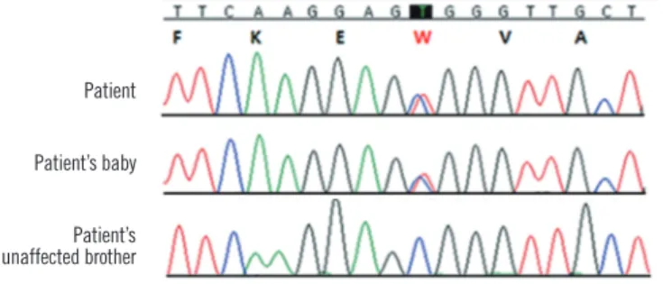
blood smears from the newborn were analyzed, which revealed elliptocytosis. Autosomal dominant inheritance was suspected, and spectrin-gene-mutation analysis of the patient and her fam- ily (affected mother, her baby, and unaffected brother) was per- formed. Genomic DNA was prepared using the QIAamp DNA Mini Kit (Qiagen, Hamburg, Germany). All coding exons and flanking intronic sequences of the spectrin, alpha, erythrocytic 1 gene (SPTA1) and spectrin, beta, erythrocytic gene (SPTB) (ref- erence cDNA sequence, NM_003126.2 and NM_000347.5) were amplified and sequenced using primers as previously described [5, 6]. In particular, the region encoding exon 2 of SPTA1 was amplified by PCR using the following forward and reverse prim- ers: 5’-GGTCCAACATGAGTAAACACCTTGACA-3’ and 5’-TCT- CACCTCTCCAACTTCATAAGGGA-3’, respectively. Direct se- quencing of the SPTA1 gene revealed a heterozygous missense mutation in exon 2, resulting in a C to T substitution at nucleotide position 121 and an amino acid change of arginine to tryptophan (c.121C>T; p.Arg41Trp) at amino acid residue 41 in the alpha l
domain in both the patient and her baby (Fig. 2). This missense mutation induces spectrin Tunis, which reduces the binding af- finity for the spectrin tetramer assembly. Spectrin Tunis was designated as αI/78 because of its location in the first alpha do- main of SPTA1 [7]. In addition, we analyzed the sequences of SLC4A1 (solute carrier family 4, anion exchanger, member 1) and EPB41 (erythrocyte membrane protein band 4.1) but found no mutations in these genes.
HE is a group of disorders characterized by the presence of elliptical erythrocytes on a peripheral blood smear. Disorders in which elliptocytosis may be prominent also include iron defi- ciency, leukemia, megaloblastic anemia, myelofibrosis, myelo- phthisic anemia, myelodysplastic syndromes, polycythemia, py- ruvate kinase deficiency, and sickle cell disease [8]. Examina- tion of family history is the most reliable method for differentiat- ing HE from other disorders, in which elliptocytosis may be prominent [9]. Biochemical and mechanical methods can be used to determine defects in erythrocyte membrane proteins, such as spectrin or protein 4.1. Sodium dodecyl sulfate-poly- acrylamide gel electrophoresis (SDS-PAGE) is used in qualitative and quantitative detection of alterations in membrane proteins.
In addition, assessment of thermal sensitivity, in vitro studies of spectrin self-association, and trypsin peptide mapping of spec- trin can assist in the diagnosis of HE [8].
Currently, many genetic studies have been designed to detect various genetic mutations in HE [9]. All spectrin mutations asso- ciated with HE are located in or near self-association sites be- tween the α- or β-chains. As a result, the corresponding subset of HE is derived from an impaired self-association process, which is a critical process for red cell deformability. Erythroid
A B
Fig. 1. (A) Marked anisopoikilocytosis, including elliptocytosis, schistocytes, and teardrop cells, on a peripheral blood smear. (B) Bone marrow aspirates showing hypercellularity and erythroid hyperplasia (Wright-Giemsa Stain, ×1,000).
Fig. 2. DNA sequence analysis of the SPTA1 gene. The patient and her baby carried a heterozygous missense mutation in exon 2 (c.121C>T; p.Arg41Trp) in the alpha 1 domain. Protein changes are indicated by red lettering.
Patient Patient’s baby Patient’s unaffected brother
Han E, et al.
SPTA1 mutation in hereditary elliptocytosis
388 www.annlabmed.org http://dx.doi.org/10.3343/alm.2013.33.5.386 spectrin α- and β-chains are encoded by the SPTA1 gene on
1q22-23 and the SPTB gene on 14q23-24.2, respectively. Many SPTA1 gene mutations are single-nucleotide substitutions, whereas others are intronic mutations that cause errors in gene splicing [10-14]. SPTA1-related HE is characterized by marked clinical, biochemical, and genetic heterogeneity. Clinically, the presentation varies from a nearly complete absence of symp- toms to transfusion-dependent hemolytic anemia. The location of the change within the spectrin α-chain and the homozygous or compound heterozygous status affect clinical manifestations.
Remarkably, both mildly and severely affected patients are found in almost all affected kindred [15].
In a review of Korean literature on HHA, we found 15 case re- ports on HE, including our case [1, 16]. Red-cell-membrane de- fects were the most common (88.6%) in HHA. Hereditary sphe- rocytosis accounted for 87.2% of red-cell-membrane defects, whereas HE accounted for 1.4% of the defects [1]. Lee et al.
[16] detected protein 4.1 deficiency in 6 Korean patients with HE by using SDS-PAGE analysis.
In this study, we identified the first SPTA1 gene mutation in a Korean family diagnosed with HE. The heterozygous c.121C>T mutation induces an amino acid change p.Arg41Trp in the α1 domain of the α-spectrin protein. Morlé et al. [7] first described this mutation and called this disease spectrin Tunis, which causes asymptomatic HE (OMIM 130600) in patients with a het- erozygous mutation. A variant was also found in a white North African man and his mother. The αI/78 variant (rs121918640) ex- hibits reduced binding affinity, generates an abnormal spectrin protein with an αI 78-kDa fragment, and results in a mutation, which partially destroys the ability of the dimer to form a tetra- mer [7]. Most spectrin mutations are private, and it is interesting that spectrin Tunis was detected in a Korean family. However, some mutations in SPTA1 are encountered more frequently than originally thought, such as the case of a mutation affecting codon α28, which contains a CpG hot spot [17]. The CpG dinu- cleotide has also been implicated in a number of band 3 gene hot spots, most of which encode arginine (CGN, N indicating any nucleotide). The mutation resulting in spectrin Tunis occurs in the CpG dinucleotide at codon 41, which also encodes argi- nine (CGG).
The patient who transmits the production-defective spectrin allele is clinically normal with unremarkable erythrocyte mor- phology because α-spectrin is normally synthesized in a 2- to 3-fold excess, and the output from a single normal α-spectrin al- lele is sufficient to maintain membrane integrity [18, 19]. Be- cause this α-spectrin variant is related to asymptomatic HE in a
heterozygous state, the outcomes may include mild elliptocyto- sis in the proband and her baby. To our knowledge, this is the first report on HE confirmed by mutation analysis in a Korean family. More data need to be collected from HE patients to un- derstand the genetic distributions and genotype-phenotype cor- relations.
Authors’ Disclosures of Potential Conflicts of Interest
No potential conflicts of interest relevant to this article were re- ported.
Acknowledegments
This study was supported by a grant from the Korea Health Technology R&D Project, Ministry of Health & Welfare, Republic of Korea (A120175).
REFERENCES
1. Cho HS, Hah JO, Kang IJ, Kang HJ, Kwak JY, Koo HH, et al. Hereditary hemolytic anemia in Korea: a retrospective study from 1997 to 2006. Korean J Hematol 2007;42:197-205.
2. Lux SE and Palek J. Disorders of the red cell membrane. In: Handin RI, Lux SE, Stossel TP, eds. Blood principles and practice of hematology.
Philadelphia: Lippincott, 1995:1701.
3. Becker PS and Lux SE. Disorders of the red cell membrane skeleton:
hereditary spherocytosis and hereditary elliptocytosis. In: Scriver CS, Beaudet AL, Sly WS, Valle D, eds. The metabolic and molecular bases of inherited disease. 7th ed. New York: McGraw-Hill, 1995:529. 4. An X and Mohandas N. Disorders of red cell membrane. Br J Haema-
tol 2008;141:367-75.
5. Costa DB, Lozovatsky L, Gallagher PG, Forget BG. A novel splicing mu- tation of the alpha-spectrin gene in the original hereditary pyropoikilocy- tosis kindred. Blood 2005;106:4367-9.
6. Dhermy D, Galand C, Bournier O, Cynober T, Méchinaud F, Tchemia G, et al. Hereditary spherocytosis with spectrin deficiency related to null mutations of the beta-spectrin gene. Blood Cells Mol Dis 1998;24:251- 61.
7. Morlé L, Morlé F, Roux AF, Godet J, Forget BG, Denoroy L, et al. Spec- trin Tunis (Sp alpha I/78), an elliptocytogenic variant, is due to the CGG- ---TGG codon change (Arg----Trp) at position 35 of the alpha I domain.
Blood 1989;74:828-32.
8. Gallagher PG. Hereditary elliptocytosis: spectrin and protein 4.1R.
Semin Hematol 2004;41:142-64.
9. Delaunay J. The molecular basis of hereditary red cell membrane disor- ders. Blood Rev 2007;21:1-20.
10. Maillet P, Alloisio N, Morlé L, Delaunay J. Spectrin mutations in heredi- tary elliptocytosis and hereditary spherocytosis. Hum Mutat 1996;8:97- 107.
11. Alloisio N, Wilmotte R, Maréchal J, Texier P, Denoroy L, Féo C, et al. A splice site mutation of alpha-spectrin gene causing skipping of exon 18 in hereditary elliptocytosis. Blood 1993;81:2791-8.
Han E, et al.
SPTA1 mutation in hereditary elliptocytosis
389
http://dx.doi.org/10.3343/alm.2013.33.5.386 www.annlabmed.org
12. Baklouti F, Maréchal J, Wilmotte R, Alloisio N, Morlé L, Ducluzeau MT, et al. Elliptocytogenic alpha I/36 spectrin Sfax lacks nine amino acids in helix 3 of repeat 4. Evidence for the activation of a cryptic 5′-splice site in exon 8 of spectrin alpha-gene. Blood 1992;79:2464-70.
13. Fournier CM, Nicolas G, Gallagher PG, Dhermy D, Grandchamp B, Lecomte MC. Spectrin St Claude, a splicing mutation of the human al- pha-spectrin gene associated with severe poikilocytic anemia. Blood 1997;89:4584-90.
14. Costa DB, Lozovatsky L, Gallagher PG, Forget BG. A novel splicing mu- tation of the alpha-spectrin gene in the original hereditary pyropoikilocy- tosis kindred. Blood 2005;106:4367-9.
15. Iolascon A, King MJ, Robertson S, Avvisati RA, Vitiello F, Asci R, et al. A genomic deletion causes truncation of α-spectrin and ellipto-poikilocy- tosis. Blood Cells Mol Dis 2011;46:195-200.
16. Lee YK, Cho HI, Park SS, Ra ER, Chang YH, Huh MN, et al. SDS-PAGE analysis of red cell membrane proteins in hereditary hemolytic anemia.
Korean J Hematol 1999;34:559-67.
17. Coetzer TL, Sahr K, Prchal J, Blacklock H, Peterson L, Koler R, et al.
Four different mutations in codon 28 of alpha spectrin are associated with structurally and functionally abnormal spectrin alpha I/74 in heredi- tary elliptocytosis. J Clin Invest 1991;88:743-9.
18. Knowles WJ, Morrow JS, Speicher DW, Zarkowsky HS, Mohandas N, Mentzer WC, et al. Molecular and functional changes in spectrin from patients with hereditary pyropoikilocytosis. J Clin Invest 1983;71:1867- 77.
19. Hanspal M and Palek J. Synthesis and assembly of membrane skeletal proteins in mammalian red cell precursors. J Cell Biol 1987;105:1417- 24.