1. Introduction
Neurogenesis comprises of cell prolifera- tion, survival and differentiation. Although neurogenesis ceases in most areas at birth, stem cells continue to generate neurons within the subventricular zone and hippocampal
dentate gyrus throughout adult life
1-6). Cell birth and neurogenesis have also been demonstrated in the dentate gyrus of the several mammals, including humans
7,8). Previous studies have shown that several factors such as glucocorticoids, estrogen, N-methyl-d-aspartate (NMDA) receptor antagonist, serotonin, aging, seizures, and environmental stimuli influence the proliferation of granular cell precursors and/or
신기환 약침이 HiB5 해마세포와 어린 Sprague-Dawley계 흰쥐의 치상회 세포생성에 미치는 영향
김연정1⋅장미현1⋅신민철1⋅임백빈1⋅정주호2⋅반건호3⋅백은경4⋅박재형4⋅ 김이화5⋅김창주1
경희대학교 의과대학 1생리학교실, 2약리학교실, 3정신과학교실, 4고천한의원, 5세명대학교 한의과대학 경혈학교실
Shenqi-wan Increases Cell Proliferation of Cultured Hippocampal Cell Line HiB5 and Dentate Gyrus of Young Sprague-Dawley Rats
Youn-Jung Kim
1, Mi-Hyun Jang
1, Min-Chul Shin
1, Baek-Vin Lim
1, Joo-Ho Chung
2, Gyun-Ho Bahn
3, Eun-Kyung Paik
4, Jae-Hyung Park
4, Ee-Hwa Kim
5, Chang-Ju Kim
1Dept. of 1Physiology, 2Pharmacology, 3Psychiatry, College of Medicine, Kyung Hee University; 4Gochun Oriental Medical Clinic; 5Dept. of Meridian & Acupoint, College of Oriental Medicine, Semyung University
Abstract
목적 : 소아의 정신적 및 신체적 발달지연에 사용되고 있는 신기환이 해마의 신경세포 생성에 미치는 영향 을 관찰하고자 하였다.
방법 : 신기환이 세포생성에 미치는 영향을 알아보기 위하여 해마 세포인 HiB5에 신기환을 처리, 배양하여 3-(4,5-dimethylthiazol-2-yl)-2,5-diphenyltetrazolium bromide (MTT) assay를 통해서 세포생성을 관찰 하였다. 또한 3주령의 Sprague-Dawley (S-D)계 흰쥐의 중완혈에 신기환 약침을 자침후 해마의 치상 회에서 bromodeoxyuridine (BrdU) immunohistochemistry를 시행하여 세포생성을 관찰하였다.
결과 : HiB5 배양세포에서는 신기환을 처리시 배양시간과 농도별로 세포생성율이 증가하였다. 또한, 대조군 의 치상회에서 BrdU 양성세포수는 128.50 ± 9.53, 1 mg/kg 신기환 자침군은 157.08 ± 10.82 및 10 mg/kg 신기환 자침군은 204.80 ± 17.68로 나타났다. 이러한 결과로 신기환은 어린 S-D계 흰쥐의 치 상회에서 세포생성을 증가시키는 것으로 사려된다.
Keywords : Shenqi-wan; MTT assay; BrdU immunohistochemistry; cell proliferation
■교신저자 : 김창주, 경희대학교 의과대학 생리학교실, Tel. 02-961-0282, Fax. 02-964-2195, E-mail: [email protected] The Journal of Korean Meridian & Acupoint
neurogenesis in the adult dentate gyrus
9-15). Proliferating cells can be identified using 5-bromo-2-deoxyuridine (BrdU), which labels cellular DNA during the synthesis, or S-phase
7,10,16). Recent evidences suggest that cell proliferation and neurogenesis may play a role in learning and the hippocampal formation is clearly recognized as being involved in learning and memory
13,15,17). Although cell proliferation and neurogenesis is not the only mechanism in either learning or recovery from damage, may be involved in learning in many cases
13,15,17,18). Post-mitotic fate of a population of constitutively proliferating cells has been shown to be cell death
19)or neuronal differentiation after migration
20). It is known that growth factors promote the migration of proliferated cells into adjacent tissues where they differentiated
21,22)and to begin generate neurons
23,24). Recently, Åberg et al.
25)showed that peripheral infusion of insulin-like growth factor-1 (IGF-1), a growth-promoting peptide hormone, increases progenitor cell proliferation and selectively induces neurogenesis in the progeny of adult neuronal progenitor cells in rat hippocampus.
Traditionally, Shenqi-wan (SW), an Oriental herbal medicine formulation, has been used for delayed mental and physical development of children and complications of diabetes
26,27). To study the medications which possess the ability of neuronal stem cells to differentiated into neurons indigenous to various brain regions offers hope for improve mental retardation and/or restorative therapies for ischemia, traumatic and degenerative brain diseases. However, there are to our knowledge no known exogenous herbal substances that selectively increase cell proliferation in the mammalian central nervous system (CNS). In this study, ability
of SW which has been used for mental and physical retardation of children in Oriental medicine to confer increase of hippocampal cell proliferation was investigated.
2. Materials and methods
2.1. Effects of SW on cultured hippocampal cell line
To investigate the effect of SW on cell proliferation in cultured hippocampal cell line HiB5, MTT aasay was performed.
2.1.1. Preparation of SW
Composition of SW is as follows:
Rehmanniae Radix 16g, Dioscoreae Radix 8g, Corni Fructus 8g, Alismatis Rhizoma 6g, Moutan Cortex Radicis 6g, Hoelen 6g, Maximowicziae Fructus 8g, Cervi Cornu 4g.
After washing, each component of SW was immersed in cold water for 12 hr, and aqueous extract from SW was made by using rotatory evaporator, and filtered by 0.45 ㎛ syringe filter before use.
2.1.2. Cell culture
The hippocampal cell line HiB5 was originally established from the hippocampus of E16 Sprague-Dawley rats. These cells proliferate at permissive temperature of 32 ℃ and differentiated into neuronal or glial phenotype at the non-permissive temperature of 39 ℃. Cells were grown in Dulbeccos Modified Eagle Medium (DMEM; Gibco BRL, Grand Island, NY, USA) supplemented with 10 % heat-inactivated fetal bovine serum (FBS;
Gibco BRL, Grand Island, NY, USA) at 32 ℃
in 5% CO
2and 95% O
2in a humidified cell
incubator and the media was changed once
every 2 day.
2.1.3. MTT assay
Cell proliferation rate was determined using the 3-(4,5-dimethylthiazol-2-yl)-2,5-dip- henyltetrazolium bromide (MTT) assay kit (Boehringer Mannheim, Mannheim, Germany) as per the manufactures protocol. HiB5 cells were culture in 96-well plates. The SW treated groups are exposed to SW at final concen- trations of 0.1 mg/ml, 1 mg/ml and 10 mg/ml (diluted with saline) for 24 hr and 48 hr, and saline of an equal volume was added to the control group. Ten ㎕ of the MTT labeling reagent was added to each well, and plates were incubated for 4 hr. After the cells were incubated in 100 ㎕ solubilization solution for 12 hr, the absorbance was measured with a microtiter plate reader (Bio-Tek, Winooski, VT, USA) at a test wavelength of 595 nm with a reference wavelength of 690 nm. The absor- bance of results was calculated as the result of the substraction of the absorbance at the reference wavelength from that of the test wavelength.
2.2. Effect of SW on hippocampal dentate gyrus cell proliferation
In order to study the effect of SW on cell proliferation in the dentate gyrus of hippocampus, the brains of Sprague-Dawley (S-D) rats (51.00 ± 1.11g, 3 weeks old) were examined by immunohistochemistry.
2.2.1 Animals and SW treatment
The rats were equally classified into three groups (n = 15): control, 1 mg/kg of SW treated and 10 mg/kg of SW treated groups. The experimental procedures were performed in accordance with the animal care guidelines of NIH and the Korean Academy of Medical
Sciences. Animals were housed under controlled temperature (20 ± 2 ℃) and lighting (07:00 - 19:00 hr) conditions and supplied with food and water ad libitum. The rats of the control group were injected intraperitoneally with BrdU (50 mg/kg; Sigma, St. Louis, MO, USA) twice (10:00, a.m. and 05:00 p.m.) per day for 3 consecutive days. And 1 mg/kg SW treated and 10 mg/kg SW treated groups were injected BrdU with 1mg/kg of SW and 10 mg/kg SW, for same period, respectively.
2.2.2. Tissue preparation
Rats were completely anesthetized using Zoletil 50 (10 mg/kg, i.m.) 2 hr after last BrdU injection. The anesthetized rats were transcardially perfused with 0.05 M phosphate buffered saline (PBS) and fixed with 4%
paraformaldehyde (PFA) in 0.1 M sodium phosphate buffer (PB) at pH 7.4. Brains were removed, postfixed in the same fixative overnight and transferred into 30% sucrose solution for cryoprotection. Coronal sections of 40 ㎛ thickness were made with a freezing microtome (Leica, Nuloch, Germany). Average 5 sections were collected from each rat.
2.2.3. BrdU immunohistochemistry
For detection of cell birth in the dentate gyrus, BrdU immunohistochemistry was performed. In brief, sections were incubated in 50% formamide-2 x SSC at 65 ℃ for 2 hr, denatured in 2 N HCl at 37 ℃ for 30 min, and washed twice in 0.1 M sodium borate, pH 8.5. The sections were then incubated with 1%
hydrogen peroxide (H
2O
2) for 15 min.
Afterwards, the sections were incubated
overnight with a BrdU-specific mouse
monoclonal antibody (1:600; Roche, Mannheim,
Germany) at room temperature, and processed
with VECTASTAIN Elite ABC Kit (Vector
Laboratories, Burlingame, CA, USA) using horseradish peroxidase. For staining, the sections were reacted with 0.02% 3,3'-diamino- benzidine (DAB) containing 0.03% H
2O
2in 0.05 M Tris-HCl (pH 7.6) for 5 min. The number of BrdU-positive cells in the dentate gyrus was counted hemilaterally in each of the selected sections, using a Zeiss microscope (Oberköchen, Germany).
2.3. Statistical analysis
Data were collected and summarized as mean ± standard error mean (S.E.M.). Results were analyzed using Students t-test and P <
0.05 was considered to represent statistical significance.
3. Results
3.1. Effect of SW on proliferation of cultured hippocampal cell line
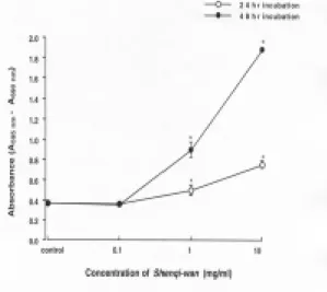
The absorbance of control cells which was not treated with SW was 0.375 ± 0.004 and absorbances of cells treated with 0.1 mg/ml, 1mg/ml and 10 mg/ml of SW for 24 hr were 0.366 ± 0.003, 0.500 ± 0.051 and 0.754 ± 0.038, respectively. The absorbances of cells treated with 0.1 mg/ml, 1mg/ml and 10 mg/ml of SW for 48 hr were 0.350 ± 0.006, 0.895 ± 0.076 and 1.881 ± 0.020, respectively. This result showed that the proliferation rate of cultured HiB5 cells was increased by incubation time- and SW concentration-dependent manners.
Figure 1. Proliferation effect of Shenqi-wan (SW) on hippocampal cell line HiB5 via MTT assay. The SW treated groups are exposed to aqueous extract of SW at final concen- trations of 0.1 mg/ml, 1 mg/ml and 10 mg/ml (diluted with saline) for 24 hr and 48 hr, while saline of an equal volume was added to the control group. * represents P < 0.05 compared to control.
3.2. Effect of SW on cell proliferation in dentate gyrus of young S-D rats
In sections obtained from each group,
BrdU-positive cells in the dentate gyrus of
hippocampus were counted via immunohisto-
chemistry.
Figure 2. Photomicrographs of BrdU-posi- tive cells in dentate gyrus of hippocampus. Brown dots repre- sent BrdU-positive cells. Control group was injected intraperito- neally with BrdU (50 mg/kg) twice per day for 3 consecutive days while 1 mg/kg SW treated and 10 mg/kg SW treated groups were injected BrdU with 1 mg/kg of SW and 10 mg/kg SW, for same period, respectively. Each of the rats used in this experiment was sacrificed 2 hr after last BrdU injection. A, control group; B, 1 mg/kg SW treated group; C, 10 mg/kg SW treated group. Scale bar represents 50 ㎛.
The mean number of BrdU-positive cells in the dentate gyrus per section was 128.50
± 9.53 for the control group. The figure was 157.08 ± 10.82 and 204.80 ± 17.68 for the 1 mg/kg and 10 mg/kg SW treated groups, respectively. In SW treated groups, the number of newly formed cells was increased compared to the control group. In this result, SW treatment appears to have resulted in increase in cell proliferation in the dentate gyrus of the young rat hippocampus.
Figure 3. Number of BrdU-positive cells of each group. SW treated groups showed increase of BrdU-positive cells in the dentate gyrus of hippocampus, especially at concentration of 10 mg/kg. * represents P < 0.05 compared to control.
4. Discussion
The hippocampus plays an important role
in specific types of learning and in the formation
of memory related to the personal exper-
ience
15,17). The results of cultured cell study
via MTT assay revealed that SW increases cell
proliferation of hippocampal cell line HiB5
significantly as time- and concentration-
dependent fashions.
Neuronal precursors of the hippocampus are known to reside in the subgranular zone of the dentate gyrus, where they proliferate and migrate continuously into the granular cell layer
12,28). In the granular cell layer, they are known to develop their characteristic cell morphology, express neuronal differentiation markers
3,12,28), and extend axonal processes toward their postsynaptic targets
29). Progenitor cells grafted into the adult brain have also been reported to develop into mature neurons, with presentation of the morphological and biochemical features characteristic of the surrounding neurons
30).
To examine the role of behavioral experience on proliferation in the hippocampus, Kempermann et al.
13)placed mice in a special environmental complex cages or in standard environmental complex cages. They found that the environmental complexity did promote the survival of the proliferating cells, as had significant more BrdU-labeled cells remaining one month later. Van Praag et al.
16)demonstra- ted that voluntary running exercise enhances surviving of new born cells similar to enrich- ment condition. It was suggested that enhanced cell proliferation in the dentate gyrus may be a compensatory adaptive responses to ischemia-associated injury and could promote functional recovery after ischemic hippocampal damage
18). Recently, acupunctures effect on cell proliferation in the dentate gyrus after transient global ischemia in gerbils was also reported
31). Considering that dentate cell proliferation and neurogenesis is increased with enriched environment and learning
15,17). The opposite order of event is also very exciting and after for example drug-induced cell proliferation. The relationship between memory and adult neurogenesis is supported
further by the fact that radiation, which is known to impair memory
32), also inhibits granular cell layer neurogenesis
33).
SW has been used for delayed mental and physical retardation traditionally and its therapeutic ability for diabetic neuropathy and nephrotic syndrome was reported
26,27). In present study, aqueous extract of SW increased cell proliferation in dentate gyrus of rats.
Understanding the factors controlling neuronal stem cell growth and differentiation may contribute to the achievement of a goal for improve learning ability and/or treatment for neurodegenerative disorders. Present results revealed that aqueous extract of SW increases cell proliferation of cultured hippocampal cell line and dentate gyrus of 3 weeks old S-D rats.
In this study, it can be suggested that therapeutic potential of SW in treating for mental retardation of children may related to this formulation contains herb which possess increase cell proliferation properties. Additio- nal studies of the mechanism of SW may therefore yield novel idea with possible implications for further therapeutic approa- ches.
5. Acknowledgement
This work was supported by Gochun