Research Article Open Access
뇌성마비 아동의 근력과 호흡기능의 상관관계
신승오⋅김난수1†
부산가톨릭대학교 대학원 물리치료학과, 1부산가톨릭대학교 보건과학대학 물리치료학과
Correlation Between Muscle Strength, Pulmonary Function and Respiratory Muscle in Children with Cerebral Palsy
Seung-Oh Shin, PT⋅Nan-Su Kim, PT, PhD1†
Dept. of Physical Therapy, Graduate School, Catholic University of Pusan
1Dept. of Physical Therapy, College of Health Sciences, Catholic University of Pusan
Received: May 10, 2016 / Revised: May 10, 2016 / Accepted: May 11, 2016
ⓒ 2016 J Korean Soc Phys Med
| Abstract |1)
PURPOSE: This study was conducted to determine correlations between grip and lower limb muscle strength and pulmonary function and respiratory muscle in children with cerebral palsy.
METHODS: Subjects were 17 children with cerebral palsy. Inclusion criteria for participation were having GMFCS from Ⅰ to Ⅲ grade and ability to independently blow into a spirometer. Pulmonary function and respiratory muscle were measured with a spirometer. All subjects performed maximal expiratory flow maneuvers using a spirometer in order to determine their forced expiratory volume in 1 second (FEV1), forced vital capacity (FVC), peak expiratory flow (PEF) and FEV1/FVC, and maximum inspiratory pressure (MIP) and maximum expiratory pressure (MEP).
†Corresponding Author : [email protected]
This is an Open Access article distributed under the terms of the Creative Commons Attribution Non-Commercial License (http://creativecommons.org/licenses/by-nc/3.0) which permits unrestricted non-commercial use, distribution, and reproduction in any medium, provided the original work is properly cited.
Muscle strength was measured in terms of grip strength and lower limb muscle strength in terms of knee extension strength with a dynamometer and manual digital muscle tester respectively. Data were analyzed using Person product correlation.
RESULTS: Grip strength significantly positively correlated with FVC (r=0.95, p<0.01), FEV1 (r=0.95, p<0.01), PEF (r=0.84, p<0.01), MIP (r=0.65, p<0.01) MEP (r=0.71, p<0.01) and lower limb strength with FVC (r=0.72, p<0.01), FEV1 (r=0.69, p<0.01), PEF (r=0.54, p<0.05), and MEP (r=0.69, p<0.01).
CONCLUSION: Grip and lower limb muscle strengths of children with cerebral palsy were positively correlated pulmonary function and respiratory muscle.
Key Words: Cerebral palsy, Grip strength, Pulmonary function, Quadriceps muscle strength, Respiratory muscle
Ⅰ. 서 론
뇌성마비는 태내 혹은 출산시 뇌의 손상으로 운동기 능 및 자세 조절 장애를 가진 소아의 대표적인 질환이다 (Bax 등, 2005). 뇌성마비 아동은 관절의 구축, 근육의 단축, 근 위축 등 근골격계의 변형 뿐만 아니라 전신의 근력 약화, 호흡근의 약화, 흉곽의 비대칭적 성장으로 호흡기능의 어려움과 호흡기계 감염률, 사망률이 높아 지게 된다(Toder, 2000). 뇌성마비 아동의 호흡은 흉부 와 복부의 호흡근이 적절하게 활성화되지 못하여 흡기 시 흡기근인 흉부근이 수축할 때 호기근인 복부근은 이완되어야 하는데 흡기근과 호기근이 함께 수축해버 리는 비정상적인 호흡패턴을 가지게 된다(Youn, 1990).
호흡시 약화된 호흡근의 보상으로 횡격막을 이용한 호 흡을 하게 되어 흡기를 위해 횡격막이 수축할 때 늑골의 하부는 바깥쪽으로 팽창하고 흉골은 강한 당김을 이기 지 못해 내려앉아 흉곽 안쪽이 함몰되어 깔때기 모양을 하게 된다(Crystal, 1997). 이러한 비정상적인 호흡 패턴 과 흉곽은 호흡근과 폐기능에 영향을 미치게 된다(Ersoz 등, 2006). 또한, 뇌성마비 아동은 폐의 실질적인 문제를 가지고 있지 않지만 호흡근의 약화로 환기의 어려움과 기도 청결 능력의 감소를 보이고 흉곽의 발육비가 정상 아동에 비해 낮으며 폐 용적이 감소된 제한성 폐질환을 나타낸다(De Troyer와 Deisser, 1981; Park, 2005).
호흡은 호흡에 관여하는 체간 근위부 근육인 복근과 흉곽의 움직임에 관여하는 내외늑간근에 의해 이루어 진다(Rutb, 1993). 호흡근들은 사지의 근육과 관련 있고 (Enright 등, 1994), 특히 상지의 악력은 신체의 전신 근력을 대표할 수 있으며(Rantanen 등, 1994) 호흡근과 높은 상관 관계가 있고(Bahat 등, 2014), 하지 근력 중 슬관절 신전근은 폐기능과 관련 있다(Ju와 Chen, 2014).
악력은 손바닥을 이용해 쥐는 힘 즉 손가락의 굴곡과 신전하는 힘을 견관절 내전, 주관절 90도 굴곡, 전완은 중립위치에서 측정한다(Kuzala와 Vargo, 1992). 견관절 주변의 견갑대(shoulder girdle)의 안정성은 악력에 영향 을 미치는데(Kobesova, 2015) 이는 견갑대 주변의 근육 은 상부 체간에 위치하여 호흡 보조근으로 사용되며 호흡시 흉곽을 움직여 복강내압을 조절하고 흡기와 호
기시 활성화 되기 때문이다(Bolser와 Reier, 1998; Derenne 등, 1978). 하지 근력은 신체의 체중 부하구조로서 체간 의 안정성과 관계 있으며(Galley와 Foster, 1987) 체간의 안정성은 복부 근육과 척추 기립근의 적절한 활성화와 협응에 의해 이루어진다. 배복부근은 호흡 보조근으로 호흡시 주동근으로 사용되는 횡격막, 늑간근, 사각근과 같이 호흡을 위해 사용된다(Neumann, 2002). 또한, 하지 근력은 보행과 독립적인 일상생활을 위한 기본적인 요 소이자 심폐기능의 필수적인 요소이다(Steven 등, 2007;
Burtner 등, 1998). 그러나 뇌성마비는 태내에서의 경험 부족과 발달 지연으로 근위부 근육의 동시 수축이 이루 어지지 않아 체간 안정성이 부족하다(Nicholson 등, 2001; Dodd 등, 2002). 이로 인해 사지의 수의적인 움직 임도 어려워지고 특히 복근의 약화는 정상발달을 위한 기능적 어려움뿐만 아니라 호흡기능의 문제를 가지게 된다(Jeon, 2007).
사지 근력과 호흡기능의 연관성에도 불구하고 호흡 에 대한 최근 선행 연구들은 주로 뇌졸중 환자 대상이거 나(Kim과 Shin, 2013) 경추 손상 환자(Kim 등, 2010) 또는 만성폐쇄성 폐질환 환자를 대상으로 하였다 (Singer 등, 2011). 태어나면서부터 호흡근의 약화를 동 반한 뇌성마비 아동을 대상으로 한 근력과 호흡기능의 상관관계를 본 연구는 전무한 실정이다. 이에 본 연구 는 호흡근의 약화를 보이는 뇌성마비 아동을 대상으로 근력과 호흡기능과의 상관관계를 알아보고자 한다.
Ⅱ. 연구 방법
1. 연구 대상의 일반적인 특성
본 연구는 U광역시에 소재하고 있는 복지관에서 물 리치료를 받는 뇌성마비 아동 17명을 대상으로 실시하 였다. 참여 대상자는 뇌성마비아 대운동 기능 분류시스 템(Gross Motor Function Classification System: GMFCS) 에서 I, II, III 수준으로 호흡기능을 측정하기 위해 연구 자의 지시를 충분히 이해하고 수행할 수 있는 아동으로 선별하였다. 또한 모든 연구 대상자 및 보호자는 실험 전에 본 연구의 목적에 대해 충분한 설명을 듣고 자발적
으로 연구 참여에 동의하였다.
2. 실험 도구
1) 폐기능 검사
폐기능 검사는 폐활량계(Pony Fx, Cosmed, Italy)를 사용하였다(Fig. 1). 정확한 폐활량 측정을 위해 대상자 가 이해할 수 있도록 충분히 설명하고 시범을 보여준 뒤 등받이가 없는 의자에 편안하게 앉고 검사 장비의 마우스피스를 최대한 입술에 밀착시켜 공기가 새지 않 도록 하고 코마개를 부착하였다. 미국 흉부 학회의 지 침에 따라 3회 이상을 반복 측정하여 재현성 있는 값 중 가장 큰 값을 선택하였다. 제한성 폐질환 유무를 확인하기 위해 노력성 폐활량(Forced vital capacity:
FVC), 폐쇄성 폐질환 확인을 위해 1초간 노력성 호기량 (Forced expiratory volume in 1 second: FEV1), 노력성 폐활량에 대한 1초간 노력성 호기량의 비율(FEV1/FVC), 기도의 저항을 확인하기 위해 최대 호기 속도(Peak expiratory flow: PEF)를 측정하였다.
Fig. 1. Spirometer
2) 호흡근 검사
호흡 근력은 최대 흡기압(Maximum inspiratory pressure:
MIP)과 최대 호기압(Maximum expiratory pressure: MEP) 을 호흡근 측정계(Pony Fx, Cosmed, Italy)를 이용하여 측정하였다. 등받이가 없는 의자에 편안하게 앉고 검사 장비의 마우스피스를 최대한 입술에 밀착시켜 공기가 새지 않도록 하고 코마개를 부착하였다. 대상자의 호흡 을 관찰하면서 잔기량에 최대한 가깝게 흡기 또는 호기 했을 때 바로 ‘시작’ 신호를 주어 즉시 최대한 깊고 빠르게 2초 이상 흡기 또는 호기를 지속하도록 하였다.
3회 이상을 반복 측정하여 재현성 있는 값 중 가장 큰 값을 선택하였다.
3) 하지 근력 측정
하지 근력은 디지털 근력 측정계(MicroFET2, Hoggan, USA)를 사용하여(Fig. 2) 뇌성마비 아동의 하지 슬관절 신전근을 측정하였다. 근력은 최소 3회 측정하여 평균 값을 사용하였다. 벽에 등을 기대지 않고 앉은 자세에 서 무릎을 90도 구부린 자세로 측정하였다. 치료사의 지시에 따라 무릎을 펴되 무릎의 관절 가동 범위가 움직 이지 않는 등척성 운동으로 측정하였다.
Fig. 2. Disital manual muscle tester
4) 악력 측정
악력 측정은 휴대용 악력계(T.T.K.5401, Takei, Japan) 를 사용하였고(Fig. 3), 측정자세는 앉은 자세에서 견관 절을 내전시키고 주관절 90도, 전완부의 손목관절을 중 립으로 위치하고(Kuzala와 Vargo, 1992) 우세손으로 3번 측정 한 후 가장 높은 값을 사용하였다(Shah 등, 2013).
Fig. 3. Digital hand grip dynamometer
3. 자료분석
모든 대상자의 일반적 특성, 악력, 하지 근력, 폐기능 과 호흡 근력은 기술통계를 이용하여 평균과 표준편차 로 구하였다. 뇌성마비 아동의 근력과 호흡기능과의 상관관계를 파악하기 위해 Pearson 상관분석법을 이용 하였고 모든 자료는 SPSS ver. 22.0 (for window) 프로그 램을 이용하여 분석하였으며, 통계학적 유의수준은 0.05로 설정하였다.
Ⅲ. 연구 결과
1. 대상자의 일반적 특성
연구에 참여한 뇌성마비 아동은 총 17명으로 남자는 7명 여자는 10명으로 일반적인 특성은 Table 1에 제시 하였다.
Mean±SD
Gender (M/F) 7/10
Age (years) 8.82±4.29 Weight (kg) 32.58±19.81 Height (cm) 123.36±22.67 Table 1. General characteristics of the research subject
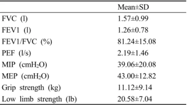
2. 뇌성마비 아동의 근력과 호흡기능의 상관관계 연구에 참여한 뇌성마비 아동의 FVC, FEV1, FEV1/FVC, PEF, MIP, MEP, 악력, 그리고 하지 근력의 평균과 표준편차는 Table 2에 제시하였다. 뇌성마비 아 동의 근력과 호흡기능의 상관관계를 분석한 결과 악력 과 FVC (r=0.95, p<0.01), FEV1 (r=0.95, p<0.01), PEF (r=0.84, p<0.01), MIP (r=0.65, p<0.01), MEP (r=0.71, p<0.01)는 각각 양의 상관관계를 가졌다. 하지 근력은 FVC (r=0.72, p<0.01), FEV1 (r=0.69, p<0.01), PEF (r=0.54, p<0.05), MEP (r=0.69, p<0.01)와 양의 상관관계 를 나타내었다(Table 3).
Mean±SD
FVC (l) 1.57±0.99
FEV1 (l) 1.26±0.78
FEV1/FVC (%) 81.24±15.08
PEF (l/s) 2.19±1.46
MIP (cmH2O) 39.06±20.08 MEP (cmH2O) 43.00±12.82 Grip strength (kg) 11.12±9.14 Low limb strength (lb) 20.58±7.04 Table 2. Measurement of pulmonary function,
respiratory and extremity muscle strength (n=17)
FVC: Forced vital capacity, FEV1: Forced expiratory volume in one second, PEF: Peak expiratory flow, MIP: Maximum inspiratory pressure, MEP: Maximum expiratory pressure
FVC FEV1 FEV1/FVC PEF (l) MIP MEP GS LS
FVC (l) 1
FEV1 (l) 0.97** 1
FEV1/FVC -2.26 -0.02 1
PEF (l) 0.81** 0.89** 0.06 1
MIP
(cmH2O) 0.65** 0.68** 0.03 0.44 1
MEP
(cmH2O) 0.71** 0.77** 0.04 0.69** 0.55* 1
GS (kg) 0.95** 0.95** -0.08 0.84** 0.65** 0.71** 1
LS (lb) 0.72** 0.69** -0.08 0.54* 0.35 0.69** 0.69** 1
Table 3. Correlation between pulmonary function, respiratory muscle and extremity muscle strength
(n=17)
*p<0.05, **p<0.01, GS: Grip strength, LS: Low limb strength
Ⅳ. 고 찰
호흡은 생명을 유지하기 위해 기본적이며 중요한 요소이지만 뇌성마비 아동은 호흡근 약화로 흉곽의 움 직임 제한, 비대칭적 성장으로 폐용적이 감소하고 호흡 량도 줄어드는 제한성 폐질환으로 호흡기능의 제한을 가진다(Kwon과 Lee, 2013; Park, 2006). FEV1/FVC(노력 성 폐활량에 대한 1초간 노력성 호기량의 비)가 70이하 이면 폐쇄성 폐질환으로 진단하는데(Swanney 등, 2008) 본 연구결과 FEV1/FVC는 81.24±15.08로 뇌성마비 아 동은 기류의 제한을 가지는 폐쇄성 폐질환이 아님을 알 수 있었다. 또한 뇌성마비 아동은 관절의 구축, 근육 의 단축, 근 위축 등 근골격계의 변형뿐만 아니라 전신 의 근력 약화, 호흡근의 약화, 뇌 손상으로 비정상적인 반사, 그리고 근 긴장도의 이상을 보이게 된다(Bax 등, 2005; Toder, 2000). 이런 이론적 근거를 바탕으로 본 연구는 뇌성마비의 대표적인 증상인 근력의 약화와 호 흡기능과의 상관관계를 알아보고자 하였다.
본 연구 결과 악력은 FVC, FEV1, PEF, MIP, MEP와 높은 상관관계를 나타내었다. Shah 등(2013)은 악력이 증가하면 FVC, FEV1가 증가하는 양의 상관관계를 나 타낸다고 하여 본 연구 결과와 일치한다. Jeon (2014)은 뇌성마비 아동을 대상으로 흉곽의 움직임에 영향을 미 치는 체간근 강화 운동을 통해 FVC, FEV1, PEF가 증가 했다고 보고하였는데 이는 체간의 근력이 뇌성마비 아 동의 흉곽과 관련 있으며 폐기능에 영향을 주었다고 할 수 있다.
악력은 손바닥을 이용해 쥐는 힘 즉 손가락의 굴곡과 신전 힘을 견관절의 내전, 주관절은 90도 굴곡, 전완은 중립위치에 측정한다(Kuzala와 Vargo, 1992). 견관절 주 변의 견갑대(shoulder girdle)의 안정성은 악력에 영향을 미치게 된다(Kobesova, 2015). 견갑대 주변의 근육은 상부 체간에 위치하고 호흡 보조근으로 호흡시 흡기와 호기에 사용된다(Bolser와 Reier, 1998).
FVC의 측정은 최대한 공기를 들이마신 후 빠르고 세게 공기를 불어내는데 이는 상부 체간근에 의해 움직 이는 흉곽과 관계 있다(Derenne 등, 1978). Czaplinski 등(2006)은 상부 체간근이면서 흡기 보조근으로 작용
하는 흉쇄유돌근(Sternocleidomastoid)이 흡기시 흉곽을 위로 들어올려 폐 용적을 넓혀 FVC에 영향을 미친다고 하였다. 상부 체간근은 흉곽을 움직여 폐기능에 영향을 미치고 악력의 증가에도 도움을 줄 수 있다(Kobesova, 2015). 따라서 뇌성마비 아동에게 실시하는 체간근 강 화 운동은 체간의 안정성, 상지 기능 개선과 균형 능력 뿐만 아니라(Choi 등, 2013; Lee와 Kim, 2011) 폐기능에 도 영향을 미쳐 호흡기능 개선에도 도움을 줄 수 있을 것이라 생각한다.
또한 악력이 증가하면 MIP, MEP가 증가하는 양의 상관관계를 나타내었는데 이는 앞서 설명한 바와 같이 악력의 증가는 체간 안정성이 뒷받침 되어야 하기 때문 에 체간 근력과 관계 있다. 체간의 안정성은 횡격막, 내외늑간근, 내외복사근, 복직근의 동시수축으로 이루 어지는데 이는 호기와 흡기를 담당하는 호흡근이다 (Knox와 Evan, 2002). 특히 복직근은 호흡시 호기에 관 여함으로 체간 근력이 증가하면 MEP가 증가하는 양의 상관관계를 나타낸다(Davies와 Klein, 1990). 호흡시 호 기근인 복직근의 근력 증가는 뇌성마비 아동의 기침 능력 향상에도 영향을 미쳐 기도의 이물질이나 분비물 을 외부로 배출해 폐렴이나 합병증을 예방할 수 있을 것이다.
본 연구 결과 하지 근력도 FVC, FEV1, PEF, 그리고 MEP와 양의 상관관계를 나타내었다. 하지 근력과 폐기 능, 호흡근과의 상관관계는 앞서 설명한 악력과의 관계 와 같이 체간의 안정성과 관련 있지만 상관계수에서 볼 수 있듯이 악력보다는 낮은 수치를 나타냈다. 이는 하지 근력은 호흡에 직접 관여하는 상부 체간보다는 하부 체간에 의해 영향을 받기 때문이다(Hodges와 Richardson, 1997). 하지의 움직임 즉 이동 혹은 보행을 하기 위한 움직임을 할 때 체간의 안정성이 뒷받침되지 않고서는 원위부 근육의 수의적이고 의미 있는 움직임 을 할 수 없다(McGill 등, 2003). 체간의 안정성은 복부 근육과 척추 기립근의 적절한 활성화와 협응에 의해 이루어지는데 이들은 호흡시 주동근으로 작용하는 횡 격막, 늑간근, 사각근과 같이 사용된다(Neumann, 2002).
또한, 하지 근력 중 슬관절 신전근인 대퇴사두근은 일 상생활능력에서 중요한 역할을 할 뿐만 아니라 호흡기
능과도 밀접한 관련이 있다(Swallow 등, 2007). 대퇴사 두근의 약화는 움직임에 대한 피로도를 증가시키고 자 주 사용하지 않은 근육으로 일상생활 동작의 어려움과 전신 근육의 약화를 초래하여 호흡시 약화된 호흡근을 과다하게 사용하여 호흡근의 피로감을 더하여 호흡기 능과 폐의 환기 능력에도 영향을 미친다(Dempsey, 2006a;
Seymour 등, 2010; Van't 등, 2004). 따라서 하지 근력의 증가는 일상생활의 활동량과 호흡근의 근력을 증가할 수 있고 호흡기능에도 영향을 미치게 된다(Dempsey, 2006b). 이는 본 연구 결과 하지근력이 FVC, FEV1, PEF, MEP와 양의 상관관계를 나타낸 결과와 일치한다. 이는 하지 근력이 보행과 독립적인 일상생활을 하기 위해 기본적인 요소이기도 하지만 심폐기능과 운동기능의 필수적인 요소라고 할 수 있다(Steven 등, 2007; Burtner 등, 1998). 그러므로 뇌성마비 아동을 대상으로 하지 근력의 향상은 운동수행 능력, 일상생활동작과 호흡기 능 개선의 긍정적인 효과를 가져다 줄 수 있을 것이라 생각한다(Schlough 등, 2005; Maltais 등, 2000).
Enright 등(1994)은 노인을 대상으로 사지의 근력이 호흡기능과 상관관계를 가진다고 보고하였고 뇌성마 비 아동을 대상으로 한 본 연구의 결과와 일치하였다.
노인과 뇌성마비 아동의 호흡 패턴이 일치하지 않음에 도 불구하고 이와 같은 결과는 사지의 근력은 체간의 근력을 대변하고 이는 폐기능과 호흡근에도 영향을 미 칠 수 있다고 할 수 있다.
본 연구는 뇌성마비 아동에게 폐기능, 호흡근 검사는 아동의 협조를 구하기 어렵고 검사자의 숙련도와 인내 심을 요구함에도 불구하고 재현성 있는 측정 값을 이용 해 뇌성마비 아동의 악력, 하지 근력과 호흡기능의 상관 관계를 파악하였다. 하지만, 연구에 참여한 대상자의 수가 적었고 뇌성마비의 유형, 나이, 성별에 따른 상관관 계를 파악하는 지속적인 연구가 더 필요할 것이다.
Ⅴ. 결 론
본 연구는 뇌성마비 아동 17명을 대상으로 악력, 하 지 근력이 호흡기능과 상관관계가 있는지 알아보고자
하였다. 그 결과 악력이 증가하면 FVC, FEV1, PEF, MIP, MEP가 증가하는 양의 상관관계를 나타내었고 하지 근력은 FVC, FEV1, PEF, MEP가 양의 상관관계를 나타내었다. 이러한 연구 결과를 통해 뇌성마비 아동의 근력은 호흡기능과 관계 있음을 확인하였고, 향후 일상 생활동작에 필요한 상지 근력과 하지 슬관절 신전근 등 다양한 근력 강화 프로그램 개발을 통해 뇌성마비 아동의 호흡기능 개선에도 긍정적 영향을 줄 수 있을 것으로 기대된다.
References
Bahat G, Tufan A, Ozkaya H, et al. Relation between hand grip strength, respiratory muscle strength and spirometric measures in male nursing home residents.
Aging Male. 2014;17(3):136-40.
Bax M, Goldstein M, Rosenbaum P, et al. Proposed definition and classification of cerebral palsy. Dev Med Child Neurol. 2005;47(8):571-6.
Bolser DC, Reier PJ. Inspiratory and expiratory patterns of the pectoralis major muscle during pulmonary defensive reflexes. J Appl Physiol (1985). 1998;85(5):
1786-92.
Burtner PA, Qualls C, Woollacott MH. Muscle activation characteristics of stance balance control in children with spastic cerebral palsy. 1998;1879-2219.
Choi YC, Park SJ, Lee MH, et al. The effects of trunk muscle strengthening exercises on balance performance of sitting posture and upper extremity function of children with spastic diplegic cerebral palsy. J Korean soc phys med. 2013;8(1):117-25.
Crystal RG. The lung: Scientific foundations: Lippincott-Raven.
1997.
Czaplinski A, Yen AA, Appel SH. Forced vital capacity (fvc) as an indicator of survival and disease progression in an als clinic population. J Neurol Neurosurg Psychiatry. 2006;77(3):390-2.
Davies PM, Klein-Vogelbach S. Right in the middle: Selective trunk activity in the treatment of adult hemiplegia:
Springer Berlin Heidelberg. 1990.
De Troyer A, Deisser P. The effects of intermittent positive pressure breathing on patients with respiratory muscle weakness. American Review of Respiratory Disease.
1981;124(2):132-7.
Dempsey JA, Romer L, Rodman J, et al. Consequences of exercise-induced respiratory muscle work. Respir Physiol Neurobiol. 2006a;151(2-3):242-50.
Dempsey JA. Is the healthy respiratory system (always) built for exercise. The Journal of Physiology. 2006b;576(2):
339-40.
Derenne JP, Macklem PT, Roussos C. The respiratory muscles:
Mechanics, control, and pathophysiology. Part iii.
Am Rev Respir Dis. 1978;118(3):581-601.
Dodd KJ, Taylor NF, Damiano DL. A systematic review of the effectiveness of strength-training programs for people with cerebral palsy. Arch Phys Med Rehabil.
2002;83(8):1157-64.
Enright PL, Kronmal RA, Manolio TA, et al. Respiratory muscle strength in the elderly. Correlates and reference values.
Cardiovascular health study research group. Am J Respir Crit Care Med. 1994;149(2):430-8.
Ersoz M, Selcuk B, Gunduz R, et al. Decreased chest mobility in children with spastic cerebral palsy. Turk J Pediatr.
2006;48(4):344-50.
Galley PM, Foster AL. Human movement. London: Churchill Livingston. 1987.
Hodges PW, Richardson CA. Contraction of the abdominal muscles associated with movement of the lower limb.
Phys Ther. 1997;77(2):132-42.
Jeon JY. Effects of the trunk muscle strengthening exercise and using the trunk belt on pulmonary function and trunk control ability for children with spastic cerebral palsy. Master's Degree. Daejeon University. 2014.
Jeon SC. Respiratory muscle strength and cough capacity in patients with amyotrophic lateral sclerosis. Master's
Degree. Yonsei University. 2007.
Ju C, Chen R. Factors associated with impairment of quadriceps muscle function in chinese patients with chronic obstructive pulmonary disease. PLoS One. 2014;9(2):
e84167.
Kim JS, Shin WS. The effects of respiratory muscle strengthening training on pulmonary function and gait ability in subacute stroke patients. J Korean soc phys med. 2013;8(4):489-96.
Kim MK, Cho MS, Hwang BG. The efficacy of pulmonary rehabilitation using air stacking exercise in cervical cord injured patients. J Korean soc phys med. 2010;
5(4):597-604.
Knox V, Evans AL. Evaluation of the functional effects of a course of bobath therapy in children with cerebral palsy: A preliminary study. Dev Med Child Neurol.
2002;44(7):447-60.
Kobesova A, Dzvonik J, Kolar P, et al. Effects of shoulder girdle dynamic stabilization exercise on hand muscle strength. Isokinetics and Exercise Science.
2015;23(1):21-32.
Kuzala EA, Vargo MC. The relationship between elbow position and grip strength. Am J Occup Ther. 1992;46(6):
509-12.
Kwon YH, Lee HY. Differences of respiratory function in children with spastic diplegic and hemiplegic cerebral palsy, compared with normally developed children.
J Pediatr Rehabil Med. 2013;6(2):113-7.
Lee EJ, Kim JS. The changes of gross motor function and balance ability in children with spastic diplegic cerebral palsy by trunk muscle strengthening exercise : Single group repeated easure study. J Korean soc phys med. 2011;6(2):189-97.
Maltais F, LeBlanc P, Whittom F, et al. Oxidative enzyme activities of the vastus lateralis muscle and the functional status in patients with copd. Thorax.
2000;55(10):848-53.
McGill SM, Grenier S, Kavcic N, et al. Coordination of muscle
activity to assure stability of the lumbar spine. J Electromyogr Kinesiol. 2003;13(4):353-9.
Neumann DA. Kinesiology of the musculoskeletal system:
Foundations for physical rehabilitation: Mosby. 2002.
Nicholson JH, Morton RE, Attfield S, et al. Assessment of upper-limb function and movement in children with cerebral palsy wearing lycra garments. Dev Med Child Neurol. 2001;43(6):384-91.
Park JH. Chest wall growth patterns in children with quadriplegic cerebral palsy. Master's Degree. Yonsei University.
2005.
Park YR. The effects of trunk muscle training by ball exercise program on respiration in children with spastic cerebral palsy. Master's Degree. Daegu University. 2006.
Rantanen T, Era P, Kauppinen M, et al. Maximal isometric muscle strength and socioeconomic status, health, and physical activity in 75-year-old persons. J Aging Phys Act. 1994;2(3):206-20.
Rutb sk. Normal development of functional motor skills. 1993.
Schlough K, Nawoczenski D, Case LE, et al. The effects of aerobic exercise on endurance, strength, function and self-perception in adolescents with spastic cerebral palsy: A report of three case studies. Pediatr Phys Ther. 2005;17(4):234-50.
Seymour JM, Moore L, Jolley CJ, et al. Outpatient pulmonary rehabilitation following acute exacerbations of copd.
Thorax. 2010;65(5):423-8.
Shah S, Nahar P, Vaidya S, et al. Upper limb muscle strength
& endurance in chronic obstructive pulmonary disease.
The Indian Journal of Medical Research. 2013;138(4):
492-6.
Singer J, Yelin EH, Katz PP, et al. Respiratory and skeletal muscle strength in copd: Impact on exercise capacity and lower extremity function. Journal of cardiopul- monary rehabilitation and prevention. 2011; 31(2):
111-9.
Stevens VK, Coorevits PL, Bouche KG, et al. The influence of specific training on trunk muscle recruitment patterns in healthy subjects during stabilization exercises. Manual therapy. 2007;12(3):271-9.
Swallow EB, Reyes D, Hopkinson NS, et al. Quadriceps strength predicts mortality in patients with moderate to severe chronic obstructive pulmonary disease. Thorax.
2007;62(2):115-20.
Swanney MP, Ruppel G, Enright PL, et al. Using the lower limit of normal for the fev1/fvc ratio reduces the misclassification of airway obstruction. Thorax.
2008;63(12):1046-51.
Toder DS. Respiratory problems in the adolescent with developmental delay. Adolesc Med. 2000;11(3):
617-31.
Van't Hul A, Harlaar J, Gosselink R, et al. Quadriceps muscle endurance in patients with chronic obstructive pulmonary disease. Muscle Nerve. 2004;29(2):267-74.
Youn BY. The training effect of respiration and articulatory function in cerebral palsied children. Master's Degree.
Daegu University. 1990.