초기 다계통위축증에 대한 뇌관류 Single Photon Emission Computed Tomography 의 임상적 유용성
이종윤 *·송인욱*·정성우*
정용안
†·양영순
‡가톨릭대학교 의과대학 신경과학교실*·
핵의학과학교실†, 보훈공단 중앙보훈병원
신경과‡
Clinical Usefulness of 99mTc-Hexamethyl Propylene Amine Oxime Perfusion Single Photon Emission Computed Tomography for Early Phase Multiple System Atrophy
Jong-Yoon Lee, M.D.*, In-Uk Song, M.D.*, Sung-Woo Chung, M.D.*, Yong-An Chung, M.D.
†, YoungSoon Yang, M.D.
‡Departments of Neurology* and Nuclear Medicine†, College of Medicine, The Catholic University of Korea, Seoul;
Department of Neurology‡, Veterans Hospital, Seoul Medical Center, Seoul, Korea
Background: Clinical diagnosis of multiple system atrophy (MSA) relays on signs and symptoms that are often difficult to identify particularly at early stage. Indeed neuropathological studies have demonstrated that Parkinson variant of MSA (MSA-P) is the first cause of misdiagnosis in a cohort of patients present- ing with parkinsonian features. But accurate diagnosis of these disorders is important for deciding on treatment, appropriate advice and prognosis since atypical parkinsonian disorders are characterized by poor response to dopaminergic treatment and more rapid disease progression. Therefore, we conducted this study to investigate difference of perfusion Single Photon Emission Computed Tomography (SPECT) in patients with the early phase of MSA-P using SPM program. Methods: We recruited consecutively 21 patients with MSA-P and 48 age-matched healthy controls. All subjects underwent Tc-99m HMPAO per- fusion SPECT and this perfusion images were analyzed. Results: For MSA-P, only hypoperfusion was seen in the middle frontal gyrus of left frontal lobe, superior frontal gyrus of right frontal lobe, precentral gyrus of left frontal lobe, middle frontal gyrus of right frontal lobe and precentral gyrus of right frontal lobe with respect to healthy subjects. Conclusions: We cautiously assume that perfusion SPECT may offer significant advantages compared to other imaging techniques in the assessment of neuronal degenera- tion in MSA-P and may help the clinician in the diagnostic characterization of patients presenting with atypical parkinsonism.
Received: May 16, 2014 Revision received: May 28, 2014 Accepted: May 28, 2014
Address for correspondence In-Uk Song, M.D.
Department of Neurology, Incheon St. Mary’s Hospital, The Catholic University College of Medicine, 56 Dongsu-ro, Bupyeong-gu, Incheon 403-720, Korea
Tel: +32-280-5010
E-mail: [email protected] Key Words: Multiple system atrophy, Tc-99m HMPAO perfusion SPECT, Frontal lobe
서 론
특발파킨슨병과 다계통위축증은 대표적인 파킨슨증을 일으키 는 질환이다[1]. 다계통위축증은 임상적으로 파킨슨증상과 소뇌증 상, 자율신경이상증상과 추체외로증상을 동반하는 질환이다[2]. 이 러한 다계통위축증의 병리소견은 흑질줄무늬체도파민로(nigros- triatal dopaminergic pathway), 꼬리핵(caudate)과 조가비핵(putamen), 창백핵(globus pallidus), 뇌간(brain stem), 소뇌 그리고 척수에서 신 경퇴행성 변화를 보여주는 질환으로 특발파킨슨병과 마찬가지로
확진은 뇌조직검사뿐이므로 다계통위축증도 임상증상 및 신경학 적 검사를 중심으로 추정 진단을 내릴 뿐이다[2, 3]. 다계통위축증 의 조기 진단은 특발파킨슨병에 비해 진행이 빠르고 약물에 반응 을 하지 않아 조기의 증상 조절 및 예후에 대한 명확한 설명이 반드 시 필요하다. 특발파킨슨병의 경우는 도파민전달체를 이용한 뇌기 능 영상기법과 iodine-123-meta-iodobenzylguanidine (I-123 MIBG) 신 티그라피 등으로 인하여 조기진단에 많은 도움을 받고 있으나 다계 통위축증에 대해서는 이러한 영상기법이 아직까지 나와 있지 않는 실정이다[4]. 하지만 뇌자기공명사진 촬영으로 다계통위축증을 시
© 2014 Korean Dementia Association
This is an Open Access article distributed under the terms of the Creative Commons Attribution Non-Commercial License (http://creativecommons.org/licenses/by-nc/3.0) which permits unrestricted non-commercial use, distribution, and reproduction in any medium, provided the original work is properly cited.
사하는 소견들이 보고되고는 있으나 어느 정도 진행된 단계에서의 소견이므로 초기 다계통위축증에서는 적용되기 힘들다. 물론 이전 연구들에 의하면 statistical parametric mapping (SPM) 을 이용한 flu- orine-18 fluorodeoxyglucose positron emission tomography (FDG-PET) 영상으로 인해 특발파킨슨병과 다계통위축증의 특징적인 소견을 보였다고 하나 이는 경제적인 문제 등으로 인하여 일반적으로 사용 하기 어려운 실정이다[5, 6]. 이에 반해 뇌관류 brain perfusion single- photon emission computed tomography (SPECT)은 일반적으로 많이 유용되는 검사로서 뇌혈류를 technetium을 붙인 Hexamethyl-pro- phylene-amine-oxime 또는 ethyl cysteinate dimer를 이용하여 많이 활용되고 있으며, 최근 신경퇴행성질환에 있어서 뇌혈류량에 따른 기능 향상을 평가하는 데 많이 이용되고 있다[7, 8]. 특발파킨슨병에 대해서는 두정엽, 후두엽, 기저핵 등에서 관류저하를 보이는 특징적 인 소견이 보고되고 있으나 뇌관류 SPECT의 다계통위축증 소견에 대해서는 아직까지 확립되지 않았을 뿐만 아니라 이에 대한 연구도 미미한 실정이다[6, 7, 9]. 더욱이 다계통위축증은 파킨슨증의 임상 증상을 동반하기 때문에 조기에 감별진단이 더욱 어려운 실정이다.
하지만 다계통위축증은 특발파킨슨병에 비해 진행이 빠르고 약물 에 반응을 하지 않아 조기의 증상 조절 및 예후에 대한 명확한 설명 이 반드시 필요하다. 따라서 다계통위축증의 뇌관류 SPECT 소견을 객관적인 방법인 SPM 프로그램을 이용하여 정상군과 비교 분석하 여 조기진단에 도움이 되고자 본 연구를 시행한다.
방 법
본 연구는 2012년 1월부터 12월까지 본원 신경과에 내원한 파킨 슨증 양상을 주로 보이는 다계통위축증의 파킨슨 변형(Parkinson variant of multiple system atrophy, MSA-P) 환자를 대상하였고, 본원 임상윤리심의위원회의 승인을 받고 진행하였다. 모든 연구 대상자 들은 Gilman 등[2]에 의해 제안된 추정 다계통위축증 진단기능에 맞는 환자를 선택하였다. 또한 초기 환자만을 선정하기 위해 Modi- fied Hoehn-Yahr stage (H-Y stage) 3 미만 환자만을 선택하였고, 본원 123I-n-fluoropropyl-2b-carbomethoxy-3b-(4-iodophenyl) nortropane positron emission tomography (FP-CIT PET)상에서도 도파민 전달체 의 흡수가 저하되지 않은 환자만을 선택하였다. 뇌자기공명사진상 에서 뇌의 구조적 병변이나 허혈성 병변 등이 있는 환자는 본 연구 에서 제외하였다. 또한 임상적으로 의미 있는 신장, 간장, 심혈관계, 호흡기계, 뇌혈관계, 내분비계 질환 혹은 기타 심각한 진행형 신체 질환이 있는 경우, 우울증이나 정신과적 장애를 가졌거나 과거력을 가진 경우, 2차성 파킨슨증을 일으킬 만한 약물 및 알코올의 남용 력이 있는 경우는 본 연구에서 제외하였다. 이들 선택된 환자군은 나이와 성별을 맞춘 정상군과 비교 분석되었으며, 모든 대상군은
뇌관류 SPECT를 안정영상으로 촬영하였다.
99mTc-HMPAO SPECT 영상 획득
뇌영상 연구를 위해 사용한 단일광자방출 전산화 단층촬영(Sin- gle Photon Emission Computed Tomographic, SPECT)을 시행했다. 영 상획득은 6° 간격으로 360° 회전하여 저에너지 고해상도 조준기(low energy high resolution collimator)를 사용하여 60프레임을 얻는다. 안 정상태에서의 영상을 얻기 위해 925 MBq의 99mTc-HMPAO를 정맥 주사하고, 주사한 후 20분 경과 후 약 20-30분 동안 스캔하였다. 촬영 하는 동안 피검자는 편안한 상태를 유지하며, 머리의 움직임을 최 소로 하였으며 촬영실은 조용하고 조명을 어두운 상태로 유지했다.
영상분석 및 SPM
SPECT 영상은 표준다이콤(DICOM) 형태로 변환되어 마이크로 소프트윈도우 기반의 컴퓨터에 저장되었다. 각각의 파일은 분석을 위해 Analyze형식으로 변환된 후 모든 영상을 SPECT 표준지도 위 에 공간 정규화했다. 뇌피질 및 내부 구조의 미세한 차이는 저주파 함수들로 미리 정의된 기저함수(basis function)들의 가중합으로 나 타내는 선형화된 비선형적(linearized nonlinear) 방법을 사용하여 제거했다. 신호 대 잡음비를 높이고 미처 보정되지 않은 뇌 피질의 차이를 극복하기 위하여 공간정규화가 된 영상들을 16 mm의 full width at half maximum (FWHM)을 갖는 가우시안 커널(Gaussian kernel)로 중첩적분(convolution)했다. 이러한 전처리 과정으로서의 편평화(smoothing)는 통계 분석 결과에서 크기가 작은 덩어리의 생 성을 줄이므로 위양성 결과를 보정하고 고주파의 잡음을 제거해서 예민도를 높이는 역할을 한다. 그리고 각 복셀이 가지는 국소 계수 는 뇌피질내의 전체계수와 개인적인 내변이 그리고 불규칙한 잡음 의 선형결합(일반 선형 모델: general linear model)으로 나타내어진 다는 가정 하에 선형회귀분석을 이용하여 각 복셀에서의 이러한 변수들의 가중치를 찾아낸 후 국소 계수의 차이만을 추출했다.
양 대상군의 기본 자료에 대한 통계 분석은 SPSS ver. 17.0 (SPSS Inc., Chicago, IL, USA)을 이용하여 연속 변수는 독립 t-test을 시행하 였고, 비연속 변수에 대해서는 Pearson chi-square analysis를 사용하 였다. 뇌 영상 사진에 대한 분석은 복셀별로 통계 분석하였다. 복셀 별로 각각의 SPECT군 사이의 국소 계수 차이에 대한 귀무 가설을 검증하고자 뇌 영역 내의 모든 복셀에 대해 t-test를 시행하고 용이 한 해석을 위해 t값들을 표준 정규 분포로 변환하여 Z값으로 나타 냈다. 이는 위에서 언급한 일반 선형 모델을 기본 모델로 하고, 각 변 수의 효과가 Student’s t 통계를 따른다고 보아 그 유의성을 검정하는 것으로 이렇게 각 복셀에서 구해진 t값들은 그 자체가 뇌 기능 영상
이 포함된 3차원 공간상에서 또 다른 하나의 확률 변수(random variable)로서 행동한다고 생각하고, 그 분포는 고정 가우시안 랜덤 영역(stationary Gaussian random field)을 따른다고 가정하였다. 이러 한 가정하에서 각 복셀의 t값에 대한 통계적 유의성은 그 값을 Z값 [Z = (t-m)/SD; m, 평균; SD, 표준편차]으로 변환하여 가우시안 랜덤 영역의 행동과 비교함으로써 얻을 수 있다. 통계 분석의 결과를 해 석하고 유의한 관류 차이와 덩어리 크기를 갖는 영역의 Z값들을 magnetic resonance imaging 표준 지도 위에 표현하여 표준 지도 상 에서의 좌표와 해부학적 이름, 브로드만 영역 등을 표시해 주었다.
또한 각 복셀의 Z값에 대한 통계적 매개 변수 영상(statistical para- metric image)을 구성하고 3차원으로 렌더링(rendering)한 표준 지도 위에 투사하여 관류 차이를 보이는 부위를 쉽게 알아볼 수 있게 한 다. 이상의 과정은 Matlab, ver. 5.3 (Mathworks Inc., Natick, MA, USA) 상에서 구현된 SPM2 소프트웨어(statistical parametric mapping soft- ware, Institute of Neurology, University College of London, London, UK)를 이용하였다.
결 과
다계통위축증 환자 중에서 파킨슨증상의 변형(MSA-P)을 가진
환자 21명과 48명의 정상군과의 평균 연령 및 성별의 의미 있는 차 이는 없었다. MSA-P의 평균 H-Y stage는 2.14± 0.63이고 유병기간은 23.39±14.64개월을 보였다(Table 1).
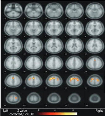
뇌관류 SPECT 분석상에서는 MSA-P를 가진 환자군과 정상군을 비교했을 때 좌측 전두엽의 중간전두이랑(middle frontal gyrus of left frontal lobe; BA 6), 우측 전두엽의 상전두이랑(superior frontal gyrus of right frontal lobe), 좌측 전두엽의 전중심이랑(precentral gyrus of left frontal lobe; BA 4), 우측전두엽의 중간전두이랑(middle frontal gy- rus of right frontal lobe; BA 6) 그리고 우측 전두엽의 전중심이랑(pre- central gyrus of right frontal lobe)에서 MSA-P에서 관류저하 소견을 통계적으로 의미 있게 보였다(Table 2, Fig. 1).
고 찰
특발파킨슨병과 MSA-P의 임상적인 특징은 파킨슨증 양상을 중 심으로 나타나기 때문에 초기 단계에서는 감별진단하기가 매우 어 렵다[7, 9, 10]. 특히 MSA-P가 특발파킨슨병에 비해 약물에 반응을 보기가 어려울 뿐만 아니라 예후도 좋지 않음에도 불구하고도 진 단 및 뇌기능에 대한 연구는 특발파킨슨병에만 초점이 맞추어져 있 는 실정이다. 이전 초기 단계에서 임상적으로 MSA-P와 비슷한 소견 을 보이는 특발파킨슨병에 대한 뇌관류를 분석한 연구에서는 정상 Table 1. Demographic and clinical characteristics of all subjects
MSA-P Healthy control p value
Number of subject 21 48
Age 70.38± 6.94 66.23± 11.84 0.257
Gender (male/female) 8/13 11/37 0.089
Duration after diagnosis (months)
19.52± 13.68
Duration after symptom (months)
23.39± 14.64
Hoehn & Yahr stage 2.14± 0.63
MMSE 27.92± 0.91
Levodopa dose/day (mg) 387.50± 246.09 Dopamine agnoists/day (mg)* 1.09± 1.42
*All patients have taken only pramipexole.
MSA-P, Parkinson variant of multiple system atrophy; MMSE, mini-mental state ex- amination.
Table 2. Areas with significantly decreased regional cerebral blood flow in the MSA-P patients compared with the healthy controls
kE T Z x, y, z Brain areas
1703 10.19 6.41 -22, 2, 60 Left Frontal Lobe, Middle Frontal Gyrus, BA 6 1703 8.31 5.76 2, 8, 54 Right Frontal Lobe, Superior Frontal Gyrus 1703 7.02 5.21 -34, -24, 60 Left Frontal Lobe, Precentral Gyrus, BA 4 687 7.60 5.47 24, -10, 62 Right Frontal Lobe, Middle Frontal Gyrus, BA 6 687 7.46 5.41 32, -10, 60 Right Frontal Lobe, Precentral Gyrus Height threshold, T= 6.25, corrected p= 0.01; Extent threshold, k= 50 voxels.
kE, Expected voxels per cluster.
Left Z value
corrected p< 0.001 Right
Fig. 1. Comparisons of patients with MSA-P compared with the healthy controls show cerebral regions (yellow color) with decreased regional cerebral blood flow in trans-axial MR fused images.
군에 비해 전전두엽(prefrontal lobe)과 외측 전두엽(lateral frontal lobe)중심의 전두엽과 렌즈 핵(lentiform nucleus)에서 주로 뇌관류저 하를 보인다고 보고하면서 이들 영역에 비해 정도는 약하지만 측두 엽과 두정엽, 후두엽에서도 약간의 관류저하를 보고하고 있다[7, 11- 13]. 따라서 이들 보고들은 이미 잘 알려진 특발파킨슨병에서의 전 두엽 기능저하를 보이는 뇌관류 소견이라고 볼 수 있겠다. 이에 반 해 MSA-P는 신경병리학 연구에 의하면 올리브교뇌소뇌계(olivo- pontocerebellar system)보다는 흑질줄무늬체(nigrostriatal system)를 주로 침범하는 양상을 보인다고 알려져 있다[3, 14]. 하지만 MSA-P 의 뇌관류에 대한 최근 연구들에 의하면 임상적으로 소뇌증상은 거의 보이지 않음에도 불구하고 소뇌에서 주로 저관류소견을 보이 면서 전두엽, 섬이랑, 조가비핵 그리고 시상에서 저관류소견을 보인 다고 보고하였다[8, 9, 14]. 하지만 본 연구에서는 전두엽의 저관류소 견이 이전의 연구결과와 비슷한 소견을 보였으나 소뇌에서는 정상 군에 비해서 의미 있는 저관류소견을 보이지 않아 이전의 보고들과 다른 소견을 보였다. 본 연구의 이러한 결과는 아마도 이전의 연구 들과는 달리 본 연구는 초기 단계의 MSA-P 환자의 뇌관류 소견만 을 분석하였기에 기인되었을 것이라고 생각된다. 따라서 저자들은 본 연구의 결과 및 이전의 연구들을 통해서 MSA-P의 초기단계의 증상 발현시기에는 전두엽 영역의 침범으로 시작하여 증상이 진행 하면서 소뇌를 포함한 다른 뇌영역으로 퇴행성 변화가 퍼져나간다 는 추정을 할 수 있었다. 또한 본 연구에서는 특발파킨슨병에서 저 관류를 보이는 후두엽에서는 어떠한 비정상적인 뇌관류소견을 보 이지 않아 특발파킨슨병과 MSA-P의 초기단계에서도 두 질환을 감 별할 수 있는 뇌관류 소견이 될 수도 있을 것이라는 추정을 할 수 있 었다. 하지만 이러한 추정에 대해서는 향후 두 질환을 가진 환자들 간의 뇌관류에 대한 비교분석이 반드시 필요하다고 생각한다.
결론적으로 본 연구는 뇌관류 SPECT을 통해서 정상군에 비해 MSA-P를 가진 환자군에서 의미 있는 뇌관류 변화의 소견을 알아 보기 위한 연구로서 이를 통해 정상군보다 임상적 초기 단계의 MSA-P라 할지라도 전두엽 중심으로 의미 있는 저관류 소견을 보인 것을 알 수 있었다. 따라서 본 연구의 결과를 기초로 초기 단계의 MSA-P 환자의 진단에 도움이 되는 영상기법으로써 perfusion SPECT이 유용하게 사용될 수 있을 가능성을 본 저자들은 제시할 수 있었다. 따라서 이러한 영상 검사를 통해 상기 질환의 초기 감별 에 도움이 될 수 있다면 초기 단계에 각각 질환에 맞는 적절한 치료 및 예방을 가능하게 되어 향상된 삶을 영위하는 데 기여할 수 있으 리라 생각된다. 더욱이 뇌관류 SPECT은 뇌자기공명 사진이나, 뇌 PET 사진같이 고가의 검사가 아니므로 임상적으로도 쉽게 사용하 여 두 질환의 초기 감별 진단에 적용할 수 있으리라 생각된다. 하지 만 본 연구의 결과를 좀 더 명확히 하고 객관화하기 위해서는 많은 수의 환자를 통한 다기관 연구 및 추적 검사를 통한 뇌 관류에 대한
연구가 향후에 반드시 진행되어야 할 필요성이 있다고 주장하는 바 이다.
참고문헌
1. Hughes AJ, Daniel SE, Kilford L, Lees AJ. Accuracy of clinical diagnosis of idiopathic parkinson’s disease: a clinico-pathological study of 100 cases.
J Neurol Neurosurg Psychiatry 1992; 55: 181-4.
2. Gilman S, Low PA, Quinn N, Albanese A, Ben-Shlomo Y, Fowler CJ, et al. Consensus statement on the diagnosis of multiple system atrophy. J Neurol Sci 1999; 163: 94-8.
3. Wenning GK, Tison F, Ben Shlomo Y, Daniel SE, Quinn NP. Multiple system atrophy: a review of 203 pathologically proven cases. Mov Disord 1997; 12: 133-47.
4. Hughes AJ, Ben-Shlomo Y, Daniel SE, Lees AJ. What features improve the accuracy of clinical diagnosis in parkinson’s disease: a clinicopatho- logic study. Neurology 1992; 42: 1142-6.
5. Antonini A, Leenders KL, Vontobel P, Maguire RP, Missimer J, Psylla M, et al. Complementary pet studies of striatal neuronal function in the dif- ferential diagnosis between multiple system atrophy and parkinson’s dis- ease. Brain 1997; 120(Pt 12): 2187-95.
6. Juh R, Kim J, Moon D, Choe B, Suh T. Different metabolic patterns anal- ysis of parkinsonism on the 18f-fdg pet. Eur J Radiol 2004; 51: 223-33.
7. Feigin A, Antonini A, Fukuda M, De Notaris R, Benti R, Pezzoli G, et al. Tc-99m ethylene cysteinate dimer spect in the differential diagnosis of parkinsonism. Mov Disord 2002; 17: 1265-70.
8. Cilia R, Marotta G, Benti R, Pezzoli G, Antonini A. Brain spect imaging in multiple system atrophy. J Neural Transm 2005; 112: 1635-45.
9. Matsui H, Udaka F, Miyoshi T, Hara N, Tamura A, Oda M, et al. Brain perfusion differences between parkinson’s disease and multiple system atrophy with predominant parkinsonian features. Parkinsonism Relat Disord 2005; 11: 227-32.
10. Tang CC, Poston KL, Eckert T, Feigin A, Frucht S, Gudesblatt M, et al.
Differential diagnosis of parkinsonism: a metabolic imaging study using pattern analysis. Lancet Neurol 2010; 9: 149-58.
11. Van Laere K, Santens P, Bosman T, De Reuck J, Mortelmans L, Dierckx R. Statistical parametric mapping of (99m)tc-ecd spect in idiopathic par- kinson’s disease and multiple system atrophy with predominant parkin- sonian features: correlation with clinical parameters. J Nucl Med 2004;
45: 933-42.
12. Paschali A, Messinis L, Lyros E, Constantoyannis C, Kefalopoulou Z, Lakiotis V, et al. Neuropsychological functions and rcbf spect in parkin-
son’s disease patients considered candidates for deep brain stimulation.
Eur J Nucl Med Mol Imaging 2009; 36: 1851-8.
13. Firbank MJ, Molloy S, McKeith IG, Burn DJ, O’Brien JT. Longitudinal change in 99mtchmpao cerebral perfusion spect in parkinson’s disease
over one year. J Neurol Neurosurg Psychiatry 2005; 76: 1448-51.
14. Kimura N, Hanaki S, Masuda T, Hanaoka T, Hazama Y, Okazaki T, et al. Brain perfusion differences in parkinsonian disorders. Mov Disord 2011; 26: 2530-7.