HIGHLIGHTS
• A subdural hemorrhage (SDH) is a common neurological disease with benign outcome.
• Delayed extensive white matter injury of SDH was described in this case.
• The corticospinal tract integrity assessed by diffusion tensor imaging would be useful for SDH.
• Physicians should be aware of a possible delayed effect in treatment of SDH.
Brain Neurorehabil. 2019 Sep;12(2):e15 https://doi.org/10.12786/bn.2019.12.e15 pISSN 1976-8753·eISSN 2383-9910
Case Report
Received: Jun 24, 2019 Revised: Aug 14, 2019 Accepted: Aug 16, 2019 Correspondence to Hye Jung Park
Department of Rehabilitation Medicine, National Traffic Injury Rehabilitation Hospital, 260 Jungang-ro, Yangpyeong 12564, Korea.
E-mail: [email protected] Seong Hoon Lim
Department of Rehabilitation Medicine, St.
Vincent’s Hospital, College of Medicine, The Catholic University of Korea, 93 Jungbu-daero, Paldal-gu, Suwon 16247, Korea.
E-mail: [email protected]
Kyoung Bo Lee, Sang Cheol Yoon, Joon Sung Kim, Bo Young Hong, Jung Geun Park, Won Jin Sung, Hye Jung Park, Seong Hoon Lim
Delayed Extensive White Matter Injury Caused by a Subdural Hemorrhage
and Role of Corticospinal Tract Integrity
Brain & NeuroRehabilitation
Copyright © 2019. Korea Society for Neurorehabilitation i
ABSTRACT
A subdural hemorrhage (SDH) is a common disorder with usually good prognosis. Most SDHs resolve with or without with minimal sequelae. We present a case report of a patient with SDH, who had delayed extensive white matter injury with disruptions of corticospinal tracts (CSTs) by diffusion tensor imaging (DTI) and showed abysmal prognosis, despite long-term rehabilitation. A 62-year-old man with an SDH underwent burr hole trephination for hematoma removal. Within 7 days, the hemorrhage diminished. At 12 weeks after the onset, the patient's weakness did not improve, and a follow-up magnetic resonance imaging revealed extensive leukomalacia, especially in the white matter. The DTI for CST revealed severe injury of CST integrity. He did not re-gain muscle strength and functional independence, despite 3 months of inpatient rehabilitation. This case describes SDH with delayed extensive white matter injury and exceptional poor prognosis and urges caution in that the SDH may induce very variable functional recovery. Besides, DTI for CST would be useful in predicting the long-term functional prognosis in extensive white matter injury.
Keywords: Subdural hemorrhage; Corticospinal tracts; Diffusion tensor imaging;
Leukomalacia; White matter injury
INTRODUCTION
A subdural hemorrhage (SDH) is a common neurological disease in elderly patients [1].
Most SDHs resolve with or without minimal sequelae. In participants over than 90 years, SDHs are associated with a poor outcome [2]. In addition, a poor SDH outcome is related to initial neurological status and treatment options [2,3]. The typical clinical and radiological course of a patient with an SDH may be urgent in the acute stage, followed by postoperative recovery; it rarely progresses to the chronic stage.
In the present case report, we describe a patient with an SDH who was treated surgically and resolved radiologically but who experienced delayed extensive leukomalacia and showed radiologically worse findings in the chronic stage.
Case Report
Received: Jun 24, 2019 Revised: Aug 14, 2019 Accepted: Aug 16, 2019 Correspondence to Hye Jung Park
Department of Rehabilitation Medicine, National Traffic Injury Rehabilitation Hospital, 260 Jungang-ro, Yangpyeong 12564, Korea.
E-mail: [email protected] Seong Hoon Lim
Department of Rehabilitation Medicine, St.
Vincent's Hospital, College of Medicine, The Catholic University of Korea, 93 Jungbu-daero, Paldal-gu, Suwon 16247, Korea.
E-mail: [email protected] Copyright © 2019. Korea Society for Neurorehabilitation
This is an Open Access article distributed under the terms of the Creative Commons Attribution Non-Commercial License (https://
creativecommons.org/licenses/by-nc/4.0) which permits unrestricted non-commercial use, distribution, and reproduction in any medium, provided the original work is properly cited.
ORCID iDs Kyoung Bo Lee
https://orcid.org/0000-0002-7652-1393 Sang Cheol Yoon
https://orcid.org/0000-0002-4148-609X Joon Sung Kim
https://orcid.org/0000-0001-7457-593X Bo Young Hong
https://orcid.org/0000-0001-9290-6173
Kyoung Bo Lee ,1 Sang Cheol Yoon ,1 Joon Sung Kim ,1 Bo Young Hong ,1 Jung Geun Park ,1 Won Jin Sung ,1 Hye Jung Park ,2 Seong Hoon Lim 1
1Department of Rehabilitation Medicine, St. Vincent's Hospital, College of Medicine, The Catholic University of Korea, Seoul, Korea
2Department of Rehabilitation Medicine, National Traffic Injury Rehabilitation Hospital, Yangpyeong, Korea
Delayed Extensive White Matter Injury Caused by a Subdural Hemorrhage
and Role of Corticospinal Tract
Integrity
Jung Geun Park
https://orcid.org/0000-0002-9298-9850 Won Jin Sung
https://orcid.org/0000-0001-5251-3631 Hye Jung Park
https://orcid.org/0000-0002-6731-4376 Seong Hoon Lim
https://orcid.org/0000-0002-5475-4153 Funding
This research was supported by Basic Science Research Program through the National Research Foundation of Korea (NRF) funded by the Ministry of Science and ICT (grant number:2017R1E1A1A01074324) Conflict of Interest
The authors have no potential conflicts of interest to disclose. No commercial party having a direct financial interest in the results of the research supporting this article has or will confer a benefit upon the authors or upon any organization with which the authors are associated.
CASE REPORT
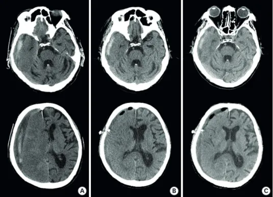
A 62-year-old man presented with slowly progressive dysarthria and left hemiplegia for 1 week without definite acute trauma. He was an alcoholic without any medical history such as high blood pressure or diabetes mellitus; however, his initial brain computed tomography (CT) scan showed an SDH (Fig. 1). Subsequently, the patient underwent burr hole trephination for hematoma removal. Brain CT scans performed on postoperative days 3 and 7 revealed that the SDH had regressed (Fig. 1), and the patient was referred to the rehabilitation department. On day 14, brain magnetic resonance imaging (MRI) showed additional SDH regression (Fig. 2A). At 12 weeks after the onset, the patient's weakness and functional status did not improve. The follow-up MRI revealed extensive leukomalacia, especially in the white matter (Fig. 2B). The diffusion tensor imaging (DTI) for corticospinal tracts (CSTs) revealed severe injury of CST integrity (Table 1, Fig. 3). DTI was performed using a 3.0-T MRI (MAGNETOM® Verio; Siemens, Erlangen, Germany) equipped with a six-channel head coil [4]. The data were acquired in the form of single-shot spin-echo echo-planar images, with axial slices covering the whole brain across 76 interleaved slices of 2.0 mm thickness (no gap); repetition time (TR)/echo time (TE) = 14,300/84 ms; field of view = 224 × 224 mm2; matrix 224 × 224; voxel size 1 × 1 × 2 mm3; number of excitations
= 1. Diffusion sensitizing gradients were applied in 64 noncollinear directions with a b-value of 1,000 ms/mm2. The b=0 images were scanned before the acquisition of the diffusion-weighted images, with 65 volumes in total [5]. Fiber tracking was based on the fiber assignment continuous tracking algorithm and multiple regions of interest (ROIs) approach using DTI-Studio. The selection of ROI, termination criteria, and the acquisition of normalized fractional anisotropy (FA) values were proceeded by the same way of previous research [5]. He did not re-gain muscle strength and functional independence, despite 3 months of inpatient rehabilitation. The Mini-Mental Status Examination score was 18 out of
2/5 https://doi.org/10.12786/bn.2019.12.e15
Delayed Injury of White Matter and CST in SDH Brain & NeuroRehabilitation
https://e-bnr.org
A B C
Fig. 1. Brain computed tomography scans over time. (A) Day of onset. (B) Postoperative evaluation on day 4 after onset. (C) Day 7 after onset.
30, and his modified Barthel Index score was estimated to be 45 on a 100-point scale; thus, the patient was returned to the nursing care unit.
DISCUSSION
Two clinical aspects are emphasized in the present case report: the occurrence of extensive injury in the chronic stage with extensive white matter injury and relatively preserved gray matter. The
A B
Fig. 2. Brain magnetic resonance images over time. (A) Two weeks after onset. (B) Twelve weeks after onset.
Table 1. FN, FA (mid-pons), and FA (pontomedullary junction) values of the CST in case
Variables Normalized FN FA (mid-pons) FA (pontomedullary junction)
Normalized values 0.017 0.390 0.460
Values are the normalized values (affected/non-affected).
CST, corticospinal tract; FA, fractional anisotropy; FN, fiber number.
A B
Rt Lt Rt Lt
Fig. 3. The diffusion tensor tractography images of the CST in patient and control. (A) CST of case. (B) CST of control (age, sex matched). The left CST was shown as red, and the right CST as yellow.
CST, corticospinal tract.
patient did not have a history of brain injury or medical comorbidity. He was an alcoholic, his functional status was independent, and organic brain damage was not evident. To date, late extensive white matter injury has not been reported, and, to the best of our knowledge, this is the first description of late extensive white matter injury caused by an SDH. We traced the pathophysiology for delayed extensive white matter injury, and severe motor weakness, the CST injury assessed by DTI in chronic phase would be the explanation for his clinical progress.
An SDH does not always have a benign course, especially in elderly persons older than 70, 85, or 90 years [2,6,7]. Typical surgical evaluations such as burr holes or craniotomy induce good clinical outcomes [3]. The patient in this case was not very old and received surgical treatment.
At 2 weeks after surgery, imaging studies showed resolution of the SDH. MRI at 12 weeks after the onset had worsened than MRI at 2 weeks after the onset; therefore, delayed brain injury may explain the poorer outcome of SDHs. Physicians should be aware of a possible delayed effect and not disregard neurological changes in the chronic stage of an SDH.
The white matter injury occurred by various mechanisms in SDH. Hematoma itself compressed the CST and induced distortion of the CST. Thus, the reversibility of the FA by DTI was related to the distortion of the pyramidal tract and vasogenic edema due to the expansion of the hematoma in SDH [8]. In addition, secondary insult after SDH was associated with significant brain swelling and stimulated a refractory rise intracranial pressure (ICP). In traumatic SDH complicated by secondary insult, brain swelling is exacerbated by surgical evacuation [9]. An important contributor to the neurological injury associated with SDH is the ischemic damage which is caused by raised ICP producing impaired cerebral perfusion. The removal of the SDH results in the immediate reversal of global ischemia accompanied by an abrupt reduction in the mass lesion and an ensuing reperfusion injury [10].
A previous study found that, in patients with a corona radiata injury, the integrity of the CST, as assessed by DTI obtained during the early stage, appears to be helpful in predicting motor outcomes on the affected side [11]. Most previous reports have shown that damage to the CST is useful for predicting poor outcome of motor function in patients with stroke [11-13]. A recent study demonstrated that the CST integrity assessed by DTI, would reflect the hand function up to 12 months after stroke [5]. A previous case report showed CST integrity assessed by DTI may be a useful add-on study for patients with diffuse axonal injury after traumatic brain injury [14]. In this case, the CST integrity assessed by DTI also reflect the motor function and disability in chronic phase in patient with SDH. The CST integrity assessed by DTI would be useful for patients with poor recovery in acute or chronic stage of SDH, despite usual benign course of SDH.
In conclusion, although an SDH is a common neurological disorder and usually has a benign course, delayed white matter injury can occur. Physicians should be aware of the possible delayed effects of SDHs in patients who do not recover or recover slowly. These concerns play an important role in predicting prognosis and establishing a long-term plan for patients and caregivers.
REFERENCES
1. Kudo H, Kuwamura K, Izawa I, Sawa H, Tamaki N. Chronic subdural hematoma in elderly people: present status on Awaji Island and epidemiological prospect. Neurol Med Chir (Tokyo) 1992;32:207-209.
PUBMED | CROSSREF
4/5 https://doi.org/10.12786/bn.2019.12.e15
Delayed Injury of White Matter and CST in SDH Brain & NeuroRehabilitation
https://e-bnr.org
2. Stippler M, Ramirez P, Berti A, Macindoe C, Villalobos N, Murray-Krezan C. Chronic subdural hematoma patients aged 90 years and older. Neurol Res 2013;35:243-246.
PUBMED | CROSSREF
3. Almenawer SA, Farrokhyar F, Hong C, Alhazzani W, Manoranjan B, Yarascavitch B, Arjmand P, Baronia B, Reddy K, Murty N, Singh S. Chronic subdural hematoma management: a systematic review and meta- analysis of 34,829 patients. Ann Surg 2014;259:449-457.
PUBMED | CROSSREF
4. Kim Y, Lim SH, Park GY. Crossed cerebellar diaschisis has an adverse effect on functional outcome in the subacute rehabilitation phase of stroke: a case-control study. Arch Phys Med Rehabil 2019;100:1308-1316.
PUBMED | CROSSREF
5. Yoo YJ, Kim JW, Kim JS, Hong BY, Lee KB, Lim SH. Corticospinal tract integrity and long-term hand function prognosis in patients with stroke. Front Neurol 2019;10:374.
PUBMED | CROSSREF
6. Miranda LB, Braxton E, Hobbs J, Quigley MR. Chronic subdural hematoma in the elderly: not a benign disease. J Neurosurg 2011;114:72-76.
PUBMED | CROSSREF
7. Mulligan P, Raore B, Liu S, Olson JJ. Neurological and functional outcomes of subdural hematoma evacuation in patients over 70 years of age. J Neurosci Rural Pract 2013;4:250-256.
PUBMED | CROSSREF
8. Yokoyama K, Matsuki M, Shimano H, Sumioka S, Ikenaga T, Hanabusa K, Yasuda S, Inoue H, Watanabe T, Miyashita M, Hiramatsu R, Murao K, Kondo A, Tanabe H, Kuroiwa T. Diffusion tensor imaging in chronic subdural hematoma: correlation between clinical signs and fractional anisotropy in the pyramidal tract. AJNR Am J Neuroradiol 2008;29:1159-1163.
PUBMED | CROSSREF
9. Sawauchi S, Beaumont A, Signoretti S, Tomita Y, Dunbar J, Marmarou A. Diffuse brain injury complicated by acute subdural hematoma and secondary insults in the rodents: the effect of surgical evacuation. Acta Neurochir Suppl (Wien) 2002;81:241-242.
PUBMED
10. Yokobori S, Nakae R, Yokota H, Spurlock MS, Mondello S, Gajavelli S, Bullock RM. Subdural hematoma decompression model: a model of traumatic brain injury with ischemic-reperfusional pathophysiology: a review of the literature. Behav Brain Res 2018;340:23-28.
PUBMED | CROSSREF
11. Cho SH, Kim DG, Kim DS, Kim YH, Lee CH, Jang SH. Motor outcome according to the integrity of the corticospinal tract determined by diffusion tensor tractography in the early stage of corona radiata infarct. Neurosci Lett 2007;426:123-127.
PUBMED | CROSSREF
12. Rosso C, Valabregue R, Attal Y, Vargas P, Gaudron M, Baronnet F, Bertasi E, Humbert F, Peskine A, Perlbarg V, Benali H, Lehéricy S, Samson Y. Contribution of corticospinal tract and functional connectivity in hand motor impairment after stroke. PLoS One 2013;8:e73164.
PUBMED | CROSSREF
13. Seo JP, Do KH, Jung GS, Seo SW, Kim K, Son SM, Kim YK, Jang SH. The difference of gait pattern according to the state of the corticospinal tract in chronic hemiparetic stroke patients.
NeuroRehabilitation 2014;34:259-266.
PUBMED
14. Shin HE, Suh HC, Kang SH, Seo KM, Kim DK, Shin HW. Diagnostic challenge of diffusion tensor imaging in a patient with hemiplegia after traumatic brain injury. Ann Rehabil Med 2017;41:153-157.
PUBMED | CROSSREF