Infection &
Chemotherapy
http://dx.doi.org/10.3947/ic.2013.45.3.335 Infect Chemother 2013;45(3):335-338 pISSN 2093-2340 · eISSN 2092-6448
Received: February 19, 2013 Revised: May 29, 2013 Accepted: May 29, 2013 Corresponding Author : Sang-Ho Choi, MD, PhD
Departments of Infectious Diseases, Asan Medical Center, University of Ulsan College of Medicine, 88 Olympic-ro 43-gil, Songpa-gu, Seoul 138-736, Korea Tel: +82-2-3010-3304, Fax: +82-2-3010-6970
E-mail: [email protected]
This is an Open Access article distributed under the terms of the Creative Commons Attribution Non-Commercial License (http://creativecommons.org/licenses/by-nc/3.0) which permits unrestricted non-commercial use, distribution, and repro- duction in any medium, provided the original work is properly cited.
Copyrights © 2013 by The Korean Society of Infectious Diseases | Korean Society for Chemotherapy
www.icjournal.org
Brain Abscess Caused by Enterococcus avium : A Case Report and Review of the Literature
So-Youn Park
1, Ki-Ho Park
1, Young Hyun Cho
2, and Sang-Ho Choi
1Departments of 1Infectious Diseases, and 2Neurological Surgery, Asan Medical Center, University of Ulsan College of Medicine, Seoul, Korea
Brain abscesses can be highly lethal if appropriate treatment is not administered, and reports of such an abscess caused by En- terococcus avium are very rare. Here,we report a case of a 48-year-old man presenting with chronic otitis media. He initially presented with a headache and right otalgia. An otoscopic evaluation performed on the day of admission showed exudation of fresh pus from the right ear. Brain magnetic resonance imaging demonstrated a hypodense area in the right temporoparietal lobe, suggestive of a brain abscess. A culturing of the ear discharge and brain abscess aspirate proved of E. avium infection. Fol- lowing stereotactic aspiration of the brain abscess and proper antimicrobial treatment, the patient recovered completely. In this report, we also review and discuss the available medical literature on previous cases of E. avium infection associated with brain abscess.
Key Words: Brain abscess, Enterococcus avium, Otitis media
Case Report
Introduction
Brain abscesses results from a focal suppurative process within the brain parenchyma, and brain abscesses account for 1–8% of all intracranial space-occupying lesions [1]. Brain ab- scesses remain a potentially fatal infection despite the avail- ability of improved diagnostic imaging modalities and recent advancements in the management of brain abscess, with a 8–25% mortality rate [1, 2].
Enterococci are indigenous to the normal intestinal flora of humans, and most clinical infections are caused by either En-
terococcus faecalis or E. faecium [3]. E. avium, formerly known as “group Q streptococcus”, is one of the 23 known Enterococcus species and is pathogenic to humans [4, 5].
However, E. avium is rarely associated with brain abscess.
Here, we report of a patient presenting with a brain abscess, from which E. avium was isolated as the only pathogen. Addi- tionally, we identified and reviewed the English language arti- cles on similar previous cases published between 1967 and 2012, using the following keywords: “brain abscess”, “group Q streptococcus”, and “Enterococcus avium”.
Park SY, et al. • Brain abscess caused by Enterococcus avium www.icjournal.org
336
Case Report
A 48-year-old man with a 13-day history of headache and right otalgia was admitted to our tertiary medical center. He was diagnosed with type 2 diabetes mellitus 3 years earlier and was thereafter treated with oral hypoglycemic agents up until the current presentation. � e patient had chronic suppu- � e patient had chronic suppu-�e patient had chronic suppu- rative otitis media for several years. Physical examination showed that his verbal output was reduced and his neurologic status was 11 on the Glasgow Coma Scale. �e patient’s tem- perature was 36.8°C, blood pressure was 140/100 mmHg, and pulse was 80 beats/min. Otoscopic evaluation revealed the exudation of fresh pus from the right ear. Laboratory studies revealed an elevated leukocyte count of 18,500/mm3 and an elevated C-reactive protein level of 18.9 mg/dL. Serological tests were negative for human immunodeficiency virus (HIV), hepatitis B, and hepatitis C. Brain magnetic resonance imag- ing (MRI) showed the presence of a 3.5-cm, rim-like enhanc- ing lesion in the right temporoparietal lobe with perilesional edema (Fig. 1). Obliteration of the right mastoid air cells with sclerotic changes was noted, and its communication to the right mastoid was suspected. The patient underwent stereo- tactic aspiration of the brain abscess and treated with paren-treated with paren-paren- teral antibiotics, including ceftriaxone (2 g every 12 hours) and metronidazole (500 mg every 6 hours). Elective surgery
was scheduled, but a follow-up computed tomography (CT) scan showed significant decreases in the extent of brain ab- scess due to abscess drainage through the communication site of the right ear. �erefore, the scheduled surgery was can- celled.
On day 6 of hospitalization, E. avium was isolate from ear discharge and aspirates of brain abscess culture. Species iden-Species iden- tification and antimicrobial susceptibilities of E. avium were determined using the MicroScan Pos Breakpoint Combo Pan- el Type 28 (Siemens, CA, USA). Ampicillin (2 g every 4 hours) was subsequently administered to the patient. Antibiotic sus- ceptibility testing of the isolates showed that the pathogen was susceptible to penicillin, ampicillin, imipenem, glycopep- tides, fluoroquinolones, and high level of aminoglycosides.
Parenteral antibiotic therapy consisting of ceftriaxone, metro- nidazole, and ampicillin was continued for 4 weeks. After par- enteral antibiotic therapy, the patient’s neurologic deficits im- proved, and he was discharged in a stable condition with oral antibiotics. Oral antibiotic therapy comprising amoxicillin (500 mg every 8 hour) and levofloxacin (750 mg every 24 hours) was continued for 8 weeks. Compared the images of the initial MRI and the follow-up CT performed 1 month later, the abscess seemed to be completely resolved. �ree months after diagnosis, the patient was full recovered without any re- full recovered without any re-without any re- sidual neurologic deficits.
Figure 1. MRI scan of the brain shows a 3.5-cm, round, rim-like enhancing lesion in the right temporoparietal lobe with perilesional edema. (A) Axial T1- weighted MRI shows a thin, ring-like, hyperintense lesion in the right temporoparietal region, which was presumed to be due to the paramagnetic effects of the abscess capsule. (B) T2-weighted axial MRI shows the hypointense rim of the abscess with a large area of high signal intensity, which is consistent with edema.
A B
http://dx.doi.org/10.3947/ic.2013.45.3.335 • Infect Chemother 2013;45(3):335-338
www.icjournal.org 337
Discussion
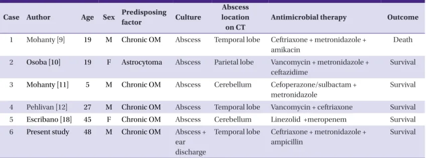
Enterococci are generally regarded as the normal intestinal flora [6]. Over the past 2 decades, however, enterococci have been identified as one group of pathogens responsible for sev- eral nosocomial infections; E. faecalis and E. faecium account for up to 90% of the responsible clinical isolates [6, 7]. In a pre- vious study, streptococci were identified as the most common bacteria identified in cultures in patients with brain abscesses, with E. faecalis and E. faecium accounting for only 5% of all isolates [1, 8]. E. avium is rarely reported as a pathogen in hu- mans. �us far, only 6 cases of brain abscesses caused E. avi- um have been reported, including those reported in the pres- ent study [9–13]. The main characteristics of these cases are summarized in Table 1. To the best of our knowledge, this case report is the first to report a brain abscess caused by E. avium in Korea.
�e pathogenic mechanism of brain abscess formation in- volves either contiguous spread from the focus of infection, hematogenous dissemination to the brain from a distant fo- cus, or head trauma. In most cases, the brain abscess was occured due to contiguous spread from the middle ear, mas-due to contiguous spread from the middle ear, mas- toid cells, or paranasal sinuses. Carpenter et al. reported that more than 40% of brain abscesses in adult patients are oto- genic in origin [14]. In their study, abscesses were located in the temporal lobe (54% of patients), cerebellum (44%), or both locations (2%). In our case review of brain abscess due to E. avium (Table 1), we found that 5 of 6 patients presented with chronic otitis media, and only 1 patient, who was previ- only 1 patient, who was previ-only 1 patient, who was previ- ously diagnosed with astrocytoma, did not present with otitis media [10]. �ese findings suggest that brain abscesses caused
by E. avium infection are predominantly otogenic in origin.
Treatment of brain abscess is done by a team approach; ade-rain abscess is done by a team approach; ade- done by a team approach; ade- a team approach; ade- quate abscess drainage and appropriate antimicrobial therapy are most important for proper treatment. In patients with bac- terial brain abscess, intravenous antimicrobials have has tra- ditionally been administered for 6–8 weeks [15]. Shorter courses (3–4 weeks) of parenteral therapy may be adequate for patients who have undergone surgical excision of the ab- scess. In the present case, follow-up CT showed a significant decrease in the extent of brain abscess, owing to abscess drainage through the communication site. Thus, parenteral antibiotic therapy was continued for 4 weeks. Attending phy- Attending phy-Attending phy- sicians need to consider E. avium infection in such cases, so that effective therapy can be initiated as soon as possible.
In 1984, Collins et al. successfully proposed the renaming of
“Streptococcus avium” to “Enterococcus avium” due to the findings of various DNA-DNA and DNA-ribosomal RNA stud- ies [5]. Although E. faecalis and E. faecium are the 2 most common species responsible for enterococcal infections, our findings and other reports demonstrate an emerging trend of E. avium infection in humans. Only a few clinical manifesta- tions caused by E. avium have been reported thus far, includ- ing bacteremia, endocarditis, osteomyelitis, and splenic ab- scess [4, 16–18]. Majority of these patients additionally presented with immunocompromising conditions or serious systemic disease. However, only 1 patient with E. avium brain abscess was in an immunocompromised state at diagnosis (Table 1, Case 2). Virulence traits are believed to permeate En- terococcus species to various degrees, thereby enhancing their disease-causing abilities [19]. However, factors that influence the virulence potential of E. avium are not well understood.
Table 1. Reported cases of brain abscess caused by Enterococcus avium Case Author Age Sex Predisposing
factor Culture
Abscess location on CT
Antimicrobial therapy Outcome
1 Mohanty [9] 19 M Chronic OM Abscess Temporal lobe Ceftriaxone + metronidazole + amikacin
Death
2 Osoba [10] 19 F Astrocytoma Abscess Parietal lobe Vancomycin + metronidazole + ceftazidime
Survival
3 Mohanty [11] 5 M Chronic OM Abscess Cerebellum Cefoperazone/sulbactam + metronidazole
Survival
4 Pehlivan [12] 27 M Chronic OM Abscess Temporal lobe Vancomycin + ceftriaxone Survival 5 Escribano [18] 45 F Chronic OM Abscess Cerebellum Linezolid +meropenem Survival 6 Present study 48 M Chronic OM Abscess +
ear discharge
Temporal lobe Ceftriaxone + metronidazole + ampicillin
Survival
M, male; F, female; OM, otitis media; CT, computed tomography.
Park SY, et al. • Brain abscess caused by Enterococcus avium www.icjournal.org
338
Recently, Na et al. [4] suggested that E. avium seems to be more virulent than E. gallinarum and E. casseliflavus, but less virulent than E. faecium. Further studies are needed to define the pathogenesis and virulence of E. avium.
Although E. avium is not commonly associated with brain abscesses, it should be considered as an etiological agent in the future, especially in patients diagnosed with chronic otitis media.
References
1. Prasad KN, Mishra AM, Gupta D, Husain N, Husain M, Gupta RK. Analysis of microbialetiology and mortality in patients with brain abscess. J Infect 2006;53:221-7.
2. Hakan T, Ceran N, Erdem I, Berkman MZ, Göktaş P. Bacte- rial brain abscesses: an evaluation of 96 cases. J Infect 2006;52:359-66.
3. Koch S, Hufnagel M, �eilacker C, Huebner J. Enterococ- cal infections: host response, therapeutic, and prophylac- tic possibilities. Vaccine 2004;22:822-30.
4. Na S, Park HJ, Park KH, Cho OH, Chong YP, Kim SH, Lee SO, Sung H, Kim MN, Jeong JY, Kim YS, Woo JH, Choi SH.
Enterococcus avium bacteremia: a 12-yearclinicalexperi- ence with 53patients. Eur J ClinMicrobiol Infect Dis 2012;31:303-10.
5. Patel R, Keating MR, Cockerill FR 3rd, Steckelberg JM.
Bacteremia due to Enterococcus avium. Clin Infect Dis 1993;17:1006-11.
6. Prakash VP, Rao SR, Parija SC. Emergence of unusual spe- cies of enterococci causing infections, South India. BMC Infect Dis 2005;5:14.
7. Jett BD, Huycke MM, Gilmore MS. Virulence of enterococ- ci. Clin Microbiol Rev 1994;7:462-78.
8. Tattevin P, Bruneel F, Clair B, Lellouche F, de Broucker T,
Chevret S, Bédos JP, Wolff M, Régnier B. Bacterial brain abscesses: a retrospective study of 94 patients admitted to an intensive care unit (1980 to 1999). Am J Med 2003;115:
143-6.
9. Mohanty S, Dhawan B, Kapil A, Das BK, Pandey P, Gupta A.
Brain abscess due to Enterococcus avium. Am J Med Sci 2005;329:161-2.
10. Osoba AO, Kutub H, Waliuddin A, Sharab MO. Enterococ- cus avium. An unusual cause of cerebral abscess. Neuro- sciences (Riyadh) 2005;10:297-300.
11. Mohanty S, Kapil A, Das BK, Dhawan B. Enterococcus avi- um cerebellar abscess. Neurol India 2006;54:108-9.
12. Pehlivan Y, Toy MA, Karaoglan I, Namiduru M, Buyukhati- poglu H. Enterococcus avium cerebral abscess. Intern Med 2007;46:1280.
13. Escribano JA, Solivera J, Vidal E, Rivin E, Lozano J. Oto- genic cerebellar abscess by Enterococcus avium, a very rare infectious agent. J Neurol Surg A Cent Eur Neurosurg 2013 Feb 20. [Epub ahead of print].
14. Carpenter J, Stapleton S, Holliman R. Retrospective analy- sis of 49 cases of brain abscess and review of the literature.
Eur J ClinMicrobiol Infect Dis 2007;26:1-11.
15. Mandell, Bennett, and Dolin. Principles and Practice of Infectious Diseases. 7th ed. PA: Elsevier; 2010;1274 16. Mirzoyev Z, Anavekar N, Wilson F, Uslan D, Baddour L,
Mookadam F. Enterococcus avium endocarditis. Scand J Infect Dis 2004;36:876-8.
17. Cottagnoud P, Rossi M. Enterococcus avium osteomyelitis.
Clin Microbiol Infect 1998;4:290.
18. Farnsworth TA. Enterococcus avium splenic abscess: a rare bird. Lancet Infect Dis 2002;2:765.
19. Mundy LM, Sahm DF, Gilmore M. Relationships between enterococcal virulence and antimicrobial resistance. Clin- Microbiol Rev 2000;13:513-22.