성인 혈구탐식증후군 환자들의 원인과 사망에 관련된 요인
김혜인
1· 김신우
1· 장현하
1· 이종명
1· 김능수
1· 권기태
2· 류성열
3· 허지안
4경북대학교 의과대학 내과학교실1, 대구파티마병원 내과2, 계명대학교 의과대학 내과학교실3, 영남대학교 의과대학 내과학교실4
Submitted: December 23, 2011 Revised: February 27, 2012 Accepted: February 28, 2012
Correspondence to Shin-Woo Kim, M.D.
Department of Internal Medicine, Kyungpook National Uni- versity Hospital, 50 Samduk-dong 2-ga, Jung-gu, Daegu 700- 721, Korea
Tel: +82-53-420-6525, Fax: +82-53-426-2046 E-mail: ksw2kms@knu.ac.kr
Infect Chemother 2012;44(2):51-55 pISSN 2093-2340 eISSN 2092-6448
www.icjournal.org
Causes and Risk Factors of Mortality in Adult Patients with Hemophagocytic Syndrome
Background: Hemophagocytic syndrome (HS) is a distinct clinical entity characterized by high fever and hemophagocytosis with histiocytosis in tissue biopsy. We seldom encounter patients who suffer from unexplained, persistent fevers. Although there have been many studies about childhood HS, studies about adult HS are relatively rare. The causes and prognoses of HS in adults were evaluated in this study. We focused on infection-related HS.
Material and Methods: We enrolled 41 adult patients with HS retrospectively from four hospitals in Kyungbuk province and Daegu city. The patients were diagnosed by bone marrow or liver biopsy, either of which showed hemophagocytosis with histiocytosis and had clinical findings consistent with HS. We explored the etiologies, clinical symptoms, laboratory findings, treatments, and outcomes of each case.
Results: The most common cause of HS was infection, such as the Epstein-Barr virus (EBV) or Mycobacterium tuberculosis. Old age and malignancy-associated HS had a poor prognosis. The overall mortality rate was 17.1%. Most patients survived after conservative therapy and the control of underlying diseases, in contrast to previous studies that showed a poor prognosis of infection-associated HS.
Conclusions: A proper investigation is crucial to determine the cause of HS in patients who have unexplained persistent fever and hemophagocytosis with histio- cytosis in their tissue. Cases of infection-related HS are common, but physicians should consider undiagnosed malignancy that may be related to a poor prognosis.
Treatments appropriate to the causes are important for better outcomes in adult HS.
Key Words: Hemophagocytic lymphohistiocytosis, Infection, Prognosis
Hye-In Kim1, Shin-Woo Kim1, Hyun-Ha Chang1, Jong- Myung Lee1, Neung-Su Kim1, Ki-Tae Kwon2, Seong-Yeol Ryu3, and Ji-An Hur4
1Department of Internal Medicine, School of Medi cine, Kyungpook National University, 2Department of Internal Medicine, Daegu Fatima Hospital, 3Depart ment of Inter- nal Medicine, Keimyung University Dongsan Medical Centers, and 4Department of Internal Medicine, Yeung- nam University Hospital, Daegu, Korea
서론
임상에서 불명열은 발열의 원인을 찾는 사람들에게 늘 도전적 주제가 되고 있다[1- 3]. 대개는 검사를 통하여 원인을 밝히는 경우가 많으나 최근 시행된 외국 연구에서 약 10-30%가 원인을 찾지 못한 불명열로 진단되었고[4, 5], 국내 연구에서도 약 10-30%
정도에서는 원인을 찾지 못한다고 보고된 바 있다[6-8]. 혈구탐식 증후군(hemopha- gocytic syndrome, HS)은 원인을 쉽게 찾을 수 없는 지속적인 열을 보이는 경우가 많 아 불명열이나 지속적인 발열의 원인을 찾기 위한 조사를 할 경우 이 질환을 한 번쯤 염
This is an Open Access article distributed under the terms of the Creative Commons Attribution Non-Commercial License (http://creativecommons.
org/licenses/by-nc/3.0) which permits unrestricted non-commercial use, distribution, and reproduction in any medium, provided the original work is properly cited.
Copyright © 2012 by The Korean Society of Infectious Diseases | Korean Society for Chemotherapy
두에 두어야 한다[9-11].
혈구탐식 증후군은 골수, 간 또는 림프절 등에 혈구포식이 발생하면 서 지속적인 발열과 범혈구감소증, 간비종대, 황달, 혈액응고장애 등을 특 징으로 하는 질환이며, 혈구탐식 림프조직구증식증(hemophago cytic lymphohistiocytosis)으로 불리기도 한다[12, 13]. 혈구탐식 증후군은 아 직까지 확립된 치료방법은 없고[14-16], 혈구탐식 증후군의 사망률은 감 염성 질환에 의한 경우 20-42%로 보고되며, 림프종과 같은 종양 및 비감 염성 질환과 관련되어 발생한 경우 거의 100%에 이른다[17].
혈구탐식 증후군은 이처럼 사망률이 높고 확립된 치료방법이 없는 질 환이므로 예후에 대한 분석이 중요할 것으로 생각된다. 국내에 성인의 혈구탐식 증후군의 임상적 특징 및 예후인자에 대한 몇몇 증례보고는 있었으나[18-23], 다수의 성인 환자들을 대상으로 한 연구는 1995년에 16명[24], 1996년에 78명[25]예 외에 보고된 바가 없으며, 이 연구들은 주로 혈구탐식 증후군의 원인이나 임상적 특징에 관한 내용으로 예후인 자에 대한 조사는 기술하지 않고 있다. 이에 저자들은 혈구탐식 증후군 을 진단 받은 41명의 성인 환자를 대상으로 원인 및 임상 증상, 혈액검사 소견, 치료 후의 경과를 분석하여 성인 혈구탐식 증후군의 임상적 특징 과 예후에 대해 파악하고자 하였다.
대상 및 방법
혈구탐식 증후군은 유전적인 원인에 의해 발생하는 일차성 혈구탐 식 증후군과 감염이나 종양 등의 질환에 의한 이차성 혈구탐식 증후군 으로 구분되는데[26], 본 연구는 소아에서 흔히 발생하는 일차성 혈구 탐식 증후군을 제외한 41명의 이차성 혈구탐식 증후군의 성인환자만 을 대상으로 하였다. 2003년 7월부터 2009년 9월까지 총 6년 2개월 동 안 대구, 경북지역의 3차 병원 3곳과 2차 병원 1곳에서 입원환자들의 전 산자료 및 골수조직 검사 데이터베이스 등을 이용하여 자료를 수집하였 고, 혈구탐식 증후군의 진단 기준은 다른 원인이 밝혀지지 않은 7일 이 상의 발열, 간비 종대, 임파선 종대 등의 증상이 있으면서 골수 또는 간, 임파선 조직검사에서 혈구탐식증을 나타낸 경우로 하였으며, 진단 당시 의 임상적 특징, 검사소견, 기저원인질환, 치료 및 경과, 사망원인 등을 후향적으로 조사하였다.
인구학적인 변수로 연령, 성별, 입원기간을 조사하였으며 입원기간 은 환자가 내원한 날로부터 퇴원 또는 사망한 날짜까지로 정의하였다.
원인질환에 대하여 감염, 종양, 자가면역질환, 유전질환으로 구분하였 고 감염의 종류는 바이러스, 세균, 진균, 기생충, 결핵균 등에 대해 조 사하였고, 기저질환, 가족력의 유무에 대해서 조사하였으며 원인질환 의 진단방법은 조직 검사, 혈청학적 검사, 배양검사, 중합효소연쇄반 응(polymerase chain reaction, PCR), 컴퓨터 단층촬영(computer tomography)로 구분하여 조사하였다. 주소(chief complaint), 발진, 간비 종대, 임파선 종대, 신경학적 증상 및 혈액검사 결과를 수집하였고, 이중에서 혈색소, 혈소판, 알부민은 증상 발현 후 치료시작 전 최저의 수 치, 나머지는 증상발현 후 치료 시작 전 최고의 수치를 기록하였다. 치료 방법과 사망여부 및 사망원인에 대하여 조사하였다. 경과관찰기간은
입원일로부터 마지막 추적시점 및 사망까지 걸린 기간으로 하였다.
1. 통계적 분석
기술적 통계를 기본적으로 시행하였으며, 사망과 관계된 인자에 대해 불연속변수에 대해 Chi-square test을, 연속변수에 대해서는 t-test을 단변량분석으로 사용하였다. 사망여부와 관련하여 단변량 분석에서 P 값이 0.2 미만인 변수와 임상적 의미를 고려한 60세 이상의 고령환자에 대하여 Cox 회귀 분석을 시행하였다. Kaplan-Meier 곡선을 그리고 유 의한 변수에 대해 log-rank 검정을 시행하였다. SPSS version 15.0 프로 그램(SPSS for Windows, SPSS, Chicago, IL, USA)을 이용하였으며, 통 계적 의미부여는 P 값 <0.05로 하였다.
결과
1. 인구학적 특성과 임상적 특징
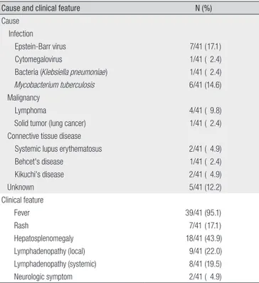
환자들의 평균 나이는 51.4±15.5세 였으며, 남자 환자가 30명(73.2%) 이었고, 여자 환자가 11명(26.8%) 이었으며 평균 입원기간은 24.4±18.1 일 이었다. 골수조직 검사에서 혈구탐식증을 보여 진단된 환자는 40명 (97.6%) 이었으며 1명은 골수와 간조직에서 모두 혈구탐식증을 나타 내었다. 원인질환에 대하여 11명(44.0%)은 조직검사, 7명(28.0%)은 혈 청검사, 1명(4%)은 배양 검사, 4명(16%)은 중합효소연쇄반응, 2명(8%) 은 전산화 단층촬영으로 진단 되었으며, 조직검사로 원인 규명을 한 경 우가 가장 많았다. Ebstein-Barr virus (EBV) 감염은 41명 중 24명에서 검사가 시행되었고, 7명(17.1%)은 혈청검사에서 급성 EBV 감염에서 보 이는 항체(EBV VCA IgM) 양성으로, EBV 감염과 연관된 혈구탐식 증 후군으로 진단되었다. 결핵균 감염은 6명(14.6%)에서 확인되었으며 이 중 2명은 폐 결핵, 2명은 임파선 결핵, 2명은 파종성 결핵으로 확인되었 다. 거대세포바이러스(cytomegalovirus, CMV) 감염은 41명 중 14명 에서 검사가 시행되었으며, 1명(2.4%)에서 급성기에 보이는 항체(CMV IgM)의 양성으로 CMV 감염과 연관된 혈구탐식 증후군으로 진단되었 다. 세균성 감염 역시 1명(2.4%)에서 진단되었으며, 원인균은 Klebsiella pneu moniae로 혈액배양에서 확인되었다. 20명(48.7%)의 환자들이 기저질환을 가지고 있었고, 이들 중 7명(35.0%)은 만성 간질환을 동반 하였다. 환자들의 혈구탐식증의 원인 질환 및 임상 증상은 Table 1에 요 약하여 나타내었다. 입원 전 발열의 기간은 평균 14.7±13.0일 이었으며 체온은 평균 39.0±1.2℃이었다.
2. 검사결과
Ferritin이 8,887.9±11,732.0 ng/dL, lactate dehydrogenase가 1,624.0±1355.0 U/L로 증가되어 있었으며 주요한 혈액 검사결과는 Table 2에 요약하여 나타내었다.
3. 치료방법과 경과
25명(61.0%)은 원인질환을 확인하여 그에 대한 치료를 시행할 수 있 었다. 전체 환자의 사망률은 17.1% (7/41)이었으며, 이중에서 혈구탐식
증후군 그 자체에 의한 사망이 5명(5/7, 71.4%), 기저질환의 악화로 인한 사망이 2명(2/7, 28.6%) 이었고, 혈구탐식 증후군 자체로 인해 사망한 환자 중에서 1명(20%)은 급성 신부전, 4명(40%)은 다장기 부전으로 인 해 사망하였다. 전체 환자들의 총 경과관찰 기간은 평균 246.8±344.8 일 이었다. 단변량 분석에서 원인질환 중 EBV 감염 및 결핵은 사망과 유 의한 연관성을 나타내지는 않았다(P=1, P=1) (Table 3). 60세 이상의 고 령은 단변량 분석에서는 통계적으로 유의하지 않았으나(P=0.392), 다 변량 분석에서는 유의하였다(P=0.031). 원인질환으로서 종양은 사망 과 유의한 연관성이 있었다(단변량분석 vs. 다변량분석, P=0.03 vs. P=
0.025). 사망과 관련된 환자들의 임상적 특징은 Table 3, Table 4 에 나타 내었다. Fig. 1에서는 종양 유무에 따른 환자군별 생명곡선을 나타내었 다(log-rank test, P<0.001).
고찰
혈구탐식 증후군은의 진단은 1991년 Henter 등이 진단기준을 제시 한 바 있으나[12], 광범위하게 수용되는 기준은 없으며[13, 27], Henter 등이 제시한 기준이 병리 기전을 명확히 반영하지는 않는 것으로 생각 된다. 또한 실제 임상에서는 범혈구 감소증이나 ferritin, 섬유소원의 상 승이 혈구탐식 증후군에 특이적인 검사 결과는 아니며 sIL-2 receptor 를 측정하는데 제한이 있어 이 진단 기준을 임상에 적용하는 데는 무리 가 있다. 본 연구에서는 혈구탐식 증후군의 주된 증상인 7일 이상의 원 인 불명의 발열, 간비 종대, 임파선 종대 등의 임상 증상을 만족하고 골 수 또는 간, 임파선 조직검사에서 혈구탐식증을 나타내는 경우를 진단 기준으로 삼았으며, 혈구탐식 증후군 외의 다른 대안적 진단을 고려할 필요를 발견하지는 못하였다. 또한 혈구탐식 증후군을 보이는 질환 중 Table 1. Causes and Clinical Features of 41 Patients with Hemophagocytic
Syndrome
Cause and clinical feature N (%)
Cause Infection
Epstein-Barr virus 7/41 (17.1)
Cytomegalovirus 1/41 ( 2.4)
Bacteria (Klebsiella pneumoniae) 1/41 ( 2.4)
Mycobacterium tuberculosis 6/41 (14.6)
Malignancy
Lymphoma 4/41 ( 9.8)
Solid tumor (lung cancer) 1/41 ( 2.4)
Connective tissue disease
Systemic lupus erythematosus 2/41 ( 4.9)
Behcet's disease 1/41 ( 2.4)
Kikuchi's disease 2/41 ( 4.9)
Unknown 5/41 (12.2)
Clinical feature
Fever 39/41 (95.1)
Rash 7/41 (17.1)
Hepatosplenomegaly 18/41 (43.9)
Lymphadenopathy (local) 9/41 (22.0)
Lymphadenopathy (systemic) 8/41 (19.5)
Neurologic symptom 2/41 ( 4.9)
Table 2. Laboratory Findings Regarding 41 Patients with Hemophagocytic Syndrome Mean±standard deviation
WBC (/mm3) 5,497±4,657
Absolute neutrophil count (/mm3) 3,598±3,978
Hemoglobin (g/dL) 9.2±2.3
Platelet (/mm3) 65,907±75,830
Aspartate aminotransferase (IU/L) 223±224
Alanine aminotransferase (IU/L) 126±140
Total bilirubin (mg/dL) 3.6±9.7
Direct bilirubin (mg/dL) 2.7±8.6
Ferritin (ng/dL) 8,888±11,732
Fibrinogen (mg/dL) 260±138
Lactate dehydrogenase (U/L) 1,624±1,355
Triglyceride (mg/dL) 191±102
Table 3. Patients Characteristics Associated with Death of 41 Cases with Hemo- phagocytic Syndrome by Univariate Analysis
ORa (95% CIb) P-value
Age (>60 yrs) 2.7 (0.51-14.06) 0.392
Male Sex N.D.c 0.159
Ebstein Barr virus 1.17 (0.09-15.46) 1
Tuberculosis 0.93 (0.09-9.51) 1
Malignancy 11.63 (1.47-92.14) 0.03
Chemotherapy (No.) 1.33 (0.13-13.23) 1
Steroid medication (No.) 1.42 (0.27-7.34) 1
Absolute neutrophil count (/mm3) <1,000 1.07 (0.18-6.52) 1
Hemoglobin (g/dL) <8 0.92 (0.15-5.57) 1
Platelet (/mm3) <100,000 N.D.c 0.309
Fibrinogen (mg/dL) ≥350 0.87 (0.17-4.52) 1
aOdds ratio, bconfidence interval, cnot defined
Figure 1. Survival curve of 41 patients with hemophagocytic syndrome (log-rank test between malignancy group vs. non-malignancy: P<0.001).
Table 4. Patients Characteristics Associated with Death of 41 Cases of Hemophagocytic Syndrome Multivariate Analysis by Cox Regression Hazard Model
Parameter P-value Hazard Ratio 95% Hazard Ratio limit
Age (≥60yr) 0.031 6.36 1.18-34.24
Male sex 0.965 N.D.a N.D.a
Malignancy 0.025 6.55 1.27-33.95
anot defined
악성 조직구증가증(malignant histiocytosis)은 골수 등에서 다른 조 직 소견(식세포 단일클론의 심한 증식 소견)을 보이며 조직구 분화도에 따라 특수한 표지자를 나타내는 종양세포를 확인함으로써 진단되므로 [28, 29], 본 증례들에 포함되지 않는다고 판단된다.
성인을 대상으로 한 본 연구에서 혈구탐식 증후군의 원인으로서 기 존에 알려진 바와 같이 감염이 가장 흔하였으며, 두번째로 악성 종양이 많았다. 1996년 Lee 등이 발표한 78명의 혈구탐식 증후군 환자를 대상 으로 한 연구에서도 43명(55.1%)에서 악성질환을 동반하거나 감염증 이 발견되었다[25]. 그러나 주목할 만한 점은, 일본의 한 연구에서 799 명의 혈구탐식 증후군 환자들 중 단지 2명만이 결핵으로 인한 것이었고 [30, 31], 1998년 감염 관련 혈구탐식 증후군 33예 중에서도 바이러스 와 세균감염이 흔한 원인이었으나 결핵의 빈도는 2예(6.1%)에 지나지 않은데 반해[32], 본 연구에서는 결핵이 감염에 의한 혈구탐식 증후군 의 원인 중 두번째로 흔하게 발견되었다는 것이다. 이차성 혈구탐식 증 후군의 주된 치료는 기저 질환을 치료하는 것으로, 본 연구결과에서 결 핵과 같이 완치가 가능한 원인이 적지 않다는 점을 감안할 때 혈구탐식 증후군의 원인 규명이 예후 향상에 중요함이 강조되어야 하겠다.
혈구탐식 증후군의 임상증상은 발열과 비장비대가 흔하며, 간비대, 임파선 비대, 황달, 피부발진 등의 증상을 동반한다고 알려져 있다[12, 13, 33]. 본 연구에서도 다른 문헌에서와 같이 대다수의 환자에서 발열 과 간비 종대가 가장 흔한 증상 이었으며, 피부발진(17.1%) 및 신경학적 증상(4.9%)은 이전의 연구보다 다소 흔하게 나타났다. 혈액 검사에서는 특히 LDH 및 ferritin의 상승이 현저하였다. 그러나 이러한 혈액 검사 결 과들은 진단 기준에 포함되기도 하나, 진단에 특이적인 요소는 아니므 로 조직 검사 등을 확정적 검사의 필요성을 판단하는 한 지표로 사용하 는 것이 좋을 것으로 생각된다. 특히 ferritin의 증가는 다른 보고에서도 진단의 단서가 될 수 있음을 시사하였다[34, 35].
혈구탐식 증후군의 진단 되고, 원인 질환이 밝혀지면 이에 대한 적절 한 치료를 시작해야 한다[25]. 감염에 의한 혈구탐식 증후군은 대개 선 행 감염의 조절과 대증치료로 60-70%에서 회복이 되나, EBV와 연관된 경우는 거의 대부분 사망한다고 보고되고 있다[13, 33, 36, 37]. 이 연구 에서는 EBV 연관 혈구탐식 증후군이 가장 흔하였고, 사망률이 이전의 연구보다 높지는 않으나 증례의 수가 적어 판단에 제한점이 있다. 2003 년 Dhote 등의 연구에서는 혈색소 수치의 감소, 혈소판 수치의 감소, 높 은 ALP, 빌리루빈, ferritin 및 높은 연령 등이 사망률의 증가와 관련이 있 는 것으로 보고되었다[38]. 반면 본 연구에서는 60세 이상의 고령 외에 도 악성 종양과 연관된 경우 사망률이 높은 것으로 나타났는데, 고령 또 는 악성 종양을 진단 받은 환자는 전신 상태가 좋지 못하고 면역저하가 동반되어 있을 가능성이 있으므로, 이러한 요소가 사망률 증가에 영향 을 미친 것으로 생각된다.
본 연구는 또한 대상자의 수가 많지 않은 후향적 연구이며, 성인 혈구 탐식 증후군의 발생 빈도자체가 매우 적어 소규모의 환자들을 대상으 로 분석한 결과로서 이것을 일반화 하기는 어려움이 있으며, 앞으로 혈 구탐식 증후군 환자들에 임상적 특징에 대해서 전향적 연구나 조사가 필요할 것으로 생각된다.
결론적으로, 감염과 악성 종양이 혈구탐식 증후군의 가장 흔한 원인
이며 이들의 사망률이 높다는 것을 알 수 있었다. 우리 연구에서도 EBV 감염 연관 혈구탐식 증후군이 가장 흔한 원인이었고, 두번째로는 결핵 이 많은 원인을 차지 하였다. 사망률은 이전 보고와 같이 높지는 않았으 며, 악성 종양관 연관된 혈구탐식 증후군 환자와 고령의 환자들에서 높 은 사망률과 관련성이 있었다. 악성 종양, 고령이 아닌 성인 혈구탐식 증 후군의 경우 원인에 대한 정확한 조사와 이에 따른 적절한 치료를 통해 좀 더 개선된 임상경과를 유도할 수 있을 것으로 생각된다.
References
1. Petersdorf RG, Beeson PB. Fever of unexplained origin: report on 100 cases. Medicine (Baltimore) 1961;40:1-30.
2. Cunha BA. Fever of unknown origin: clinical overview of classic and current concepts. Infect Dis Clin North Am 2007;
21:867-915.
3. Cunha BA. Fever of unknown origin: focused diagnostic approach based on clinical clues from the history, physical examination, and laboratory tests. Infect Dis Clin North Am 2007;21:1137-87.
4. Zhiyong Z, Bingjun L, Xiaoju L, Xinjian F, Ping F, Wenya W.
Fever of unknown origin: a report from China of 208 cases. Int J Clin Pract 2003;57:592-6.
5. Mourad O, Palda V, Detsky AS. A comprehensive evidence- based approach to fever of unknown origin. Arch Intern Med 2003;163:545-51.
6. Kee SY, Jo YM, Kim JY, Choi WS, Jeong HW, Jung SJ, Kim SB, Hyun JJ, Hwang BY, Cheong HJ, Kim WJ. Etiology of adult patients with fever of unknown origin (FUO) observed in a university hospital in Korea from 1998-2003. Infect Chemo- ther 2005;37:127-32.
7. Kim YK, Kim MS, Lee KS, Huh AJ, Yeom JS, Hong SK, Chang KH, Song YG, Kim JM. A comparison of causes of fever of unknown origin between the 1980s and the 1990s. Korean J Med 2001;61:546-52.
8. Oh MD, Baik KR, Song YW, Choe KW. A clinical study on 55 patients with fever of undetermined origin. Korean J Infect 1993;25:1-8.
9. Levy L, Nasereddin A, Rav-Acha M, Kedmi M, Rund D, Gatt ME. Prolonged fever, hepatosplenomegaly, and pancytopenia in a 46-year-old woman. PLoS Med 2009;6:e1000053.
10. Han B, Yang Z, Yang T, Gao W, Sang X, Zhao Y, Shen T. Diagno- stic splenectomy in patients with fever of unknown origin and splenomegaly. Acta Haematol 2008;119:83-8.
11. Albrecht H, Schäfer H, Stellbrink HJ, Greten H. Epstein-Barr virus--Associated hemophagocytic syndrome. A cause of fever of unknown origin in human immunodeficiency virus infection. Arch Pathol Lab Med 1997;121:853-8.
12. Henter JI, Elinder G, Ost A. Diagnostic guidelines for hemo- phagocytic lymphohistiocytosis. The FHL Study Group of the
Histiocyte Society. Semin Oncol 1991;18:29-33.
13. Fisman DN. Hemophagocytic syndromes and infection.
Emerg Infect Dis 2000;6:601-8.
14. Risdall RJ, McKenna RW, Nesbit ME, Krivit W, Balfour HH Jr, Simmons RL, Brunning RD. Virus-associated hemopha- gocytic syndrome: a benign histiocytic proliferation distinct from malignant histiocytosis. Cancer 1979;44:993-1002.
15. Chandra P, Chaudhery SA, Rosner F, Kagen M. Transient histiocytosis with striking phagocytosis of platelets, leuko- cytes, and erythrocytes. Arch Intern Med 1975;135:989-91.
16. Risdall RJ, Brunning RD, Hernandez JI, Gordon DH. Bacteria- associated hemophagocytic syndrome. Cancer 1984;54:2968- 72.
17. Lee GR, Bithell TC, Foerster J, Athens JW, Lukens JN, eds.
Wintrobe's clinical hematology. 9th ed. Philadelphia: Lea &
Febiger; 1993;2:2189-91.
18. Kang SK, Chun SB, Jung M, Ryoo YK, Choi KS, Kim JW. A case of infection-associated hemophagocytic syndrome asso- ciated with CMV mononucleosis. Infect Chemother 1993;25:
387-91.
19. Kim HW, Choi BS, Kim JH, Shin YM, Lee SJ, Kim SR, Jun JB. A case of death due to hemophagocytic lymphohistiocytosis accompanied by scrub typhus. Infect Chemother 2010;42:266- 70.
20. Kim JW, Yoo JH, Park YH, Kahng JM, Kim JS, Song DS, Chung HY, Choi JH, Kang JH, Shin WS, Kang MW. Epstein-Barr virus- associated hemophagocytic syndrome confirmed by in situ hybridization : case report and review of the related literature.
J Korean Soc Chemother 1998;16:369-75.
21. Oh JH, Park JH, Hwang SY, Lee SH, Kim SL, Kim JY, Lee CH, Chung JS, Lee EY, Cho KJ. A Case of Kikuchi's disease ac- companied by hemophagocytic lymphohistiocytosis. Infect Chemother 2004;36:185-8.
22. Ryoo HO, Kim GH, Koo DS, Chun CH. A case of infection- associated hemophagocytic syndrome (IAHS). Infect Chemo- ther 1993;25:71-7.
23. Yang CW, Kwak SM, Lee CM, Park ES, Chae SL, Shin WS, Kang MW, Bang BK. A case of Rickettsia-associated pancytopenia and hemophagocytosis. Infect Chemother 1992;24:215-20.
24. Kim IH, Lee JT, Ahn JS, Jang YH, Heo DS, Bang YJ, Park S, Kim BK, Cho HI, Kim NK. Reactive hemophagocytic syndrome: a clinical study of 16 cases. Korean J Hematol 1995;30:397-405.
25. Lee SY, Chi HS. Hemophagocytic histiocytosis: analysis of underlying causes and hematological findings. Korean J Clin Pathol 1996;16:435-46.
26. Yoon HJ, Hwang SH, Park YW, Kim YA, Lee SS. Successful treatment with intravenous immunoglobulin and etoposide in a systemic lupus erythematosus patient with hemo-
phagocytic syndrome. Korean J Med 2006;70:S363-7.
27. Larroche C, Mouthon L. Pathogenesis of hemophagocytic syndrome (HPS). Autoimmun Rev 2004;3:69-75.
28. Shukla N, Kobos R, Renaud T, Teruya-Feldstein J, Price A, McAllister-Lucas L, Steinherz P. Successful treatment of refractory metastatic histiocytic sarcoma with alemtuzumab.
Cancer 2011 [In press].
29. Schmidt D. Malignant histiocytosis. Curr Opin Hematol 2001;
8:1-4.
30. Su NW, Chen CK, Chen GS, Hsieh RK, Chang MC. A case of tuberculosis-induced hemophagocytic lymphohistiocytosis in a patient under hemodialysis. Int J Hematol 2009;89:298- 301.
31. Ishii E, Ohga S, Imashuku S, Yasukawa M, Tsuda H, Miura I, Yamamoto K, Horiuchi H, Takada K, Ohshima K, Nakamura S, Kinukawa N, Oshimi K, Kawa K. Nationwide survey of hemophagocytic lymphohistiocytosis in Japan. Int J Hematol 2007;86:58-65.
32. Ryu DR, Yeom JS, Chang KH, Hong SK, Park YS, Choi YH, Song YG, Yang WI, Yoo NC, Hahn JS, Kim JM. Two cases of infection- associated hemophagocytic syndrome with review of lite- rature. Korean J Infect Dis 1998;30:470-7.
33. Jeong JI, Park CS, Jeon WJ, Chae HB, Park SM, Yoon SJ, Shin KS. A case of viurs-associated hemophagocytic syndrome.
Korean J Med 2008;75:322-6.
34. Esumi N, Ikushima S, Hibi S, Todo S, Imashuku S. High serum ferritin level as a marker of malignant histiocytosis and virus- associated hemophagocytic syndrome. Cancer 1988;61:2071- 6.
35. Fardet L, Coppo P, Kettaneh A, Dehoux M, Cabane J, Lambotte O. Low glycosylated ferritin, a good marker for the diagnosis of hemophagocytic syndrome. Arthritis Rheum 2008;58:1521- 7.
36. Chen RL, Lin KH, Lin DT, Su IJ, Huang LM, Lee PI, Hseih KH, Lin KS, Lee CY. Immunomodulation treatment for childhood virus-associated haemophagocytic lymphohistiocytosis. Br J Haematol 1995;89:282-90.
37. Janka G, Imashuku S, Elinder G, Schneider M, Henter JI.
Infection- and malignancy-associated hemophagocytic syn- dromes. Secondary hemophagocytic lymphohistiocytosis.
Hematol Oncol Clin North Am 1998;12:435-44.
38. Dhote R, Simon J, Papo T, Detournay B, Sailler L, Andre MH, Dupond JL, Larroche C, Piette AM, Mechenstock D, Ziza JM, Arlaud J, Labussiere AS, Desvaux A, Baty V, Blanche P, Schaeffer A, Piette JC, Guillevin L, Boissonnas A, Christoforov B. Reactive hemophagocytic syndrome in adult systemic disease: report of twenty-six cases and literature review.
Arthritis Rheum 2003;49:633-9.