. - .
366 Chapter 6: How Cells Read the Genome: From DNA to Protein FR
An
IVa
()f
dill
One mati
StfI
and
It.'m
trail
\crH.
sag-c I a tr
s}JIlI
differ for h.
amin diary lhat a
J
gnJUI alc4
\(J \
hltllld a'dull
St.'COIl
p/wrc nude acid0
Ih afc\\' ofmil Ih('~i s ,lI1dIt i dalemi
~CA
':iCC GCG
GC~
Summary
Given the importance of nuclear subdomains in RNA processing, it might have been expected that pre-mRNA splicing would occur in a particular location in the nucleus, asitrequiresnUlllerous RNA and protein components. Ilowcver, the assembly of splicing components on pre-mRNA is co-transcriptional; thus, splicing must occur al manylocations along chromosomes. Allhough atypical mammalian cell may be expressing on the order of 15,000 genes, transcription and RNA splicing may be localized to only several thousand sites in the nucleus.
These sites themselves are highly dynamic and probably result from the associ- ation of transcription and splicing components to create small "assembly lines"
with a high local concentration of these components. Interchromatin granule clusters-which comain stockpiles of RNA-processing components-are often observed next to sites of transcription, as though poised to replenish supplies.
Thus, the nucleus seems to be highly organized into subdomains, with snR Ps, snoHNPs, and other nuclear components moving between them in an orderly fashion according to the needs of the cell (see Figure 6-48; also see Figure 4-69).
Before the synthesis of a particular protein call begin, the corres/JOlu!illg mR A molecule mustbe producedbytranscription. Bacteria contain a single type of R :A polymerase (the enzyme that carries ollt the transcription ofDNA i11l0 UNA). An mUNA molecllle is produced when this enzyme initiates transcriptionata promoter. synthe- sizes the UNA bychain elongation, stalls transcriptionat a terminfllor. al/d releases voth the DNA template and the completed mRNA molecule. III eucmyotic cells, the process oftranscription is rnllch more complex, and there are three UNA polymerases- polymerase I, II, and III-llwt are related ello/Ulionarily to one another and co the vac·
terial polymerase.
RNA polymerase/Isy11lhesizes eucaryotic mRNA. This enzyme requires a series of allditional proteins, the general transcription factors, to initiate transcription on a purifiell DNA template, and still more proteins (including chromatin-remodeling complexes and histone-modifying enzymes) to initiate transcription on its chromatin templates inside the cell.
During the elongation phase of transcription, the nascent UNA undergoes three types of processing el'ents: a special tludeotide is added to its S enll (capping), itltroll sequellces are removed from tile ,-nilldle of the RNA molecllie (splicing), and the:.r end of the UNA is generated (cleavage anll polyadenyilitiofl). Each of these processes is ini·
titHed vy proteins that trauel along with RNA polymerase II by bil/lling to sites on its 101/g, e.xtended C-terminal tail. Splicing is unusual i" thm many ofits key steps are car·
ried alit by specializel/ RNA molecules rather than proteins. Properly processed mHNAs are passed through nuclear pore complexes into the cytosol, where they are trans/med i",o protein.
For some genes. UNA is tI,e final product. In eucaryotes. these genes are usually transcribed by either RNA polymerase I or RNA polymerase III. RNA polymerase I makes the ribosomal RNAs. After their synthesis as a large precursor. the rRNAs are chemically modified, cleaved, and assembled into the two ribosomal subunits ill the nucleoills-a distinct sllbnuclear structure that also helps to process some srnal/er UNA-protein cornplexes in the cell. Additional subnuclear structures (including Cajlll bodies and interchromatin granule clusters) are sites where componellis involved in UNA processing are assembled. storell, and recycled.
FROM RNA TO PROTEIN
In the preceding section we have seen that the final product
or
some genes is an IlNA molecule itself. such as those present in thc snll Ps and in ribosomcs.Jlowever, most genes in a cell produce mHNA molecules thai serve as interme- diaries on the pathway to proteins. In this section we examine how the cell COI1-
verts the information carried in an mRNA molecule into a protein molecule. This feat of translation was a focus of attention of biologists in the late 1950s, when it
RNA TOPROTEIN 367
po"l'das the "coding problem": how is the information in a linear sequence t:'Qtides in R A translated into the linear sequence of a chemically quite 1~t of units-the amino acids in proteins? This fascinating question ted great excitement among scientists at the time. Ilere was a cryp·
<et up by nature that, after more than 3 billion years of evolution, could . besol\'ed by one of the products of e\ollltion-human beings. And
\'d. not only has the code been cracked step by step, but in the year 2000 the nure of the elaborate machinery by which cells read this code-the ribo-
was finally revealed in atomic detail.
AnmRNA
Sequence Is Decoded in Sets of Three Nucleotidesce an mRNA has been produced bytranscription and processing, the infor- 'Kinpresent in its nucleotide sequence is used to synthesize a protein. Tran-
?lIonis simple to understand as a means of information transfer: since DNA R\Aare chemically and struclllrally similar, the 0 A can act as a direct late for the synthesis of RNA by complementary base-pairing. As the term iprioll signifies. it is as ifa message written out by hand is being con- ,say. into a rypewrillcn lext. The language itself and the form of lhe mes-
" not change. and the symbols used are closely related.
. :omrast, Ihecon\Crliion oflhe information ill H A into prolein represents rranslation of the information into another language that uses quite different '!areover. since there are only 4 differenl nucleotides in mR A and 20 en! types of amino acids in a protein, this lranslation cannot bc accounted
~adirect one-to-one correspondence between a nucleotidc in H A and an
!lOacidin protein. The nucleotide sequcnce of a gene, lhrough lhe intefme- ltr)'ofmRNA, is translated into the amino acid scquence of a prolein by rules tarelmown as the genetic code. This code was deciphered in the early 1960s.
The sequenceof nucleotides in the mHNA molecule is read in consecutive upsof three. R A is a linear polymer of four different nucleotides, so there 4x4x 4=64 possible combinations of three nucleotides: the triplets AAA,
~ WG, and so on. Ilowever, only 20 different amino acids are commonly in proteins. Either some nucleotide triplets are never used. or the code is ,.jant and some amino acids are specified by more than one triplet. The poSSibility is, in fact. the correct one, as shown by the completely deci- genetic code in figure 6-50. Each group of three consecutive lides in R A is called a codon, and each codon specifies either one amino
t,-a stopto the translation process.
[higenetic code is used lIniversally in all present-day organisms. Although ,light dilTerences in the code have been found, these are chiefly in the DNA
~I:lochondria. Mitochondria have their own transcription and protein syn- lIsystems that operate quite independently from those of the resl of the cell,
;ditis understandable (hal Iheir slllall genomcs have been ablc 10 accollllllo- minor changestothe code (discussed in Chapter 14).
>{,A lIUA AGC
>{" UUG AGlI
(GA GGA CUA CCA lICA ACA GUA
(G( GGC ALIA CllC CCC UCC ACC GUC UAA
(GG GAC AAC UGC GAA CAA GGG CAC AlIC CllG AAA UlIC CCG lICG ACG lIAC GUG UAG
(GU GAU AAU UGlI GAG CAG GGU CAU AUlI CUU AAG AUG UUU CCU UCU ACU UGG UAU GlIU lIGA
Asp Asn Cy> Glu Gin Gly H, Ie Leu Ly> M~t Pile P<o 50, Th' T,p Ty' Val .top
0 N C E Q G H L K M f P S W y V
Figure 6-50 The genetic code. The standard one-letter abbreviation for each amino acid is presented below its three-letter abbreviation (see Panel3-1,pp.128 - 129,for the full name of each amino acid and its structure). By convention, coelom are always written with the 5'-terminal nucleotide to the left. Note that most amino acids are represented by more than one codon, and that there are some regularities in the set of codons that specifies each amino aCid. Codons for the same amino acid tend to contain the same nucleotides at the first and second positions, and vary at the third position. Three codons do not specify any amino acid but act as termination sites (stop codons), signaling the end of the protein-coding sequence.
One codon AUG-acts both as an initiation codon, signaling the start of a protein-coding message, and also as the codon that specifies methionine.
368 Chapter 6: How Cells Read the Genome: From DNA to Protein FRO,
tRW
LikeI
allowl
mera~
larger additi intror pre-rr uses ;:
Trimn its clo
crss('<
ity-co , I maWr cleoti(
sho\vl produ pairin ate mI racy""
regie vvith gle-s acid
~
is, se This amm codo tRNA ing0
wahL why' nucl(
to fit mole' one s amm
v.,
Thr LeuFigure6-51The three possibleread~
frames in protein synthesis. In the process of translating a nucleotide sequence(blue)into an amino acid sequence(red),the sequence of nucleotides in an mRNA molecule is from the 5' end to the 3' end in consecutive sets of three nucleotides.
principle, therefore, the same RNA sequence can specify threecomplet~
different amino acid sequences, depending on the reading frame. In reality, however, only one of these reading frames contains the actual message.
3 ~~~~[' -Gln-A,g--Tyr-H
s'
~~~~
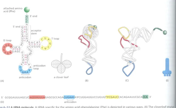
aclover leaf anticodon
A 3'end C C A 5'end G~Ce"G
1
GJ=1C acceptor
D loop
g~~
stem e e u A T loop;; ~~~~_ G
CUCG CCUGUG T C
lllil III. U 'II
GAGCG
8
e"'G'"
e"'GA~U
G
"e
Ap 'I' anticodon A loop UGAAY
~
attached amino acid (Phe)
The coelons in an mRNA molecule do not direclly recognize the amino acids they specify: the group of three nucleotides does not, for example, bind directly to the amino acid. Rather, the translation of mRNA into protein depends on adaptor molecules thaI can recognize and bind both to the codon and, at another site on their surface,tothe amino acid. These adaptors consist of a SCi of small RNA molecules known as transfer RNAs (tRNAs), each about 80 Ilucleotides in length.
We saw earlier in this chaptcr thaI RNA molecules can fold into precise three-dimensional structures, and the tHNA molecules provide a striking exam·
pie. Four shon segments of the folded tRNA are double-helical, producing a molecule that looks like a cloverleaf when drawn schematically (Figure1>-52).
For example, a5'-GCUC-3'sequence in one pan of a polynucleotide chain can form a relatively strong association with a 5'·GAGC-3' sequence in another region of the same molecule. The cloverleaf undergoes further foldingto form a compact L-shaped structure that is held together by additional hydrogen bonds between different regions of the molecule.
Two regions of unpaired nucleotides situated at either end of the L-shaped molecule are crucial to the hmctioll of tRNA in protein synthesis. One of these
tRNA Molecules Match Amino Acids to Codons in mRNA
In principle, an RNA sequence can be translated in anyone of three differ- ent reading frames, depending on where the decoding process begins (Figure 1>-51). Ilowever, only one of the three possible reading frames in an mRNA encodes the required protein. We see later how a special punctuation signal at the beginning of each RNA message sets the correct reading frame at the start of prOlcinsynthesis.
(A) (8) (C) IE)
5' GCGGAUUUAGCU (D)
AGCGCCAGACUGAAY,A'I'CUGGAGGUCCUGUGPIICGAUCCACAGAAUUCGCA C 3'
~
anticodon
Figure6-52A tRNA molecule. A tRNA specific for the amino acid phenylalanine {Ph e) is depicted in various ways. (A) The cloverleaf structu showing the complementary base-pairing(red lines)that creates the double-helical regions of the molecule. The anticodon is the sequence three nucleotides that base-pairs with a codon in mRNA. The amino acid matching the codon/anticodon pair is attached at the 3' end ofthe tRNA. tRNAs contain some unusual bases, which are produced by chemical modification after the tRNA has been synthesized. For example, bases denoted\II(pseudouridine-see Figure6-43)and 0 (dihydrouridine-see Figure6-55)are derived from uracil. (8 andC)Views ofthe L-shaped molecule, based on x-ray diffraction analysis. Although this diagram shows the tRNA for the amino acid phenylalanine, all othert have similar structures. <CGCA> (D) The linear nucleotide sequence of the molecule, color-coded to match (A), (8), and (C). (E) The tRNAicoo use in this book.
RNATO PROTEIN 369
tRNA
u
d
n
bforms the anticodon, a set of three consecutive nucleotides that pairs the complementarycodon in an mR A molecule. The other is a short sin- -stranded region at the 3' end of the molecule; this is the site where the amino 'dthat matches the codon is attached to the tRNA.
\\'eha\'e seen in the previous sectionthatthe genetic code is redundant; that
~,raldifferent codons can specify a single amino acid (see Figure6-50).
redundancy implies eitherthatthere is more than one tRNA for many of the acids or that some tRNA molecules can base-pair with mOTethan one
rJ.In fact, bothsituations occur. Some amino acids have more than one
\and some tR As areconstructed so that they require accurate base-pair- ,only at the first two positions of the codon and can tolerate a mismatch (or bb/e)at the third position (Figure6-53). This wobble base-pairing explains so many of the alternative codons for an amino acid differ only in their third
"tide (see Figure6-50). In bacteria, wobble base-pairings make it possible t the20amino acids to their61 codons with as few as 31 kinds of tllNA 1fUIe5.The exact number of different kinds of til As, however, differs from lpecies10 the nexl. For example, humans have nearly 500 tllNA genes but.
19them, only 48 different anticodons are represented.
tlINAs Are Covalently Modified Before They Exit from the Nucleus molt other eucaryotic IlNAs. tRNAs are covalently modified before they are
ed to exit from the nucleus. Eucaryotic tllNAs are synthesized by RNA poly- Ill, Both bacterial and eucaryotic til As are typically synthesized as precursor tRNAs, which are then trimmed to produce the mature tRNA. In ion, some tRNA precursors (from both bacteria and eucaryotes) cOlllain mns that must be spliced out. This splicing reaction differs chemically from . ·mRNAsplicing; rather than generating a lariat intermediate, tRNA splicing a ,ut·and·paste mechanism that is catalyzed by proteins (Figure 6-54).
'Oingand splicing both require the precursor tRNAtobe correctly folded in .erleafconfiguration. Because misfolded tRNA precursors will not be pro- properly, the trimming and splicing reactions are thought to act as qual- Jnuol steps in the generation of tR As.
llItRNAs are modified chemically-nearly I in 10 nucleotides in each tureIRNAmolecule is an altered version of a standard G, U, C, or A ribonu- lide, Over50 different types of tRNA modifications are known; a few are 'inFigure6-55. Some of the modified nucleotides-most notably inosine, ICedby the deamination of adenosine-affect the conformation and base- Igofthe anticodon and thereby facilitate the recognition of the appropri- R\Acodon by the tR A molecule (see Figure6-53). Others affect the accu- .ith which the tllNA is attached to the correct amino acid.
5'
bacteria
mRNA 5'
wobble position
3'
ure
e of
he ,the e tRNA<
.con we
Figure 6-53 Wobble base-pairing between codons and anticodons. If the nucleotide listed in the first column is present at the third, or wobble, position of the codon, it can base-pair with any of the nucleotides listed in the second column. Thus, for example, when inosine(I)is present in the wobble position of the tRNA anticodon, the tRNA can recognize anyone of three different codons in bacteria and either of two codons in eucaryotes.
The inosine in tRNAs is formed from the deamination of guanine (see Figure 6-55), a chemical modification that takes place after the tRNA has been synthesized. The nonstandard base pairs, including those made with inosine, are generally weaker than conventional base pairs. Note that codon-anticodon base pairing is more stringent at positions 1 and 2 of the codon: here only conventional base pairs are permitted. The differences in wobble base-pairing interactions between bacteria and eucaryotes presumably result from subtle structural differences between bacterial and eucaryotic ribosomes, the molecular machines that perform protein synthesis. (Adapted fromC.Guthrie andJ.Abelson, in The Molecular Biology of the YeastSaccharomyces:Metabolism and Gene Expression, pp. 487-528. Cold Spring Harbor, New York: Cold Spring Harbor Laboratory
Press, , 982,)
wobble codon possible base anticodon bases
U A,G,orl
e GorI
A Uor I
G eorU
eucaryotes
wobble codon possible base anticodon bases
U A,G.orl
C Gor I
A U
G C
370 Chapter 6: How Cells Read the Genome: From DNA to Protein
Figure6-54Structure of a tRNA-spr endonuclease docked to a precursex tRNA. The endonuclease (a four-subl..
enzyme) removes the tRNA intron (bl A second enzyme, a multifunctional ligase (not shown), then joins the two tRNA halves together. (Courtesy of Li,Christopher Trotta and John Abel
FRON
2
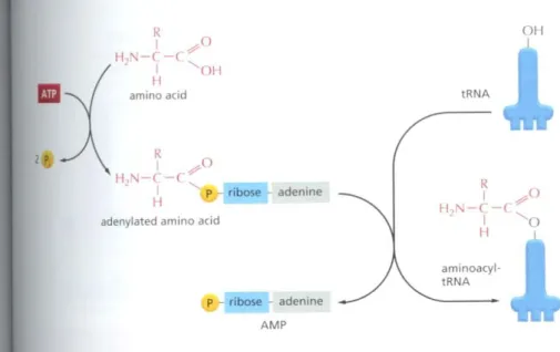
Specific Enzymes Couple Each Amino Acid to Its Appropriate tRNA Molecule
We have seen that, to read the genetic code in DNA, cells make a series of dif- ferent tRNAs. We now consider how each IRNA molecule becomes linked10Ihe one amino acid in 20 that is its appropriate partner. Recognition and attach- ment of the correct amino acid depends on enzymes called aminoacyl-tRNA synthetases, which covalently couple each amino acid to its appropriate set of tR A molecules (Figure6-56and Figure6-57).Mosl cells have a different syn- thetase enzyme for each amino acid (that is, 20 synthetases in all); one attaches glycine to all tRNAs that recognize codons for glycine, another attaches alanine toall tRNAs that recognize codons for alanine. and so all. Many bacteria, how- ever, have fewer than 20 synthetases, and the same synthetase enzyme is responsible for coupling morc than one amino acid to the appropriate lRNAs.
In these cases, a single synthetase places (he identical amino acid on two dif- ferent types of tRNAs. only one of which has an anticodon thaI matches the amino acid. A second enzyme then chemically modifies each "incorrectly"
attached amino acid so that it now corresponds to the anticodon displayed by its covalenlly linked tRNA.
1'1 oflhe ofAll and tl synth rI tant il ment ferent IRNA.
synth in the
have~
iment act ~l
specil nucle amll11
Editi
NJCO /11
H , ! ' j N
---< ~
/ ( H ,~
N N [H~P ribose
two methyl groups added to G (N,N-dimethyl G)
~
I 0 /H/I N
H 'J
~
0P~
two hydrogens added to U (dihydro U)
Sever;
carrel a01ml rect a
p
sulfur replaces oxygen in U (4-thiouridine)
p
deamination of A (inosine)
Figure6-55A few of the unusual nucleotides found in tRNA mole<ulel.
These nucleotides are producedby covalent modification of a normal nucleotide after it has been incorpor into an RNA chain. Two othertypesol modified nucleotides are shown in R 6-43.In most tRNA molecules about 1 of the nucleotides are modified (see Figure6-52).
fROM RNATO PROTEIN 371
Figure6-56Amino acid activation.
An amino acid is activated for protein synthesis by an aminoacyt-tRNA synthetase enzyme in two steps. As indicated, the energy of ATP hydrolysis is used to attach each amino acid to its tRNA molecule in a high·energy linkage.
The amino acid is first activated through the linkage of its carboxyl group directly toan AMP moiety, forming anadenylated amino acid;the linkage of the AMp, normally an unfavorable reaction, is driven by the hydrolysis of the ATP molecule that donates the AMP. Without leaving the synthetase enzyme, the AMP- linked carboxyl group on the amino acid is then transferred to a hydroxyl group on the sugar at the 3' end of the tRNA molecule. This transfer joins the amino acid by an activated ester linkage to the tRNA and forms the finat aminoacyl-tRNA molecule. The synthetase enzyme is not shown in this diagram.
011
J.
tRNA
R
I 9'0
H,N-,-,
- III J.'O
aminoacyl- tRNA adenine
Pr ribose~ adenine AMP
R
I ,pO
H "1-,-,H
I
'011amino acid
RI ",0 H."1-'-C ".
. HI p ribose
adenylated amino acid
"
Thesynthetase-catalyzed reaction that attaches the amino acid to the 3' end
. 'oletRNA is one of many reactions coupled to the energy-releasing hydrolysis . ITP (see pp. 79-81), and it produces a high-energy bond between the tRNA ';dtheamino acid.The energyofthis bond is lIsed at a later stage in protein
thesis to link the amino acid covalently to the growing polypeptide chain.
Theaminoacyl-tRNA synthetase enzymes and the tHNAs are equally impor- tInthe decoding process (Figure 6-58). This was established by an experi-
1[in which one amino acid (cysteine) was chemically converted into a dif-
".nt amino acid (alanine) ancr it already had been attached to its specific .\. When such "hybrid" aminoacyl-tRNA molecules were used for protein thesis in a cell-free system, the wrong amino acid wasinscncdat every point 'he protein chain where that tRNA was used. Although, as we shall see, cells :eseveral quality control mechanismstoavoid this type of mishap, the ex per- - ,",establishes that the genetic code is translated by two sets of adaptors that sequentially. Each matches one molecular surface to another with great
• ificity, and it is their combined action that associates each sequence of three leotides in the mR A molecule-that is, each codon-with its particular 'no acid.
Editing bytRNA Synthetases Ensures Accuracy
era! mechanisms working together ensure that the til A synthetase links the
If((amino acid to each til A. The synthetase must first select the correct tinoacid, and most synthetases do so by a two·step mechanism. First, the cor- taminoacid has the highest affinity for the active-site pocket of its synthetase
,re
1%
(A)
aminoacyl- tRNA
r--l
I 0 0 I
II --( II I
H-i R
II I I
I NH, I
L '-:_ _I
(B)
o
1 NH11 1 -
0 -P~O N'C/C"'N
6
HC'" II I,I
N'C" ",CHt~o,,1
N~ o
011o /
--
(ItI ( R amino acid
Figure6-57The structure of the aminoacyl-tRNA linkage. The carboxyl end of the amino acid forms an ester bond to ribose. Because the hydrolysis of this ester bond is associated with a large favorable change in free energy, an amino acid held in this way is said to be activated. (A) Schematic drawing of the structure. The amino acid is linked to the nucleotide at the3'end of the tRNA (see Figure6-52).(B) Actual structure corresponding to the boxed region in (A).
There are two major classes of synthetase enzymes: one links the amino acid directly to the3'-OHgroup of the ribose, and the other links it initially to the2'-OH group. In the latter case, a subsequent transesterification reaction shifts the amino acid to the3'position. As in Figure 6-56,the "Rgroup~indicates the side chain of the amino acid.
·~./
372 Chapter 6: How Cells Read the Genome: From DNA to Protein
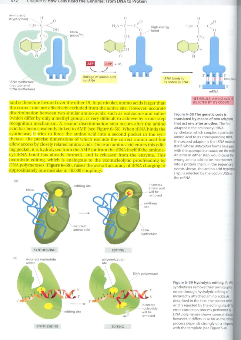
FF
eXI th' 6-' th' wi' 1'0 ret nu
mRNA S'L.-_--'3' tRNA binds to
its codon in RNA high-energy
bond
AMP +2P,
./
linkage of amino acid totRNA
tRNA ,../' (tRNATrp)
tRNA synthetase
. /
(tryptophanyl tRNA synthetase)
and is therefore favored over the other 19. In particular, amino acids larger than the correct one are effectively excluded from the active site. However, accurate discrimination between two similar amino acids, slich as isoleucine and valine (which differ by only a methyl group), is very difficult to achieve by a one-step recognition mechanism. A second discrimination step occurs after the amino acid has been covalelllly linked to AMP (see Figure&-56).When tRNA binds the synthetase, it tries to force the amino acid into a second pocket in the syn- thetase, the precise dimensions of which exclude the correct amino acid but allow accessby closely related amino acids. Once an amino acid enters this edit- ing pocket, it is hydrolyzed from theAM P (or from the tRNA itself if the aminoa- cyl-tRNA bond has already formed), and is released from the enzyme. This hydrolytic editing, which is analogous to the exonucleolytic proofreading by DNA polymerases(Figure &-59), raises the overall accuracy of tRNA charging to approximately one mistake in 40,000 couplings.
(A)
SYNTHESIZING
editing site
-
incorrect amino acid
EDITING
incorrect amino acid willbe removed synthesis site
tRNA synthetase
NET RESULT: AMINO ACID IS SELECTED BY ITS (ODON
Figure6-58The genetic code is translated by means of two adaptors that act one after another. The first adaptor is the aminoacyl-tRNA synthetase. which couples a particular amino acid to its corresponding tRNA;
the second adaptor is the tRNA mol itself, whoseanticodonforms basepail5 with the appropriatecodonon the m An error in either step would causethe wrong amino acid to be incorporated into a protein chain. In the sequenceof events shown, the amino acid tryptoph (Trp) is selected by the codonUGG00
the mRNA.
Ar
Po
lIa to mc th' grc
wi~ ce~
cm tiol an
wa
oft oft
Th
Th, To eve cor
rUJ(
«
pia: olu
(B) incorrect nucleotide added
polymerization site
SYNTHESIZING
3'
"..""~.s'
editing site
•
EDITING
DNA polymerase
3'
"'j, 5'
incorrect nucleotide willbe removed
Figure6-59Hydrolytic editing.(Al lP3 synthetases remove their own coupling errors through hydrolytic editing of incorrectly attached amino acids.As described in the text, the correct aml!lO acid is rejected by the editing site.(BiT.
error-correction process performedby DNA polymerase shows some sirnila however, it differs in50far as the re process depends strongly on a mispal with the template (see Figure 5-8).
~MRNATO PROTEIN 373
Figure 6-60 The recognition of a tRNA molecule by its aminoacyl-tRNA synthetase. For this tRNA(tRNAGln),specific nucleotides in both the
anticodon(bottom)and the amino acid-accepting arm allow the correct tRNA toberecognized by the synthetase enzyme(blue).A bound AlP molecule is yellow.(Courtesy afTorn Steitz.)
The tRNA synlhetase must also recognize the correct set of tRNAs, and 'ensireslructuralandchemicalcomplementarily betweenthesynthetase and ..tR~Aallows the synthetase to probe various feawres of the tllNA (Figure -..oJ.MaS! tRNA synthetases directly recognize the matching tllNA anticodon;
eses~llthetases contain three adjacent Ilucleotide-binding pockets, each of tuch is complementary in shape and charge to a nucleotide in (he anticodon.
mg rother synthetases, the nucleotide sequence of the acceptor stem is the key ognilion determinant. In most cases, however, the synthetase "reads" the deotides at several different positions on the tRNA.
AminoAcids Are Added to the C-terminal End of a Growing Polypeptide Chain
~A.
dlingseen that amino acids are first coupled (0tRNA molecules, we now turn rhemechanism that joins amino acids togcthcr to form proteins. The funda-
~ntalreactionof protein synthesis is the formation ofa peptide bond between ,carboxyl group at the end of a growing polypeptide chain and a free amino ouponan incoming amino acid. Consequently, a protein is synthesized step- ttrromits N·terminal end to its C-tcrminal end. Throughout the entire pro- , the growing carboxyl end of the polypeptide chain remains activated by its ,oIem auachmenttoa tRNA molecule (forming a peptidyl-lRNA). Each addi- ,disrupts this high-energy covalent linkage, but immediately replaces it with ,i<!emicallinkage on the most recently added amino acid (Figure 6-61). In this aY,each amino acid added carries with it thc activation energy for the addition the next amino acid rather than the energy for its own addition-an example me "head growth" type of polymerization described in Figure2-68.
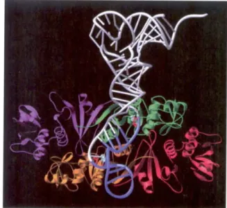
The RNA Message Is Decoded in Ribosomes
symhesis of proteins is guided by information carried by m HNA molecules.
maintain the correct reading frame and to ensure accuracy (about I mistake ry 10,000 amino acids), protein synthesis is pcrformed in the ribosome, a
"lplex catalytic machine madc from morc than 50 diffcrent protcins (the lsolllalproleills) and several HNA molecules, the ribosomal RNAs (rRNAs).
C>A typical eucaryotic cell contains millions of ribosomcs in its cyto·
~m(Figure6-62).Eucaryotic ribosome subunits are assembled at the nucle·
lS,when newly transcribed and modified rRNAs associaLe with ribosomal
Figure6-61The incorporation of an amino acid into a protein. A polypeptide chain grows by the stepwise addition of amino acids to its (-terminal end. The formation of each peptide bond is energetically favorable because the growing (-terminus has been activated by the covalent attachment of a tRNA molecule. The peptidyl-tRNA linkage that activates the growing end is regenerated during each addition. The amino acid side chains have been abbreviated as R" R2,R3,
and R4;as a reference point, all of the atoms in the second amino acid in the
polypeptide chain are shaded gray. The figure shows the addition of the fourth amino acid(red)to the growing chain.
H 0 R H H 0 R 0
I II
,1.
I I 11 .I "H,N-C-C-N-C-C-N-C-C-~-C-C
" I I t II I t "
R, H H 0 R, H H 0
I
tRNA molecule freed from
4 its peptidyl linkage
4
new peptidyl tRNA molecule attached to (-terminus of the growing polypeptide chain Oft
aminoacyl- peplJdyt tRNA attached to tRNA
(·terminusof the growing polypeptide chain
R 0
H 0 R, H H 0 4'
t" I t 4' H~-l-C
-(-C-~-C-C-N-C-C ~ I "
I t II I " H 0
R H H 0 R, 0
I
,
'al n9 he IA I
374 Chapter 6: How Cells Read the Genome: From DNA to Protein FROM
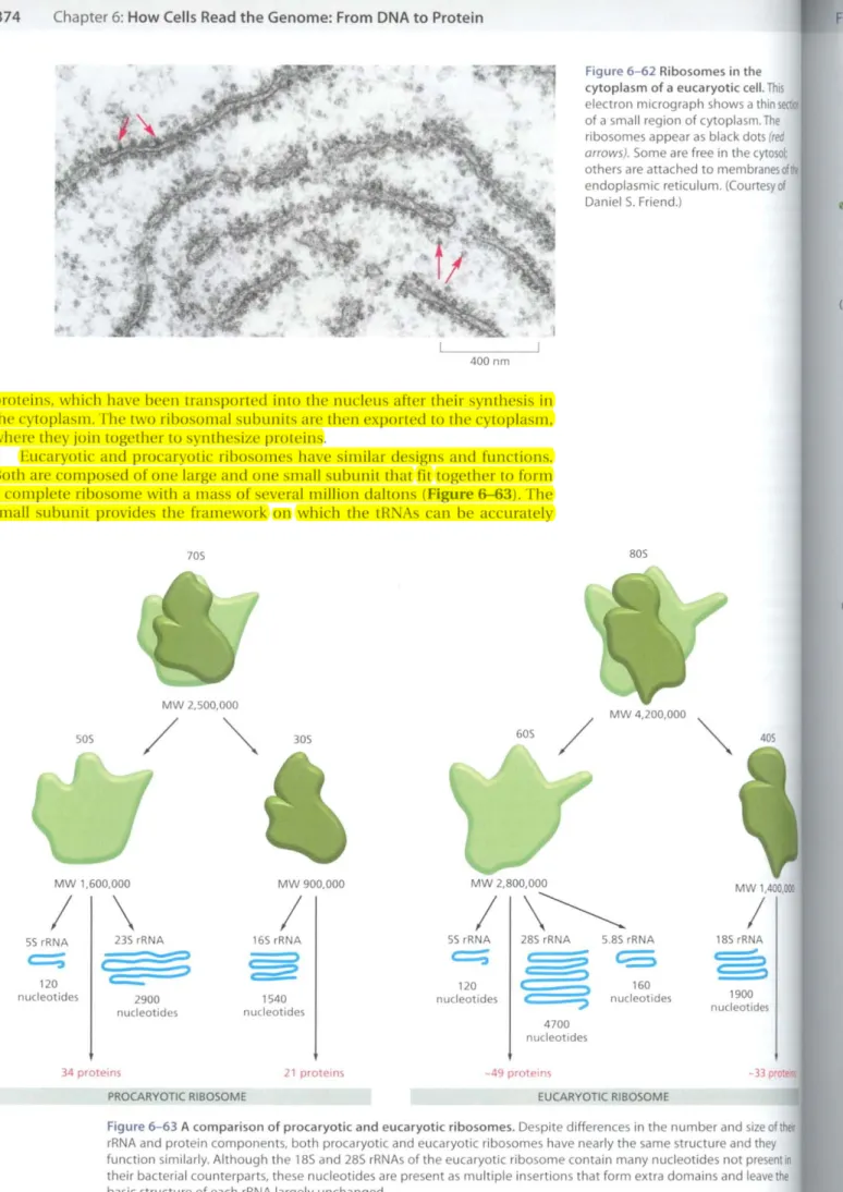
Figure 6-62 Ribosomes in the cytoplasm of a eucaryotic cell. This electron micrograph shows a thin of a small region of cytoplasm. The ribosomes appear as black dots(red arrows).Some are free in the cytosol:
others are attached to membranesol endoplasmic reticulum. ((ourtesyof DanielS. Friend.)
(A)
400nm
proteins, which have been transported into the nucleus after their synthesis in the cytoplasm. The two ribosomal subunits are then expo ned to the cytoplasm, where they join together to syJ1lhesize proteins.
Eucaryotic and procaryotic ribosomes have similar designs and fUllctions.
Both are composed of one large and one small subunit that fittogether to form a complete ribosome with a mass of several million daltons (Figure 6-63). The small subunit provides the framework 011 which the tRNAs can be accurately
70S 80S
mate eat,ll into, V are s' to in
SOlllt sequ to a(
poly!
the I then moil I sam ribo per' mell for I and A4 ~t
cod, ribo two mRI fran the
(e)
40S /
MW 4,200.000 "'-
60S " "
305 MW 2,500,000
/
Figure 6-63 A comparison of procaryotic and eucaryotic ribosomes. Despite differences in the number and size ofther rRNA and protein components, both procaryotic and eucaryotic ribosomes have nearly the same structure and they function similarly. Although the 185 and 285 rRNAs of the eucaryotic ribosome contain many nucleotides not present in their bacterial counterparts, these nucleotides are present as multiple insertions that form extra domains and leave the basic structure of each rRNA largely unchanged.
50S
MW 1,600,000 MW 900,000 MW 2,800,000 MW 1,400,001
/ \ / / \~ /
SS rRNA 23S rRNA 16S rRNA SS rRNA 28S rRNA 5.8S rRNA 18S rRNA
c::::;
(=3 ~ c::::;
~ CO §
120
c=:
120 160nucleotides 2900 1S40 nucleotides nucleotides 1900
nucleotides nucleotides nucleotides
4700 nucleotides
34 proteins 21 proteins -49 proteins -33 pro
PROCARYOTIC RIBOSOME EUCARYOTIC RIBOSOME
MRNA TO PROTEIN 375
on
(B)
large subunit small subunit
90
mRNA·
binding site (D)
tched10the cDdDns Df the mRNA (see Figure6-58),while the large subunit [cl)",the fDrmation of the peptide bonds that link the amino acids together [oapolypeptide chain (see Figure6-61).
When not actively synthesizing proteins, the two subunits of the ribosome I'I'separate. They join together on an mHNA molecule, usually near its 5' end.
initiate the synthesis of a protein. The mRNA is then pulled through the ribo- .e; as its cDdons enter [he core of the ribosome, the mRNA nucleotide
~uenceistranslatedinto an amino acidsequence usingthe tRNAs as adaptors add each amino acid in the correct sequence to the end of the growing Iypeptide chain. When a stop codon is encoullrercd, the ribosome releases finished protein, and its two subunits separate again. These subunits can n be used to start the synthesis of another protein on another mHNA lecule.
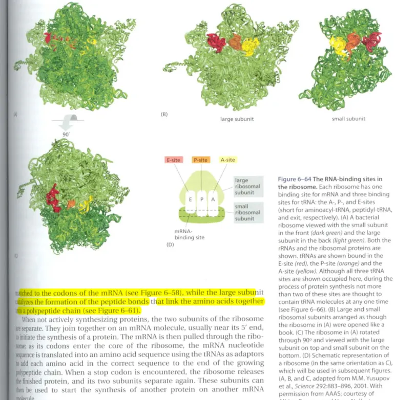
Ribosomes operate withremarkableefficiency: in one second, a singleribo- meof aeucaryotic cell adds about 2 amino acids to a polypeptide chain; the lOsomesofbacterial cells operate even faster, at a rate of about 20amino acids [second. How does the ribosome choreograph the many coordinated move- nt~required for efficient translation? A ribosome contains four binding sites [R\A molecules: one is for the mRNA and three (called the A-site, the P-site, witheE-site) are for lRNAs (Figure 6-64). A til A molecule is held lightly at the and P-sires only if its anticodon forms base pairs with a complementary
.00(allowing for wobble) on the mRNA molecule that is threaded through the )\Ome (Figure 6-65). The A- and P-sites arc close enough together for their tR~Amoleclilestobe forced toformbase pairs with adjacent codons on the R.~Amolecule. This feature of the ribosome maintains the correct reading iffieon [he mRNA.
Once protein synthesis has been initiated,each new amino acid is added to elongating chain in a cycle of reactions containing four major steps: tH A
Figure 6-64 The RNA-binding sites in the ribosome. Each ribosome has one binding site for mRNA and three binding sites for tRNA: the A·, p., and E-sites (short for aminoacyl·tRNA. peptidyl-tRNA, and exit, respectively). (A) A bacterial ribosome viewed with the small subunit in the front(dark green)and the large subunit in the back(light green).Both the rRNAs and the ribosomal proteins are shown. tRNAs are shown bound in the E-site(red),the P-site(orange)and the A-site(yellow).Although all three tRNA sites are shown occupied here, during the process of protein synthesis not more than two of these sites are thought to contain tRNA molecules at anyone time (see Figure6-66).(B) Large and small ribosomal subunits arranged as though the ribosome in (A) were opened like a book.(C)The ribosome in (A) rotated through 9oo and viewed with the large subunit on top and small subunit on the bottom. (0) Schematic representation of a ribosome (in the same orientation asC), which will be used in subsequent figures.
(A. B. andC.adapted from M.M. Yusupov et al .•Science292:883-896, 2001.With permission from AAAS; courtesy of Albion Baucom and Harry Noller.) large
ribosomal subunit small ribosomal subunit A-site
1/
P-site
E P A [-site
376 Chapter 6: How Cells Read the Genome: From DNA to Protein
E-site P·site A-site
TI
se
CtH
esc tR un(
rOl'l
eru ing intE cod Gil inC( the '>is ( so I
teln
a')$f
mel hId, corr cod, isJfl disti for \ R. ',I
reeo
newly
, ....
ae el, hl LC ell
'iiI' thl ysi st.' nb R fa' oc,
growing polypeptidechain un
STEP2
STEP4 STEP 1
Figure 6-65 The path of mRNA(b/ut through the small ribosomal subunit.
The orientation is the same as thaI in right-hand panel of Figure 6-646.
((ourtesy of HarryF.Noller, based on data in G.Z. Yusopova et aI.,Cell 106:233-241, 2001. With permission Elsevier.)
binding, peptide bond formation, large subunit and small subunit translocation.
As a result of the two translocation steps, the entire ribosome moves three nucleotides along the mRNA and is positioned to start the next cycle. (Figure 6-66).Our description of the chain elongation process begins at a point at which some amino acids have already been linked together and there is a tRNA molecule in the P-site on the ribosome, covalently joined to the end of the grow- ing polypeptide. In step I,a tRNA carrying the next amino acid in the chain bindsto the ribosomal A-site by forming base pairs with the mRNA codon posi- tioned there, so that the P-site and the A-site contain adjacent bound tRNAs. In step2,the carboxyl end of the polypeptide chain is released from the tRNA at the P-site (by breakage of the high-energy bond between the tRNA and its amino acid) and joinedtothe free amino group of the amino acid linked tothe tRNA at the A-site, forming a new peptide bond. This central reaction of protein synthe- sis is catalyzed by a /lepl/dy/Iransferasecontained in the large ribosomal sub- unit. In step 3, the large subunit moves relativeto the mRNA held by the small subunit, thereby shifting the acceptor stems of the two tllNAs to the E- and P- sites of the large subunit. In step 4, another series of conformational changes moves the small subunit and its bound mRNA exactly three nucleotides, reset- ting the ribosome so it is ready to receive the next aminoacyl-tRNA. Step J is then repeated with a new incoming aminoacyl-tRNA, and so on. <CGn'>
This four-step cycle is repeated each time an amino acid is added to the polypeptide chain, as the chain grows from its amino to its carboxyl end.
Figure 6-66 Translating an mRNA molecule. Each amino acid added to the growing end of a polypeptide chain is selected by complementary base-pairing between the anticodon on its attached tRNA molecule and the next codon on the mRNA chain. Because only one of the many types of tRNA molecules in a cell can base-pair with each codon, the codon determines the specific amino acid to be added to the growing polypeptide chain. The four-step cycle shown is repeated over and over during the synthesis of a protein. In step1.an aminoacyl-tRNA molecule binds to a vacant A·site on the ribosome and a spent tRNA molecule dissociates from the E-site. In step 2, a new peptide bond isformed. Instep 3, the large subunit translocates relative to the small subunit, leaving the two tRNAs in hybrid sites: P on the large subunit and A on the small, for one; E on the large subunit and P on the small, for theother. Instep 4, the small subunit translocates carrying its mRNA a distance of three nucleotides through the ribosome. This
"resets~the ribosome with a fully empty A-site, ready for the next aminoacyl-tRNA molecule to bind. As indicated, the mRNA is translated in the 5'-to-3' direction, and the N-terminal end of a protein is made first, with each cycle adding one amino acid to the (-terminus of the polypeptide chain.
STEP 1
1
H,N
;,t
outgoing tRNA
5'.au.e~
crilil stag.
then
alllll
then
POSI1
rodo
8th
IRNA TOPROTEIN >I,
!
incorrectly base-
r
paired tRNAs preferentially dissociateP, PROOFREADING
PROOFREADING
~9alionFactors Drive Translation Forward and Improve Its ':uracy
Figure 6-67 Detailed view of the translation cycle. The outline of translation presented in Figure 6-66 has been expanded to show the roles of two elongation factors EF·Tu and EF-G. which drive translation in the forward direction. As explained in the text, EF·Tu also provides two opportunities for proofreading of the codon-anticodon match. In this way, incorrectly paired tRNAs are selectively rejected, and the accuracy of translation is improved.
cycle of polypeptide elongation shown in outline in Figure 6-66 has an nal feature that makes translation especially efficient and accurate. '1\'1'0
• '011 factors enter and leave the ribosome during each cycle. each ',!,ing GTPto GOP and undergoing conformational changes in the pro- These factors are called EF-Tu and EF-G in bacteria, and EFI and EF2 in .oles.Under some conditionsin vitro,ribosomescanbe forced to synthe- roteins without the aid of these elongation factors and GTP hydrolysis, but
~thesisis very slow, inefficient, and inaccurate. Coupling the GTP hydrol- menchanges in the elongation factors to transitions between different otthe ribosome speeds up protein synthesis enormously. Although these states are not yet understood in detail, they almost certainly involve S[TUcture rearrangements in the ribosome core. The cycles ofelongation lociation. GTP hydrolysis, and dissociation ensure that all such changes inthe "forward" direction so that translation can proceed efficiently (Fig- ...7.
,\Ishown previously, EF-Tu simultaneously binds GTP and aminoacyl-tHNAs figure3-74). In addition to helping move translation forward, EF-Tu (EFI in .otes) increases the accuracy of translation in several ways. First, as it anincoming aminoacyl-tH A to the ribosome, EF-Tu checks whether the l-amino acid match is corrrecL Exactly how this is accomplished is not well lrOO.Accordingtoone idea. correct tH A-amino acid matches have anar- defined amnity for EF-Tu. which allows EF-Tu to discriminate. albeit
\ among many different amino acid-tRNA combinations. selectivelybring-
·'tecorrect ones with it into the ribosome. Second, EF-Tu monitors the initial radian between the anticodon of an incoming aminoacyl-tRNA and the
<Jnofthe mRNA in the A-site. Aminoacyl-tilNAs are "bent" when bound 10 the ll'-form of EF-Tu; this bent conformation allows codon pairing but prevents rporation of the amino acid into the growing polypeptide chain. Ilowever, if .Jdon-anticodon match is correct, the ribosome rapidly triggers the hydroly-
theGTP molecule, whereupon EF-Tu releases its grip on the tHNA and dis- ." from the ribosome, allmving the tH A 10 donate its amino acid for pro-
s~nthesis. But how is the "correctness" of the codon-anticodon match
;ffilThis feat is carried out by the ribosome itself through an H A-based ,:an;'m. The rRNA in the small subunit of the ribosome forms a series of ..:tgen bonds with the codon-anticodon pair that allows determination of its 'ectness (Figure6-68). In essence, the rHNA folds around the codon-anti- onpair, and its final closure-which occurs only when the correct anticodon place-triggers GTP hydrolysis. Hemarkably, this induced fit mechanism can nguish correct from incorrect codon-anticodon interactions despite the rules
",hie base-pairing summarized in Figure 6-53. From this example, as for
"plicing, one gets a sense of the highly sophisticated forms of molecular non that can be achieved solely by Il A.
1leinteractions of EF-Tu, tH A, and the ribosome just described introduce proofreading steps into protein synthesis at the initial tH A selection ,But after GTP is hydrolyoed and EF-Tu dissociates from the ribosome, isan additional opportunity for the ribosome to prevent an incorrect 'no acid from being added to the growing chain. Following GTP hydrolysis, reis a short time delay as the amino acid carried by the tH A moves into
"lionon the ribosome. This time delay is shorter for correct than incorrect n-amicodon pairs. Moreover, incorrectly matched tRNAs dissociate more
nd l.
.he
---~
- - - - . --
.~FR
The cIon the leng
call~
fune also deci ratr thes tran:
Nuc
Syn saw tion sidl' et\ no\, prol witl synl(AI Figl larg
wit~
vert stru,
Ihi doc sop whi eus Insl tim gro1 thei site cat, isp
codon anticodon
Figure 6-68 Recognition of correct codon-anticodon matches by the small subunit rRNA of the ribosome. Shown the interaction between a nucleotide 01 the small subunit rRNA and the first nucleotide pair of a correctly paired codon-anticodon; similar interactions occur between other nucleotides ofthe rRNA and the second and thirdposit~
of the codon-anticodon pair. Thesma~
subunit rRNA can form this networkof hydrogen bonds only with correctly matched codon-anticodon pairs.As explained in the text, this
codon-anticodon monitoring by thesrr subunit rRNA increases the accuracyof protein synthesis. (From J.M. Ogle etal Science 292:897-902, 2001. With permission from AAAS.)
(B)
Chapter 6: How Cells Read the Genome: From DNA to Protein 378
The Ribosome Is a Ribozyme
rapidly than those correctly bound because their interaction with the codon is weaker. Thus, most incorrectly bound tRNA molecules (as well as a significant number of correctly bound molecules) will leave the ribosome without being used for protein synthesis. All of these proofreading steps, taken together, are largely responsible for the 99.99% accuracy of the ribosome in translating RNA into protein.
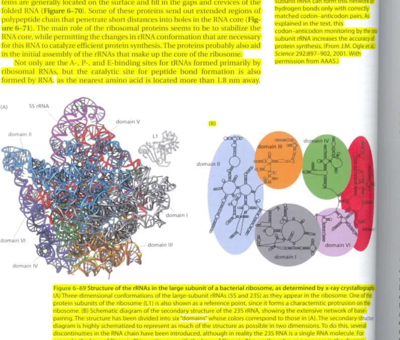
Figure 6-69 Structure of the rRNAs in the large subunit of a bacterial ribosome. as determined by x-ray crystallography (A) Three-dimensional conformations of the large-subunit rRNAs (55 and 23S) as they appear in the ribosome. One of the protein subunits of the ribosome (ll) is also shown as a reference point, since it forms a characteristic protrusion on the ribosome. (B) Schematic diagram of the secondary structure of the 23S rRNA, showing the extensive network of base·
pairing. The structure has been divided into six"domains~whose colors correspond to those in (A). The secondary·st diagram is highly schematized to represent as much of the structure as possible in two dimensions. To do this, several discontinuities in the RNA chain have been introduced, although in reality the 235 RNA is a single RNA molecule. For example, the base of Domain III is continuous with the base of Domain IV even though a gap appears in the diagram.
(Adapted from N. Ban et aI., Science 289:905-920, 2000. With permission from AAAS.)
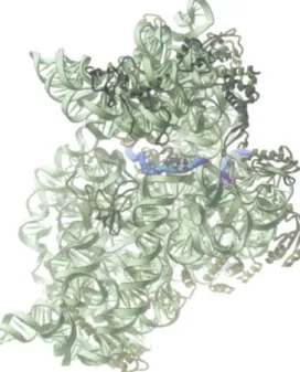
The ribosome is a large complex composed of two-thirds RNA and one-third protein. The determination, in 2000, of the entire three-dimensional conforma- tion of its large and small subunits is a major triumph of modern structural biol- ogy. The findings confirm earlier evidence that rR As-and not proteins-arc responsible for the ribosome's overall structure, its ability to position tRNAs on the mRNA, and its catalytic activity in forming covalent peptide bonds. The ribo- somal RNAs are folded into highly compact. precise three-dimensional struc- tures that form the compact core of the ribosome and determine its overall shape (Figure 6-69).
In marked contrast to the central positions of the rRNAs, the ribosomal pro·
teins are generally located on the surface and fill in the gaps and crevices of the folded RNA (Figure6-70).Some of these proteins send out extended regions of polypeptide chain that penetrate short distances into holes in the RNA core (Fig- ure 6-71). The main role of the ribosomal proteins seems to be to stabilize the RNA core, while permitting the changes in rRNA conformation that are necessary for this RNA to catalyze efficient protein synthesis. The proteins probably also aid in the initial assembly of the rRNAs that make up the core of the ribosome.
Not only are the A-, P-, and E-binding sites for tRNAs formed primarily by ribosomal RNAs, but the catalytic site For peptide bond Formation is also formed by RNA, as the nearest amino acid is located morc than 1.8 nm away.
L
9th
ROM RNA TO PROTEIN 379
s
~70location of the protein components of thebacterial large ribosomalsubunit. The rRNAs (55 and 235) are shown ingray and the ..IbuM proteins (27 of the 31 total) ingold. For convenience. the protein structures depict only the polypeptide backbones. (A) Interface small subunit, the same view shown in Figure 6-648. (8) Side opposite to that shown in (A), obtainedby rotating (A) by 1800around a axis.((JFurther slight rotation of (8) through a diagonalaxis.alloWing a view into the peptide exit channel in the center of the ,!Pe,(From N. Ban et a!.,Science 289:905-920. 2000. With permission from AAAS.l
v
II discovery came as a surprise to biologists because, unlike proteins, R A not contain easily ionizable functional groups that can be lIsedto catalyze Jphbticatcd reactions like peptide bond formation. Moreover, metal ions, tJich are often used by RNA moleClilestocatalyze chemical reactions (as dis-
l\\edlater in the chapter), were not observed at the active site of the ribosome.
IItead, it is believed that the 235 rRNA forms a highly structured pocket that, trough a network of hydrogen bonds, precisely orients the two reactants (the Mingpeptide chain and an aminoacyl-tRNA) and thereby greatly accelerates
irco\~lentjoining. In addition, the tRNA in the P site contributestothe active perhaps supplying a functional OH group that participates directly in the
)~is.This mechanism may ensure that catalysis occurs only when the tRNA voperly positioned in the ribosome .
."\ molecules that possess catalytic activity arc known as ribozymes. We
W:iulier in this chapter how other ribozymes function in self-splicing reac- 01 lor example, sec Figure 6-36). In the linal section of this chapter, we con- IIhat the ability of R A molecules to function as catalysts for a wide vari- fdifferent reactions might mean for the early evolution of living cells. For lie merely note that there is good reason to suspect that IlNA rather than , in molecules served as the lirst catalysts for living cells. If so, the ribosome, it:>R.\A core, may be a relic of an earlier time in lifc's history-when protein the>is evolved in cells that were run almost entirely by ribozymes.
Nucleotide Sequences in
mRNASignal Where to Start Protein Synthesis
initiation and termination of translation share features of the translation tion cycle described above. The site at which protein synthesis begins on mR:'-iA is especially crucial, since it sets the reading frame for the whole ngthof the message. An error of one nucleotide either way at this stage would u every subsequent codon in the mcssagetobc misread. resulting in a non- tional protein with a garblcd sequence of amino acids. The initiation step is so important because for most genes it is the last point at which the cell can 'xidewhether the mil A isto be translated and the protein synthesized; the ,tfofinitiation is thus one detcrminant of the rate at which any protein is syn-
'\~ized.We shall see in Chapter 7 that cells use several mcchan ismstoregulate
ran~lationinitiation.
Figure 6-71 Structure of the US protein in the large subunit of the bacterial ribosome. The globular domain of the protein lies on the surface of the ribosome and an extended region penetrates deeply into the RNA core of the ribosome. Thel15protein is shown in yellowand a portion of the ribosomal RNA core is shown inred.(From D. Klein, P.B. Moore and T.A. Steitz,J.Mol. Bioi.
340:141-147,2004. With permission from Academic Press.)