• 검색 결과가 없습니다.

Pleural Metastasis of Atypical Bronchial CarcinoidEugeniy Igorevich Zinchenko, M.D., Ph.D.

N/A
N/A
Protected

Academic year: 2021

Share "Pleural Metastasis of Atypical Bronchial CarcinoidEugeniy Igorevich Zinchenko, M.D., Ph.D."

Copied!
4
0
0

로드 중.... (전체 텍스트 보기)

전체 글

(1)

317 www.kjtcvs.org

KJTCVS

The Korean Journal of Thoracic and Cardiovascular Surgery

Case Report

Pleural Metastasis of Atypical Bronchial Carcinoid

Eugeniy Igorevich Zinchenko, M.D., Ph.D.

1,2

, Olga Sergeevna Maslak, M.D.

1

, Vadim Grigorievich Pischik, M.D., Ph.D.

1,2

, Aleksandr Dmitrievich Obornev, M.D., Ph.D.

2

, Aleksandr Igorevich Kovalenko, M.D.

1,2

1

Faculty of Medicine, Saint Petersburg State University;

2

Thoracic Department, L. G. Sokolov Memorial Hospital 122, Saint Petersburg, Russia

ARTICLE INFO

Received December 30, 2019 Revised January 24, 2020 Accepted February 22, 2020 Corresponding author Olga Sergeevna Maslak Tel 7-9218450993 Fax 7-9218450993 E-mail [email protected] ORCID

https://orcid.org/0000-0002-9202-8064

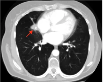
Pulmonary carcinoids are relatively rare tumors with low metastatic potential. Pleural car- cinomatosis of a bronchial carcinoid has only been reported in 4 cases. Due to the rarity of this condition, there are no guidelines for its treatment or management. We report a case of atypical carcinoid with local recurrence and pleural metastases treated by video-assist- ed thoracoscopic surgery lobectomy and total pleurectomy with photodynamic therapy after non-radical wedge resection.

Keywords: Carcinoid tumor, Neoplasm metastasis, Photochemotherapy, Pleurectomy, Video-assisted thoracoscopic surgery

Copyright

© The Korean Society for Thoracic and Cardiovascular Surgery. 2020. All right reserved.

This is an Open Access article distributed under the terms of the Creative Commons Attribution Non-Commercial License (http://creativecommons.org/licenses/

by-nc/4.0) which permits unrestricted non-commercial use, distribution, and reproduction in any medium, provided the original work is properly cited.

Case report

A 51-year-old non-smoking woman was admitted to L. G.

Sokolov Memorial Hospital 122 due to an area of consoli- dation in the middle lobe that was found on a chest com- puted tomography (CT) scan. Two years ago, chest CT was performed due to increasing dyspnea, and revealed a mid- dle lobe neoplasm (Fig. 1). The lesion was solid, centrally located, measuring 16×15 mm, with smooth margins, and caused partial atelectasis of the middle lobe with no en- larged lymph nodes. During video-assisted thoracoscopic surgery (VATS) exploration performed at another institu- tion, the neoplasm in the middle lobe was visualized, as well as multiple small yellowish foci on the parietal pleura, upon which basis pleural dissemination was suspected.

Thus, wedge resection of the middle lobe with the entire lesion and biopsy of the parietal pleura were performed.

According to the morphologic examination, an atypical carcinoid was verified, the resection margins were clear, and pleural carcinomatosis was confirmed. The patient was treated with octreotide (20 mg once in 28 days) since she had mild symptoms of carcinoid syndrome (the recur- rent feeling of blood rushing to the face). Twelve months after surgery, local recurrence was suspected based on fol- low-up CT (Fig. 2). Significant consolidation was seen in the suture line area, and several small lesions were noted

on the parietal pleura. No other signs of distant metastasis or lymph node involvement were found on chest and ab- dominal CT.

At the time of admission to our hospital, the patient had mild dyspnea. Her Charlson comorbidity index was 3. The level of chromogranin A was elevated. The patient was scheduled for VATS re-exploration of the pleural space to

Fig. 1. Computed tomography scan of the chest before the oper- ation, showing a centrally located solid lesion, measuring 16×15 mm, with smooth margins (arrow).

https://doi.org/10.5090/kjtcs.19.098 pISSN: 2233-601X eISSN: 2093-6516

Korean J Thorac Cardiovasc Surg. 2020;53(5):317-320

(2)

318 www.kjtcvs.org

KJTCVS https://doi.org/10.5090/kjtcs.19.098

prove the presence of recurrence and to evaluate the vol- ume of dissemination and the extent of the previous oper- ation. During surgery, local recurrence in the remaining middle lobe and multiple miliary yellowish foci from 2 to 5 mm in diameter on the parietal pleura were visualized, but the visceral pleura was intact (Fig. 3). Since the consolida- tion was very close to the hilum, a complete anatomic mid- dle lobectomy with lymphadenectomy was performed.

Considering the isolated involvement of parietal pleura, to- tal parietal pleurectomy was also performed, followed by photodynamic therapy (using a neodymium-doped yttri- um aluminum garnet laser as the light source, with a 30 mm diffusing fiber with wavelength of 662 nm, light dose of 100 J/cm

2

, and exposure time of 15 minutes). The total

blood loss was 100 mL, and the operative time was 255 minutes. The patient’s postoperative course was uneventful and she was discharged on postoperative day 5.

Local recurrence was proven with immunostaining (atypical cells expressing synaptophysin, CD56, thyroid transcription factor 1, cytokeratin 7, and 3%–6% of Ki67) (Fig. 4), with no metastasis in the lymph nodes at stations 12R, 11R, 10R, 7, and 2–4R. The patient took octreotide.

Nine months after the operation, no signs of disease pro- gression were found, according to CT scans and low chro- mogranin A levels. Twelve months after the operation, DOTATATE positron emission tomography/CT was per- formed, and no metabolic sites were visualized.

The study was approved by the Institutional Review

A B

Fig. 2. Computed tomography scan of the chest. (A) Local recurrence in the middle lobe (arrow). (B) Pleural focus (arrow).

A B

Fig. 3. Intraoperative photos of pleu- ral carcinomatosis. (A) Small le- sions on the parietal pleura. (B) Foci in the area of the apex.

Fig. 4. Immunohistochemical ex- amination. (A) Lung specimen with high expression of cytokeratin 7 (×100, neuroendocrine tumor of the lung). (B) Pleura specimen with high expression of CD56 (×100, pleural metastasis).

A B

(3)

319

Eugeniy Igorevich Zinchenko, et al. Pleural Carcinomatosis of Bronchial Carcinoid

www.kjtcvs.org

KJTCVS

Board of L. G. Sokolov Memorial Hospital 122 (no. 2, date 17.2.2020). The patient provided written informed consent for the publication of clinical details and images.

Discussion

Pulmonary carcinoids are quite rare tumors of the lung, accounting for approximately 0.2–2 cases per 100,000 pop- ulation [1]. They constitute only 25% of all carcinoid tu- mors [2]. During the last 30 years, the incidence of carci- noids has been reported to increase by 6% annually, but this may be explained by the evolution of immunohisto- chemical techniques, rather than a true growth in the inci- dence of carcinoids [3]. They represent 1%–2% of all inva- sive lung neoplasms, with a high predominance of typical forms [1].

Typical carcinoids have an indolent nature, with a medi- an doubling time of approximately 7 years [4]. The average 5- and 10-year overall survival rates have been reported to be 93% and 89%, respectively, and the average disease-free survival rates are 88% and 72% [2]. However, dissemina- tion occurs in 5%–10% of typical carcinoids and in 15%–

20% of atypical ones [4]. Among metastatic forms, atypical carcinoid is diagnosed in 69% of cases. Patients with meta- static lung carcinoids are twice as frequently diagnosed with carcinoid syndrome [5].

The most common sites of metastases are the liver, bone, brain, and mediastinal lymph nodes [1]. Extremely rare re- ported sites include the ovaries, pancreas, subcutaneous tissue, skin, peritoneum, thyroid gland, breast, choroid and ciliary body, spinal cord, and heart [5].

We have found 4 cases of carcinoid pleural dissemina- tion in the literature [6-8]. In all cases, the histologic fea- tures of the carcinoids were typical. Three patients were treated with lobectomy and 1 with segmentectomy [6]. All tumors were diagnosed at a non-advanced stage. The time to progression after surgery was 4, 5, 7, and 30 years [6-8].

Only one patient was diagnosed with carcinoid syndrome and was treated with metabolic radiotherapy [6]. Another patient had 2 episodes of local recurrence before dissemi- nation, treated with cryotherapy [6]. Five years later, he was diagnosed with pleural and hepatic metastases and treated with chemotherapy followed by somatostatin analogs. The patient who presented with metastases 30 years after lobec- tomy underwent surgical cytoreduction through resection of the pleural masses with the sixth and seventh ribs and pericardial resection [8]. Only 2 patients were treated with chemotherapy when pleural dissemination was confirmed [7].

The treatment guidelines for metastatic pulmonary car- cinoids are vague [1,5]. Due to their low incidence, no ran- domized trials have yet been conducted to determine an effective treatment strategy. Cytoreduction surgery is a possible treatment option [9]. Asymptomatic patients may be observed or treated with octreotide. Patients with carci- noid syndrome should be treated with somatostatin ana- logs. These drugs provide symptom control and have been reported to slow down tumor growth. For patients with progressive metastatic disease, everolimus is a recommend- ed treatment option. Chemotherapy is a possible option, but the tumor response rate is usually low. Treatment with peptide receptor radionuclide therapy may be considered in patients with somatostatin receptor-positive imaging who exhibit disease progression while taking octreotide or lanreotide.

Patients with carcinoid tumors should receive regular follow-up [10]. The observations should be made with chest CT and abdominal CT or magnetic resonance imaging ev- ery 3 to 6 months. The biochemical evaluation includes testing chromogranin A levels as a marker of tumor recur- rence every 6 months in patients with elevated levels at baseline [9]. The follow-up period should constitute at least 10 years, although according to some studies it may be prolonged for up to 20 years [5].

Photodynamic therapy is an evolving method of treating disseminated tumors that is characterized by few compli- cations. It is widely used for the treatment of pleural dis- semination of epithelial tumors. In our center, we usually perform pleurectomy and photodynamic therapy with a chlorine photosensitizer (radachlorin) in cases of pleural carcinomatosis, including thymoma dissemination and mesothelioma. Since pleural dissemination of bronchial carcinoid is an extremely rare condition, there are no pre- vious reports of photodynamic therapy use in such cases.

However, several cases of the use of photodynamic therapy to treat choroidal metastasis from pulmonary carcinoids have been reported, with good responses [11]. Considering the absence of other metastatic sites and the indolent be- havior of the tumor, we decided to perform pleurectomy and photodynamic therapy to increase the antiblastic effect of the operation.

In our opinion, photodynamic therapy is a viable treat-

ment option for the pleural dissemination of epithelial tu-

mors. Its addition to systemic therapy might prolong the

patient’s life and relieve the symptoms of tumor progres-

sion. If a patient has metastasis limited to the parietal pleu-

ra, we propose to perform pleurectomy and photodynamic

therapy to affect all sites of neoplasm spread. This proce-

(4)

320 www.kjtcvs.org

KJTCVS https://doi.org/10.5090/kjtcs.19.098

dure does not significantly increase the time or cost of the operation, and extremely rarely causes complications. We hope that the effects of photodynamic therapy on such a slowly growing tumor will prolong the patient’s life for de- cades.

Conflict of interest

No potential conflict of interest relevant to this article was reported.

ORCID

Eugeniy I. Zinchenko: https://orcid.org/0000-0002-3966-0407 Olga S. Maslak: https://orcid.org/0000-0002-9202-8064 Vadim G. Pischik: https://orcid.org/0000-0002-9602-0908 Aleksandr D. Obornev: https://orcid.org/0000-0003-4389-4756 Aleksandr I. Kovalenko: https://orcid.org/0000-0001-7997-4251

References

1. Caplin ME, Baudin E, Ferolla P, et al. Pulmonary neuroendocrine (carcinoid) tumors: European Neuroendocrine Tumor Society expert consensus and recommendations for best practice for typical and atypical pulmonary carcinoids. Ann Oncol 2015;26:1604-20.

2. Lee PC, Osakwe NC, Narula N, et al. Predictors of disease-free sur- vival and recurrence in patients with resected bronchial carcinoid tu-

mors. Thorac Cardiovasc Surg 2016;64:159-65.

3. Yao JC, Hassan M, Phan A, et al. One hundred years after “carci- noid”: epidemiology of and prognostic factors for neuroendocrine tu- mors in 35,825 cases in the United States. J Clin Oncol 2008;26:

3063-72.

4. Raz DJ, Nelson RA, Grannis FW, Kim JY. Natural history of typical pulmonary carcinoid tumors: a comparison of nonsurgical and surgi- cal treatment. Chest 2015;147:1111-7.

5. Robelin P, Hadoux J, Forestier J, et al. Characterization, prognosis, and treatment of patients with metastatic lung carcinoid tumors. J Thorac Oncol 2019;14:993-1002.

6. Bonnette P, Epardeau B, Frachon I, et al. Report of 2 cases of pleural recurrences of surgically treated bronchogenic carcinoids: diagnostic and therapeutic problems. Rev Mal Respir 1999;16:85-8.

7. Klee H, Vestring T, Bittmann I. Pleural metastases of a typical bron- chial carcinoid 7 years after lobectomy. Pneumologie 2008;62:607- 10.

8. Townshend AP, Lakshminarayanan B, Ucar AE, Chaudry ZR, Duffy JP. Rare pleural recurrence of typical pulmonary carcinoid tumor 30 years after lobectomy. Ann Thorac Surg 2007;83:1523-4.

9. Bouledrak K, Walter T, Souquet PJ, Lombard-Bohas C. Metastatic bronchial carcinoid tumors. Rev Pneumol Clin 2016;72:41-8.

10. Ma DS, Kim SA, Kim HR, Kim YH, Park SI, Kim DK. Bronchial carcinoid tumor arising from an intralobar bronchopulmonary se- questration. Korean J Thorac Cardiovasc Surg 2011;44:444-7.

11. Chu CK, El-Annan J. Photodynamic therapy for choroidal carcinoid

metastasis. Can J Ophthalmol 2018;53:e13-6.

수치

Fig. 1. Computed tomography scan of the chest before the oper- oper-ation, showing a centrally located solid lesion, measuring 16×15  mm, with smooth margins (arrow).
Fig. 2. Computed tomography scan  of the chest. (A) Local recurrence in  the middle lobe (arrow)

참조

관련 문서

Sandberg D, Lavyne M (1999) Symptomatic spinal epidural lipomatosis after local epidural corticosteroid injections:

#define 에서 인자 사용 예제 compoundassign.c... 관계와

▪ 실세계의 객체에서 불필요한 부분을 제거하여 필요한 부분만을 간결하고 이해하기 쉬운 클래스 로 만드는 작업. ▪

extracranial arteries, patients diagnosed with a transient ischemic attack or without a confirmed stroke on MRI brain DWI, patients with an atypical cause of ischemic stroke

fundamentals and mechanistic details. Synthetic mammalian prions. Evaluation of the human transmission risk of an atypical bovine spongiform encephalopathy prion strain.

The close substitutes are R&D efforts, technologies, and goods that significantly constrain the exercise of market power with respect to the relevant

One might try to solve the problem by copying the first d elements of x to a temporary array, moving the remaining n − d elements left d places, and then copying the first d

It considers the energy use of the different components that are involved in the distribution and viewing of video content: data centres and content delivery networks