메타분석을 이용한 정상 영유아의 뇌간청각유발전위의 잠시에 대한 연구
충북대학교 의과대학 재활의학교실, 예방의학교실*
황어성∙이경무∙김김헌*
– Abstract –
Estimation of Normal BAEPs Latencies from Infancy to Early Childhood Using Meta-Analysis
Eo Seong Hwang, M.D., Kyoung Moo Lee, M.D., Heon Kim, M.D.*
Department of Rehabilitation Medicine and Preventive Medicine Chungbuk National University Hospital Cheongju, Korea
Objectives: The brainstem auditory evoked potentials (BAEPs) are an useful method to evaluate func- tions from the auditory nerve to the brainstem but there are many different test methods amongst different labs that make it difficult to compare the results.
Since there are many differences associated with different test method and other reference values for the evaluation of children from the period of infancy (1~12 months) to early childhood (1 year~6 years), there is a need to establish a standardized test method with normal references. We initiated this study to overcome these difficulties.
Methods: We searched through 25 sources, which had been previously published from 1974 to 1997, concerning the brainstem auditory evoked potentials in normal children from infants to early childhood.
The search provided a common test method using all 25 sources and obtained a normal reference value of peak latencies and interpeak latencies through the meta-analysis technique by researching 7 sources stated normal reference values.
Results: After meta-analysis from 7 sources, we found normal peak latencies and interpeak latencies of a standard 70 dB stimulation intensity, 10 Hz stimulation frequency and monoaural mode.
The most common test method used active electrodes with a silver/silverchloride surface (70.6%) which was applied to the mastoid process (50%). The site for the reference electrode was the vertex (59%) and the site for the ground electrode was the contralateral mastoid process (38%) or upper forehead (29%). The interelectrode impedance was less than 5000 Ω (80%), the bandpass of filter was 100 Hz (40%)~3000 Hz (55%), the stimulation frequency was 10 Hz (71%) and the stimulation intensity was 70 dB (33%).
The total stimulation number was found to be more than 1024 and the most common stimulation mode was the monoaural mode (85%). Each test was conducted more than twice on the same ear.
Conclusion: Since there are many labs that have not established a normal reference value and a stan-
충북대학교 의과대학 재활의학교실
Address reprint requests to Eo Seong Hwang, M.D.
Department of Rehabilitation Medicine, College of Medicine, Chungbuk National University
#62 Gaesin-dong, Heungduk-gu, Cheongju 361-711, Korea
Tel : 82-43-269-6227, Fax : 82-43-266-1698, e-mail : [email protected]
서 론
뇌간청각유발전위반응은 두피전극을 통하여 통상적 으로 제 I파에서 제 V파로 표시되는 청각 전도 신경계의 활동전위를 말하며 청신경 및 뇌간내 청신경전달로의 일련의 전기적 변화를 나타낸다고 Sohmer 등(1970)1과 Jewet 등2이 처음으로 보고하였다.3
Lev 등(1972)4은 동물실험을 통하여 음자극후 10 msec 내에 발생한 5~7개의 파들이 뇌간 내의 특정한 위치에 서 각각 기원한다고 하였고, 각각의 발생기원에 대하여 Buchwald 등(1975)5과 Stockard 등(1977)6이 제 I파는 동 측 원위부의 청신경을, 제 II파는 근위부 청신경 또는 와 우신경핵을, 제 III파는 하위부 뇌교를(반대측의 상 올리 브핵도 가능), 제 IV파는 중간 또는 상부의 뇌교를(양측 외측 모대핵도 가능), 제 V파는 상위 뇌교 또는 반대측 하구핵에서 기시된다고 보고하였으며 제 VI파와 제 VII 파에 대해서는 내측슬상체와 청방선으로 추정하고 있 지만 아직 정확한 해부학적 발생장소는 밝혀내지 못하 고 있다.7
Starr 등(1975)8, Gilroy 등(1977)9과 Stockard 등(1977)10 이 뇌간청각유발전위반응의 이상과 뇌간병변과의 관계 에 대하여 보고하였고, Jewett(1972)11은 동물실험에서 연령이 증가함에 따라 각파의 잠시(잠복시간, latency)가 비례적으로 감소한다고 하였다. Lieberman 등(1972)12은 인간에 있어서도 같은 변화가 있다고 발표하여 잠시의 변화와 뇌간발달과의 관계를 설명하였고, Salamy 등
(1976)13도 뇌간청각유발전위반응에서 각 파의 발달과정
이 연령에 따라 다르다고 보고하였다.
성인과 달리 영유아기는 신경계의 성장이 급격하게 이루어지므로 뇌간청각유발전위의 참고치도 좁은 연령 구간마다 설정되어야 한다.
모든 검사실에서 참고로 삼을 수 있는 검사법을 제시 하고,영유아의 연령를 세분하여 잠시의 정상참고치를 설정하는 작업은 검사 결과의 객관적 비교와 함께 그 결 과에 대한 해석을 더욱 정확하게 할 수 있을 것이다. 메 타분석은 비교적 동일한 환경에서 여러 연구자들에 의 하여 얻어진 결과들로 더욱 숫자가 많은 가상적 모집단 을 구성하여 분석하는 방법이다. 정상 영유아를 상대로 정확한 참고치 설정을 하고자 하였으나 부모의 동의를
구하는 어려움과 인위적으로 수면을 유도해야한다는 어려움이 있어 메타분석을 이용한 간접적 연구 방법을 선택하였다.
본 연구에서는 메타분석(Kim 등, 1994)15을 이용하여 표본수를 크게 늘리는 것과 같은 효과를 얻고자 하였으 며 영유아의 정상 뇌간청각유발전위에 대하여 발표된 국내외 논문 25편을 검색하여 가장 보편적으로 사용하 는 검사방법을 제시하고자 하였다.
연구대상 및 방법 1. 연구대상
1974년부터 1997년까지 영유아의 뇌간청각유발전위 검사에 관하여 발표된 단행본과 국내외 연구 논문 25편 을 대상으로 하였다. 수집된 논문중 국내 논문은 4편
3,17,31,33국외 논문은 21편이었다(Table 1).
국외 논문은 메드라인(Pub Med)을 이용하여 “brain- stem auditory evoked potentials or auditory brainstem reponse”와 “early children or infant” 그리고 “normative data or normative reference”라는 검색어로 찾았으며 2차 적으로 단행본과 해당 논문의 참고문헌을 다시 검색하 여 관련된 참고문헌을 다시 찾았다. 국내 논문으로는 재 활의학과, 신경과, 신경외과, 소아과 학회지를 검색하였 고, 역시 2차적으로 참고문헌을 검색하여 관련된 문헌 을 다시 찾았다. 수집된 25편의 논문들에서 다룬 연구대 상은 모두가 출생 전후에 문제가 없었으며 검사 당시 청 력에 문제가 없는 건강한 영유아들이었다.
비교적 동일한 검사환경으로부터 얻어진 것만을 포함 하기 위해 25개의 논문들을 검색하여 잠시에 큰 영향을 미친다고 보고된 대상의 나이(만 6세 미만), 자극빈도(10 Hz), 자극강도(70 dB)에 있어서 큰 차이가 없는 7편(국내 논문 1편17과 국외논문 6편18-23)을 선택하였다. 2편3,18은 자 극빈도가 13~15 Hz였고, 3편18-20은 자극강도가 75~85 dB 이었으므로 기본조건을 동일한 연령별로 10 Hz, 70 dB로 보정하여 메타분석에 포함시켰다. 메타분석에 포함된 논문 7편의 영유아의 총 수는 1022명 이었다.
dardized method compared with labs in adults, the normal peak latencies and interpeak latencies suggest- ed through meta-analysis and a common test method, may be used as a valuable reference for many labs.
Key Words: Meta-analysis, Normal latency, Infancy, Early childhood, Brainstem auditory evoked potentials
2. 연구방법
1) 자극 빈도와 강도의 보정
메타분석에 포함된 논문 2편의 자극빈도 보정법은 소 아들을 상대로 자극 빈도와 정점 잠시의 관계에 대하여 연구한 Jacobson24의 결과를 인용하여 15 Hz로 자극된 값 은 제 V파의 경우 잠시에 0.006 ms/Hz의 변화값을 주어 보정하였고, 제 I파와 제 III파의 잠시에는 상대적으로 제 V파보다 자극빈도에 따른 잠시의 변화가 적으므로 0.002 ms/Hz와 0.004 ms/Hz의 변화값을 주어 보정하였 다. 또한 75~85 dB로 자극된 3편의 논문은 1991년 Jiang 등25이 소아와 성인을 상대로 자극강도와 제 V파의 잠시 사이의 상관 관계를 밝힌 결과를 이용하여 제 V파의 잠 시에 0.005 ms/dB의 변화값을 주어 보정하였고 제 I파와 제 III파의 잠시에는 상대적으로 제 V파보다 자극강도 에 따른 잠시의 변화가 적어 0.001 ms/dB과 0.003 ms/dB 의 변화값을 적용하여 자극강도를 70 dB로 보정하였다.
2) 메타분석
영유아를 상대로 뇌간청각유발전위 잠시의 범세계적 인 정상 참고치를 얻음으로서 검사실간의 비교를 가능
하게 하고 각 검사실의 정도관리에 도움이 되고자 메타 분석을 시행하였다. 메타분석을 통하여 각 검사실간 오 차의 차이를 줄이고자 하였으며, 비교적 동일한 환경에 서 얻어진 결과들을 선정하여 객관적 참고치를 제시하 고자 하였다.
Kim 등(1994)15이 발표한 메타 분석 방법을 약술하면 다음과 같다.
7개의 연구 대상자 전 수로 이루어진 가상적인 연구대 상 집단을 가정할 때 이 집단의 평균과 표준편차를 다음 과 같이 추정하였다.
여기서 ui, σ1, ni, χi, si는 각각 통합된 집단의 평균, 통합 된 집단의 표준 편차, i번째연구의 표본크기, i번째 연구 의 평균, i번째 연구의 표준 편차이다. 이 가상적인 연구 대상집단에서 잠시나 진폭의 값이 가상적인 통합된 집 단의 평균에서 표준편차의 3배 이상 벗어난 부분을 제 Table 1. Demographic Characteristics of Subjects Including Studies
No. of study Author Year of publication Reference number No. of subjects Age
01 Pierri F. 1988 028 171(M:94, F:77) 2 day~2 y
02 Jiang ZD. 1990 029 178 1 mo~6 y
03 Michael P. 1989 026 535 3 mo~3 y
04 Michael B. 1985 021 59(M:25, F:34) 3 day~18 m
05 Salamy A. 1984 118 600 1 day~4 y
06 Jacques T. 1989 030 196 4 y~14 y
07 Robert C. 1987 015 48 4 y~13 y
08 John M. 1977 027 25 17 y~33 y
09 Jiang ZD. 1990 021 80 1 day~6 y
10 Marilyn C. 1987 025 22 1 day~26 wk
11 Barbara M. 1977 021 81 6 wk~15 y
12 Christian 1996 016 72 2y~13 y
13 박은숙 1992 041 52 2 wk~6 mo
14 민정식 1987 061 243(M:122, F:121) 1 y~15 y
15 채수곤 1986 049 137(M:70, F:67) 1 day~1 y
16 김국기 1983 033 60(M:27, F:23) 9 y~62 y
17 Kurt 1974 017 25 1 day~3 y
18 Salamy A. 1976 020 158 1 day~18 mo
19 Gafni M. 1980 028 26 1 day~3 mo
20 Tand OP. 1990 017 69 3 y~13 y
21 Adelma C. 1990 013 125 1 day~5 mo
22 Lucao B. 1992 015 89 1 day~52 mo
Σni•χi
μi= Σni
Σ(ni-1)si2
σ12=
Σ(ni-1)
하여 버리고, 나머지 부분만을 취하여 하나의 새로운 대 상집단으로 재구성하였다. 이 재구성된 집단의 평균 및 표준편차는 다음과 같이 추정하였다.
여기서 μ2와 σ2는 각각 재구성된 대상집단의 평균과 표준편차이며,
-(χ-χi)2
1 2Si2
fi(χ)는 e 이다.
Si 2π
3) 검사실간 검사환경의 차이
각 검사실간의 검사방법과 기기 설정의 차이를 알아 보기 위하여 대상의 연령, 측정자세, 전극의 종류와 기 록 위치, 전극간 저항, filtering, 총 자극 횟수, 자극 mode, 수면유도 방법을 조사하여 가장 빈번히 사용되고 있는 방법을 제시하였다.
연구 결과
1. 메타 분석을 이용하여 분석한 영유아기(1m~
6y)의 정점잠시와 정점간잠시
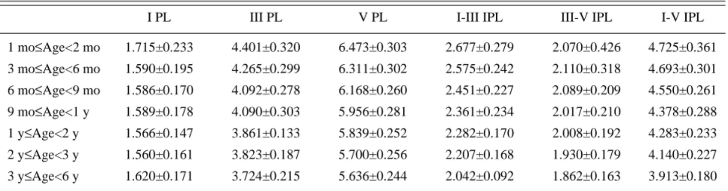
정상 영유아에서 자극 강도가 70 dB, 자극빈도가 10 Hz일 때 뇌간청각유발전위의 연령별 정점잠시와 정점 간잠시의 평균±표준편차(단위: msec)는 다음 표와 같 다(Table 2).
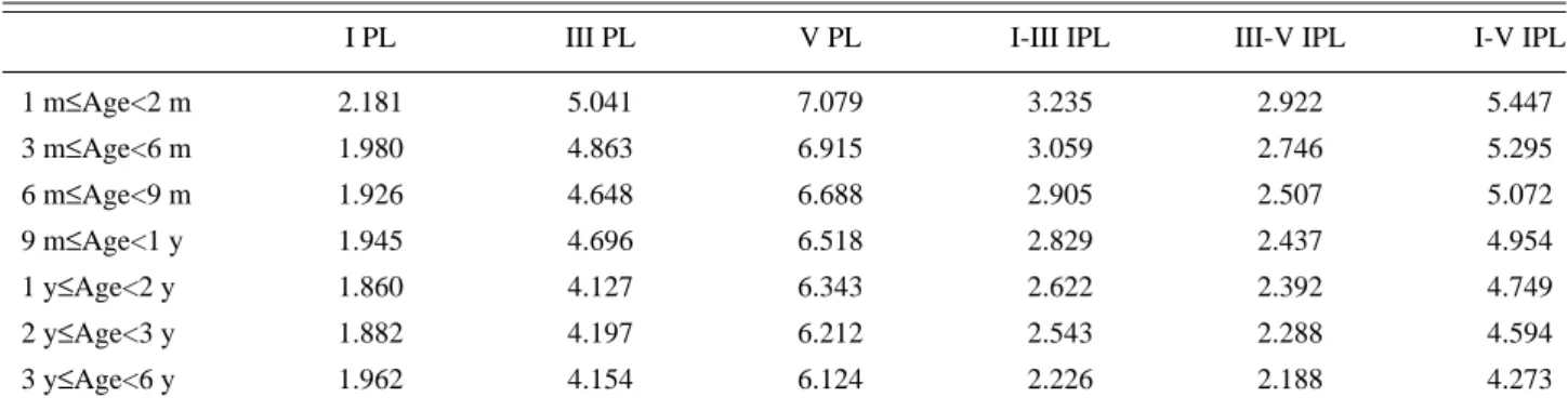
위의 결과를 이용하여 각 연령군의 평균에 표준편차 의 두배를 더하여 정상 참고치를 구하였고(Table 3), Delisa(1994)가 발표한 성인(정상 성인: 15세~51세)의 정 상 참고치와 비교하였다(Fig. 1, 2).
2. 검사실간 검사방법의 차이
1) 전극의 위치
활성전극의 위치가 밝혀진 총 22개 논문 중에서 유양 돌기가 총 11례(50%)로 가장 많은 빈도였고, 귓불이 5 례, 두정부가 4례, 윗이마가 1례였다.
기준전극의 위치는 22례 중에서 두정부가 가장 많은 13례(59%)였고 유양돌기, 윗이마, 귓불이 각각 3례였다.
접지전극의 위치는 총 21례 중에서 반대편 유양돌기 가 8례(38%), 귓불과 윗이마가 각각 6례였으며 코가 1례 였다.
2) 전극의 종류
각 검사실에서 영유아를 대상으로 사용한 전극은 sil- ver/silverchloride 재질의 표면전극이 17례 중 12례(70.58%) 로 가장 많았다. 그 외에 gold cup 전극과 grass gold disc 전 극, clip 전극이 각 1례에서 사용되었다.
3) 전극간 저항
전극간 저항치를 기술한 10례의 논문 중에서 5000 Ω 이하가 8례(80%)로 가장 많았고, 3000 Ω 이하로 설정한 경우가 2례였다.
4) Filter의 범위
Low filter의 범위는 50~300 Hz 사이에 있었고 High fil- ter는 1500~5000 Hz까지 분포하였다.
5) 자극 빈도
15례에서 10 Hz(71%)를, 2례에서 15 Hz를 사용했고, μ2= 1
Σ[ ni i∫u1+3σ1χfi(χ)d(χ)]
0.9973 Σni i
σ22= 1
Σ[ (ni i-1)∫u1+3σ1(χ-χi)2χfi(χ)d(χ)]
0.9973 Σ(ni i-1)
u1-3σ
1
u1-3σ
1
Table 2. Estimated Means and Standard Deviations of Peak Latencies and Interpeak Latencies in Infants and Early Childhood According to the Age(unit: msec)
I PL III PL V PL I-III IPL III-V IPL I-V IPL
1 mo≤Age<2 mo 1.715±0.233 4.401±0.320 6.473±0.303 2.677±0.279 2.070±0.426 4.725±0.361 3 mo≤Age<6 mo 1.590±0.195 4.265±0.299 6.311±0.302 2.575±0.242 2.110±0.318 4.693±0.301 6 mo≤Age<9 mo 1.586±0.170 4.092±0.278 6.168±0.260 2.451±0.227 2.089±0.209 4.550±0.261 9 mo≤Age<1 y 1.589±0.178 4.090±0.303 5.956±0.281 2.361±0.234 2.017±0.210 4.378±0.288 1 y≤Age<2 y 1.566±0.147 3.861±0.133 5.839±0.252 2.282±0.170 2.008±0.192 4.283±0.233 2 y≤Age<3 y 1.560±0.161 3.823±0.187 5.700±0.256 2.207±0.168 1.930±0.179 4.140±0.227 3 y≤Age<6 y 1.620±0.171 3.724±0.215 5.636±0.244 2.042±0.092 1.862±0.163 3.913±0.180 PL: peak latency
IPL: interpeak latency
나머지에서는 다양한 자극 빈도를 보였다(13 Hz, 22 Hz, 33 Hz 등).
6) 자극 강도
선택되어진 고강도 자극중 10례(33%)에서 70 dB을, 7 례(24%)에서 60 dB을 사용하였고, 5례에서90 dB, 4례에 서 80 dB을 을 사용하였다.
7) 총 자극 횟수
총 자극 횟수에 대하여 언급한 21례의 논문 중에서 1024회가 8례, 2000회가 6례, 2048회가 3례였으며 1024 회와 2048회를 동시에 사용한 논문이 2례였고, 2400회 와 1000회가 각기 1례였다.
8) Stimulation polarity
Stimulation polarity에 대하여 언급된 11례의 논문 모 두에서 rarefaction click을 이용하였다.
9) 자극 기간(Pulse duration)
14개의 논문 중 13례(92.85%)에서 100 μs로, 1례에서 60 μs로 선택하였다.
10) Stimulation mode
언급된 20례의 논문에서 17례(85%)는 monoaural stim- ulation을 3례는 biaural stimulation으로 자극하였다.
11) 소리전달 도구
언급된 21례중 17례(80.95%)는 earphone을, 나머지 4 례에서는 headphone을 사용하고 있었다.
12) 검사 자세
모든 경우 앙와위를 선택하였다.
Fig. 1. Change of estimated normal reference value(Mean±2 SD) of each peak latency of brainstem auditory evoked potentials in infancy and early childhood.
Fig. 1.* Adult value(Delisa JA: Manual of nerve conduction velocity and clinical neurophysiology, 3rd ed, Raven press, 1994:297).
Fig. 2. Change of estimated normal reference value(Mean±2 SD) of interpeak latencies about brainstem auditory evoked potentials in infants and early childhood.
Fig. 2.* Adult value(Delisa JA: Manual of nerve conduction velocity and clinical neurophysiology, 3rd ed, Raven press, 1994:297).
Table 3. Estimated Reference Values(Mean+2SD) of Peak Latencies and Interpeak Latencies in Normal Infants and Early Childhood According to the Age(unit: msec)
I PL III PL V PL I-III IPL III-V IPL I-V IPL
1 m≤Age<2 m 2.181 5.041 7.079 3.235 2.922 5.447
3 m≤Age<6 m 1.980 4.863 6.915 3.059 2.746 5.295
6 m≤Age<9 m 1.926 4.648 6.688 2.905 2.507 5.072
9 m≤Age<1 y 1.945 4.696 6.518 2.829 2.437 4.954
1 y≤Age<2 y 1.860 4.127 6.343 2.622 2.392 4.749
2 y≤Age<3 y 1.882 4.197 6.212 2.543 2.288 4.594
3 y≤Age<6 y 1.962 4.154 6.124 2.226 2.188 4.273
PL: peak latency IPL: interpeak latency m: month
13) 수면유도
수면이 이루어지지 않은 아이들은 myogenic potential 을 막기 위해 수면유도를 하였다.
수면 유도법이 언급된 12례의 논문 중 6례(50%)에서 chloral hydrate를 경구복용(45~70 mg/kg)시켰고 4례에서 는 chloral hydrate를 10%로 희석하여 0.5~1 cc/kg만큼 경 직장 투여 하였다. 그리고 각 1례에서 valium이나 prome- thazine을 사용하였다.
14) Masking noise
masking에 대하여 언급된 8례의 논문 중, 정확한 방법을 밝힌 것은 4례였다. 반대편 귀에 자극하는 강도보다 40 dB만큼 적게 사용한 것이 2례(50 %), 30 dB만큼 적게 사용 하는 것과 20 dB만큼 적게 사용하는 경우가 각 1례였다.
고 찰
성인에 비하여 영유아들을 상대로한 뇌간청각유발전 위의 검사방법은 상대적으로 표준화되지 못한 것이 사 실이었다. 또한 성장에 따른 각 연령별 정상 참고치의 제시가 요구되는 상황에서 뇌간청각유발전위검사 결과 의 검사실간 단순 비교는 아쉬움이 남아 있었다.
본 논문은 성인에 비하여 참고치의 설정이 어려운 영 유아를 상대로 좁은 연령간격을 유지하여 정상 참고치 를 설정한 것과 각 검사실의 검사방법중에서 가장 보편 적으로 사용하고 있는 방법을 확인함으로써 향후 각 검 사에서 유용하게 쓰일 수 있는 참고자료를 마련하고자 하였다.
연구 방법이 실제로 살아있는 영유아를 대상으로 하 지 않고, 간접적으로 기존에 발표된 논문을 이용하여 메 타분석을 할 수 밖에 없었던 대상 선정 과정상의 고충을 우선 밝혀두고자 한다. 연구대상을 선정할 때 성인이 아 Table 4. Electrode Setting Differences Among Collected Archives
No. of study Electrode type
Site of Site of Site of Interelectrode
active electrode reference electrode ground electrode impedance(Ω)
01 silver chloride upper
mastoid contralateral
<5000
surface forehead mastoid
02 silver disc surface earlobe midforehead earlobe <5000
03 silverchloride mastoid vertex forehead <3000
04 – earlobe vertex nose –
05 – mastoid Cz – –
06 surface mastoid vertex forehead –
07 surface mastoid vertex earlobe –
08 – Cz mastoid FPz –
09 silver disc mastoid vertex mastoid –
10 silver disc earlobe midforehead earlobe <5000
11 silver disc mastoid forehead mastoid <5000
12 gold cup mastoid vertex mastoid <5000
13 silver surface mastoid vertex mastoid <5000
14 – mastoid vertex mastoid –
15 surface mastoid Cz Fz –
16 surface Cz earlobe forehead –
17 – – – – <3000
18 disc vertex mastid mastoid –
19 grass gold disc Cz A1 A2 –
20 disc earlobe vertex earlobe –
21 silver disc Cz earlobe forehead <5000
22 clip earlobe vertex earlobe
23 – mastoid vertex mastoid <5000
닌 어린아이를 대상으로 한다는 것은 보다 어려우며, 그 것도 정상적인 어린아이를 선정하기는 더더욱 그러하 다. 왜냐하면 아무런 신체적 이상이 없는 정상적 어린아 이를 실험 대상으로 선뜻 허락할 부모는 아무도 없기 때 문이다.
지금까지 발표된 여러 논문들에서 가장 많이 사용하 는 검사방법이 가장 정확한 방법이라고 확언할 수는 없 다. 하지만 검사실마다 여러 가지 방법으로 검사가 진행 되는 환경이라면 검사실간 검사방법의 비교를 통하여 가장 보편적으로 이용되는 방법을 알아봄으로써, 향후 검사시 참고자료로 이용할 수 있고 보다 많은 검사실에 서 이런 보편적 방법을 이용한다면 검사환경의 표준화 를 이룰수 있으며, 결과의 비교도 더욱 쉽게 객관적으로 될 것이다.
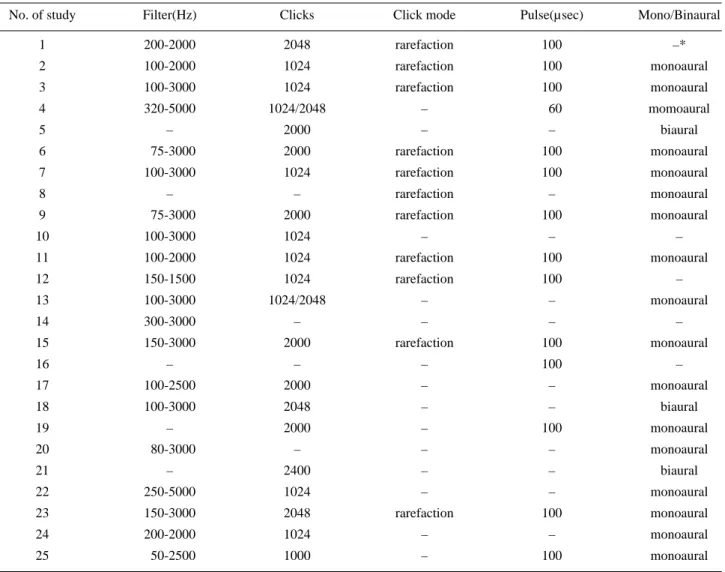
메타분석의 장점은 보다 많은 표본수를 다룰 수 있으 며 다수를 상대로 연구할 때 소요되는 시간적 경제적 비 용을 절감할 수 있고, 각 검사실간에 발생할 수 있는 편 차를 줄일 수 있다는 것이나 가상적인 거대 집단으로 통 Table 5. Characteristics of Filtering and Stimulation Method Among Collected Archives
No. of study Filter(Hz) Clicks Click mode Pulse(µsec) Mono/Binaural
1 200-2000 2048 rarefaction 100 –*
2 100-2000 1024 rarefaction 100 monoaural
3 100-3000 1024 rarefaction 100 monoaural
4 320-5000 1024/2048 – 060 momoaural
5 – 2000 – – biaural
6 075-3000 2000 rarefaction 100 monoaural
7 100-3000 1024 rarefaction 100 monoaural
8 – – rarefaction – monoaural
9 075-3000 2000 rarefaction 100 monoaural
10 100-3000 1024 – – –
11 100-2000 1024 rarefaction 100 monoaural
12 150-1500 1024 rarefaction 100 –
13 100-3000 1024/2048 – – monoaural
14 300-3000 – – – –
15 150-3000 2000 rarefaction 100 monoaural
16 – – – 100 –
17 100-2500 2000 – – monoaural
18 100-3000 2048 – – biaural
19 – 2000 – 100 monoaural
20 080-3000 – – – monoaural
21 – 2400 – – biaural
22 250-5000 1024 – – monoaural
23 150-3000 2048 rarefaction 100 monoaural
24 200-2000 1024 – – monoaural
25 050-2500 1000 – 100 monoaural
*: Not presented
Table 6. The Order of Frequency in the Use of a Examination Method in Collected Archives.
Electrode
Composition silver/silverchloride(71%), gold cup(6%)
Aactive electrode mastoid(50%), earlobe(23%), vertex(18%)
Reference electrode vertex(59%), mastoid(14%), earlobe(14%)
Ground electrode mastoid(38%), earlobe(29%), forehead(29%)
Electrode resistance < 5000 Ω(80%)
Filter 100 Hz(40%) - 3000 Hz(55%)
Stimulation frequency 10 Hz(71%), 15 Hz(9%) Stimulation intensity 70 dB(33%), 60 dB(24%),
90 dB(20%)
Stimulation mode monoaural(85%), biaural(15%)
합할 때 이 통합된 집단에 속하는 각 표본집단의 결과 도출 과정이 비교적 동일해야한다. 여러 표본집단의 모 든 연구 방법이 동일하다면 가장 이상적이나 서로 다른 연구자에 의하여 이루어지므로 어느정도 검사방법상의 차이는 존재한다. 본 연구에서도 분석의 정확성을 높히 기 위하여 결과에 영향을 끼칠 수 있는 인자들 중 많은 논문들이 공통적으로 지적하고 있는 중요한 인자(자극 강도와 빈도, 대상의 연령)만을 선별하여 그에 따른 결 과를 표본집단으로 삼아 메타분석에 이용하였다.
정상 참고치의 설정은 검사의 목적이 선별검사일 경 우는 정상치의 한계를 좁게 잡아(Mean±1SD) 의심되는 대상을 가능한 많이 포함시키는 것이 유익하며, 확진이 목적인 경우에는 정상치의 한계를 넓게 설정하여 이 범 위를 벗어난 값을 비정상 범주로 설정하는 것이 좋을 것 이다. 본 연구에서는 Mean±2SD를 사용하여 97.5 per- centile에 속하는 군을 정상 범주로 삼았으나 필요에 따 라 위에서 설명한 바와 같이 어느 정도는 정상범주를 조 정할 수 있을 것이다.
Lev 등과 Salamy 등이 지적했듯이 본 논문에서도 나 이가 증가하면서 정점잠시의 감소가 모든 파들에서 나 타났고 제 V파에서 가장 일관된 모습을 보였다. 이것은 연령이 증가하면서 신경계의 성숙이 이루어져 전도속 도가 빨라졌음을 의미하며 이중에서 가장 민감한 변화 를 보이는 제 V파의 잠시는 연령 증가에 따른 전도속도 의 변화를 가장 정확히 보여주는 것이다.
제 I파와 III파의 정점잠시가 유아기로 들어서면서 발 생한 작은 기복의 이유로는 가상집단의 모든 연령군에 표본 집단의 모든 연령군이 포함되어야 하지만 자료가 없는 일부 논문의 연령군은 메타분석에서 결측처리가 되어 기복이 발생할 수 있음을 생각할 수 있고, 제 I파는 대부분 생후 1년을 전후하여 성인의 유형에 가장 먼저 접근하기 때문에 보다 늦은 시기에 성인의 유형에 접근 하는 다른 파들과 동등하게 제 I파를 보정한 것이 만 1년 이후의 연령대에서 작은 오차를 만들 수 있었다.
Lina-Granade 등(1993),28 Gorga 등(1989)29과 Hung (1989)30은 자극 빈도와 정점간 잠시 그리고 정상 소아의 유형에서 6~9개월 이후에는 청각계 성숙(auditory matu- ration)이 말초신경보다 중추신경 수준(central level)에서 많이 일어난다고 하였다. 이런 주장은 말초신경의 성숙 도를 나타내는 제 I파보다 중추신경의 성숙도를 나타내 는 제 V파와 제 III파의 잠시가 이 시기 이후에 더욱 단 축됨을 말하며 결과적으로 정점간 잠시의 비 III-V/I-III 은 정상적으로 1 미만의 값을 갖게 된다. 동일한 검사 조 건에서 Delisa14가 발표한 성인의 정상 참고치와, 만 3~6 년에서 메타분석을 이용하여 얻은 참고치간에는 별 차 이가 없음을 알수 있다(Fig 1, 2). 이것은 논문마다 소아 의 뇌간청각유발전위 유형이 성인의 유형에 근접하는 시기에 대하여 생후 만 1년에서 4년까지 다소 시간적 차
이를 두고 이야기하고있으나 근골격계나 림프계, 생식 계 등 다른 계통보다 신경계가 가장 먼저 생후 4년에 성 인의 80%까지 성장하는 사실46을 고려하면 본 연구에서 선정한 생후 만 3~6년에는 신경계 성장이 상당히 이루 어져 성인의 값에 근접한 것이라 할 수 있다.
여러 논문에서 가장 널리 다루어진 검사방법이 이상 적인 방법이라고 단언하기는 어렵지만 아직까지 각 검 사실마다 검사법이 통일되지 않은 상황에서 보편적으 로 사용되고 있는 방법과 연령별 정상 참고치의 제시는 여러 검사실에서 보조적 척도로 사용될 수 있을 것이다.
결 론
논문 검색과 메타분석을 통하여 영유아기의 뇌간청각 유발전위의 정상 참고치를 제시하였으며 검사 방법에 대한 통계적 자료를 구하였다. 본 논문의 결과는 여러 검사실에서 유용한 참고 자료로 사용될 수 있을 것이다.
REFERENCES
01. Sohmer H, Feinmesser M: Cochlear action potentials from the external ear in man. Isr J Med Sci 1970: 6: 219-223 02. Jewett DL: Human auditory evoked potentials: possible
brainstem components detected on the scalp. Science 1970:
167: 1517-1518
03. 채수곤, 나영호, 정사준, 안창일: 영아기 청성뇌간반응의 발 달적 변화에 관한 연구. 대한소아과학회지1986: 29: 1114- 1129
04. Lev A, Sohmer H: Sources of averaged neural responses recorded in animal and human subjects during cochlear audiometry(Electrocochleogram). Arhiv fur klinische and Experimentelle Ohren-nasen-unel Kehlkopfheikunde 1972: 201: 79-90
05. Buchwald JS, Huang CM: Far field acoustic responses.
Science 1976: 189: 382-384
06. Stockard JJ: Detection and localization of occult lesions with brainstem auditory responses. Mayo Clin Proc 1977:
52: 761-769
07. Karl EM: Auditory Evoked Potentials. In: Spehlmann, Karl EM, Editors, Spehlmann,sEvoked Potential Primer, 2nd ed, Newton: Butterworth Heine mann, 1994, pp113-160 08. Starr A, Achor LJ: Auditory brainstem responses in neuro-
logical disease. Arch Neurology 1975: 32: 761-768 09. Gilroy J, Lynn GE, Ristow GE, Pellerin RJ: Auditory
evoked brainstem potentials in a case of “locked-in” syn- drome. Arch Neurology 1977: 34: 492
10. Stockard JJ, Rossiter VS: Clinical and pathologic corre- lates of brainstem auditory response abnormalities. Neu- rology 1977: 27: 316-325
11. Jewett DL, Romano MN: Neonatal development of audito- ry system potentials averaged from the scalp of rat and cat.
Brain Res 1972: 36: 101-115
12. Lieberman A, Sohmer H, Szabo G: Cochlear audiome- try(Electrocochleography) during the neonatal period. Dev Med Child Neurology 1973: 15: 8-13
13. Salamy A, McKean CM: Postnatal development of human brainstem potentials during the first year of life. Electroen- cephalogr Clin Neurophysiol 1976: 40: 418-426
14. Delisa JA: Manual of nerve conduction velocity and clini- cal neurophysiology. 1994: 297
15. Heon Kim, Soo-Hun Cho: Estimation of the geometric means and the reference values of blood lead levels among koreans. J Korean Med Sci 1994: 9: 304-312
16. 오 정근: Meta-analysis를 통한 두피기록 후경골신경 체성감 각 유발전위의 정상치에 관한 연구. 충북대학교대학원 석사 학위 논문1999
17. 박은숙, 박창일, 신정순, 조병국: 정상 영아에서의 뇌간청각 유발전위의 검사에 대한 검토. 대한재활의학회지1992: 16:
123-133
18. Michael PG, Kaminki JR, Kathryn LB, Walt J, Stephen TN: ABR from children three months to three years of age-Normal patterns of response II. J Speech Hear Res 1989: 32: 281-288
19. Michael B, Mordechai ZH, Shlomit G, Eliahu S: Maturation of auditory brainstem potentials in neonates and infants.
Intern J Pediatr Otorhinolaryngol 1985: 9: 69-76
20. Marilyn CZ, Donald EM, Judy RD: ABR characteristics in developing infants. Ann Otorhinolaryngol 1987: 96: 291- 299
21. Jacques T, Robert C: BAER - Normative study in children and adults. Electroencephalogr Clin Neurophysiol 1987:
68: 479-484
22. O,Donovan CA, Beagley HA: Latency of brainstem res- ponse in children. British J Audiol 1980: 14: 23-29 23. Tandon OP, Krishna S: Brainstem auditory evoked poten-
tials in children-A normative study. Indian Pediatr 1990:
27: 736-740
24. John TJ. Normative aspects of the pediatric auditory brain- stem response. J Otolaryngol 1985: 14: 7-11
25. Jiang ZD, Zheng MS, Sun DK, Liu XY: BAERS from birth to adulthood-Normative data of latency and interval.
Hearing Res 1991: 54: 67-74
26. Jiang ZD, Wu YY, Zheng WS, Sun DK, Feng LY, Liu XY:
The effect of click rate on latency and interpeak interval of
the BAEPs in children from birth to 6 years. Electroen- cephalogr Clin Neurophysiol 1991: 80: 60-64
27. Hecox K, Galambos R: Brainstem auditory evoked res- ponses in human infants and adults. Arch Otolaryngol 1974: 99: 30-33
28. Lina-Granade G: Maturation and effect of stimulus rate on brainstem auditory evoked potentials. Brain Dev 1993:
15(4): 263-9
29. Gorga MP: ABR from children three months to three years of age. J Speech Hear Res 1989: 32(2): 281-9
30. Hung KL: Develoment of auditory brainstem evoked response in normal Chinese children. Chung Hua Min Kuo Hsiao Erh Ko I Hseueh Hui Tsa C hih 1989: 30(1): 23-9 31. 김국기, 임영진, 김태성, 김광명, 이봉암, 임언: 유발전위반응
의 정상치. 대한의학협회지1983: 29: 865-874
32. 문혜원, 장순자, 송병두, 김연희, 김봉옥: 정상인과 만성뇌손 상환자에서의 뇌간청각유발전위의 검토. 대한재활의학회지 1989: 13: 284-295
33. 민정식, 나영호, 배종우, 정사준, 안창일: 소아기 청성뇌간반 응의 발달적 변화에 관한 연구. 대한소아과학회지1987: 30:
1387-1400
34. 신지철, 전세일, 신정순: 뇌손상환자의 예후예측에 대한 청각 유발전위의 의의. 대한재활의학회지1989: 13: 201-213 35. Barbara M, Carol SG, Robert G: BAEPs in children. Arch
Otolaryngol 1977: 103: 38-43
36. Christianto BL, Michael K, Carl S, Ibrahim D, Wolfgang JB: Measurements of BAEPs in infancy, childhood, and adolescence. Child,s Nerv Syst 1985: 1: 337-340
37. Jacques T, Robert C: BAER-normative values in children.
Elecroencephalogr and Clin Neurophysiol 1990: 77: 309- 313
38. John R: Normal variability of the BAER in young and old adult subjects. Electroencephalogr Clin Neurophysiol 1978:
44: 459-470
39. Pierri F, Rosignoli M, Almadori G, Maurizi M, Ottaviani F, Paludetti G: Effects of sex on ABRs in infancy and early childhood. Scand Audiol 1988: 17: 143-146
40. Salamy A: Maturation of the ABR from birth through early childhood. J Clin Neurophysiol 1984: 1: 293-329 41. Stephane M, Roland D, Chantal FV, Christian D: Clinical
interest of BAEPs in 72 children with inadequate language development. Intern J Neurosci 1996: 88: 261-272 42. Susan RL: BAEPs in pediatrics. In: Chiappa KH. Evoked
potentials in Clinical Medicine. 3rd ed, Philadelphia: Lip- pincott-Raven Publishers, 1997 pp269-282
43. Ulf R, Gote B, Kai P, Anders K: BAEPs in different age groups. Electroencephalog Clin Neurophysiol 1985: 62:
426-430
44. Walter CO, Geary AM: Aging and the auditory brainstem response. Audiology 1982: 21: 466-473
45. Taylor MF: Evoked Potentials in Pediatrics. In: Halliday
AM, Evoked Potentials in Clinical Testing, 2nd ed, New York: Churchill Livingstone, 1993. pp489-492
46. 홍창의: 소아과학, 개정7판, 대한 교과서, 2001, pp23-24