소아 상완골 외과 골절의 수술적 치료 결과
김동수・정국진・안종국・정병현・성열보・정형진・권칠수
인제대학교 부속 상계 백병원 정형외과학 교실
= Abstract =
The Result After Surgical Treatment
on Lateral Condyle Fracture of Humerus in Children
Dong Soo Kim, M.D., Kook Jin Chung, M.D., Jong Guk Ahn, M.D., Byung Hyun Jung, M.D., Yeol Bo Sung, M.D., Hyung Jin Chung, M.D.,
Chil Soo Kwon, M.D.
Department of Orthopedics, Sanggye Paik Hospital, Inje University
Fracture of the lateral condyle of humerus in children are common injury next to supracondylar fracture in children, account for 10 to 15% about the elbow and 18 to 20% about distal humerus fractures. This is the one fracture that can be overlooked clinically and that has a high potential for nonunion and cubitus valgus deformity.
Sixty children treated by closed or open reduction with internal fixation on the lateral condyle fracture of humerus from January 1994 to August 1997, were reviewed. Almost fractures were treated within 12 hours after injury for the purpose of prevention of further displacement and occurrence of complication.
According to Hardacre et al’s criteria, most patients showed excellent and good results. We report the result after surgical treatment on the lateral condyle fracture of humerus in children.
Key Words: Fracture, Lateral condyle, Humerus, Children
※통신저자 : 김동수
서울특별시노원구상계동761-1 (139-207) 인제대학교상계백병원정형외과학교실 Tel : (02) 950-1032
Fax : (02) 934-6342
* 본논문의요지는 1 9 9 8년도대한정형외과학회추계학술대회에서구연됨
서 론
소아에서의상완골외과골절은과상부골절다음 으로많은골절로서소아에서주관절주위골절의 1 0 내지 1 5 %를 차지하며 원위 상완골 골절의 18 내지 20% 를차지하는골절이다. 상완골외과골절은임상 적으로골절의전위가없거나적을경우에는이를간 과하기가쉬우며이로인한부적절한치료및골절의 특수성으로 인하여 불유합, 무혈성 괴사, 조기 성장 판 유합 및 지연성 척골 신경 마비와 외반주등의 불 량한결과를초래할수있는골절이다 .
이골절의 치료방법으로는 장상지석고고정의 비 수술적 방법, 경미한전위를 보이는골절은 비관혈적 정복후 경피적 내고정술, 심한전위가있는 경우에는 관혈적정복후내고정술이치료법으로사용되고있다.
골절의 특성상대부분의경우수술적 치료를요하 게되는데저자들은본원에서수술적 치료를시행한 6 0례를바탕으로소아상완골외과골절의수술적치 료후결과를문헌고찰과함께보고하는바이다 .
재료 및 방법
1 9 9 4년 1월부터 1 9 9 7년 5월까지 본 교실에서 수술 적 치료를 받았으며 1년이상의 추시가 가능했던 소 아상완골외과골절 6 0례를대상으로하였다.
성별분포는남아 39 례, 여아 21 례로 1.8 : 1의비율 을 보였고 연령분포는 1 1개월부터 1 1세 3개월로 평
균 4 . 9세였다
수상 원인은 실족 사고가 4 5례(75%), 추락 사고가 1 2례(20%), 교통사고가 3례 ( 5 % )였으며우측이 3 0례, 죄측이3 0례로같은비율을보였다.
골절의대부분은M i l c h1 0 )분류에따른제 1형이 6례 (10%), 제2형이 5 4례( 9 0 % )로 제2형이 대부분을 차지 하였으며 전례에서 모두 폐쇄성 골절이었으며 동반 손상은없었다.
수술적 치료시기는 4 3례( 7 2 % )가 1 2시간이내, 12례 ( 2 0 % )가 2 4시간이내, 5례(8%) 가 3일이내에시행되었 다. 수술법은비관혈적방법의경우경피적방법으로 관혈적 방법의 경우는 K o c h e r의 외측 J 접근법을 통 해 2 내지 3개의 K 강선을이용하였으며비관혈적정 복이 3 0례, 관혈적 정복이 3 0례의 같은 비율을 보였 으며 장상지 석고 고정 및 K -강선의 평균 제거 시기 는 6 . 3주였다. 골유합은최단 5주에서최장 1 1주로평 균 7 . 8주였다
결 과
결과판정은술후운동범위의소실여부 , 운반각의 변화와무혈성괴사및척골신경증세의동반유무를 토대로한 H a r d a c r e8 )등의기준(Table 1)을따랐다.
비관혈적 정복후 내고정을 시행했던 3 0례에서는 2 m m이상의 전위를 보이나 연골경첩에 의해 관절면 이 유지되는 1례를 포함한 모두가 우수군에 해당하 는결과를보였으며, 관혈적정복후내고정을시행했
Table 1.Evaluation of Results (by Hardacre)
Results No(%)
Excellent No loss of motion, No symptoms 57(95%)
No alteration in the carrying angle
Good Lacking no more than 15 degrees of full extension 2(3%) Incospicuously altered carrying angle
No arthritic or neurological symptoms
Poor Disabling loss of motion 1(2%)
Conspicuous alteration of the carrying angle Arthritic symptoms or ulnar neuritis Nonunion or avascular necrosis
던 3 0례중양호군에속하는술후운동범위의제한을 보인 2례와, 불량군에 속하는불유합1례의합병증이 발생하였다. 운동 범위의 제한을 보인 2례는 K -강선 의제거후 15, 20도의일시적인굴곡구축소견을보 였으나추시결과완전한운동범위의회복을보였으 며 전례에서 운반각은 정 상측 1 3 . 6도, 환측 1 3 . 9도로 의미있는 차이는 없었다. 비관혈적 정복 및 관혈적 정복모두에서척골신경마비를포함한추시중치료 를필요로하는창상감염 , 조기성장판유합 , 골주형 성, 소두과성장및외반주변형은발생하지않았다 .
고 찰
소아에서의상완골 외과골절은임상적으로골절 의전위가없거나전위의정도가불확실한경우에이 를간과하기쉬운데이경우불유합으로의발전가능 성이높으며불량한결과를초래할수있다 .
D a l l e k4 )등은상완골 원위골단은해부학적으로교 원질묶음이상완골 소두의상부에서상완골활차의
하부로향하고있다는보고를하였다. 이는전형적인 상완골외과골절의위치가골간단부에서소두의상 부를 지나 상완골 활차부위를 일부 통과 하는 S a l t e r - Harris 제 4 형 골단판 손상을 유발하는 것과 일치한 다.
M i l c h 11 3 , 1 4 )는 골절의 부위에 따라 골절선이 상완 골소두의골화중심을통과하는제 1 형과골절선이 상완골 활차까지 연장된 제 2 형으로 분류하였다.
Jacob 등1 0 )과 M c I n t y r e1 2 )는 골절의 전위 정도에 따른 분류를하였다.
또한상완골외과골절의특성상전위가심할경우 에골절편이상완골과의접하는면이골과골의접촉 보다는 골과연골의접촉이 대부분을 이루기때문에 골유합의가능성이떨어지며, 골절판이대부분연골 로 구성되어있고골은 골간단의일부에만있으므로 혈류의공급이부족하여골절의유합에 불리한환경 을형성하게되어불유합, 무혈성괴사등의합병증이 발생할가능성이높다.
상완골골절은골절의특성상관절내골절로관절 액이골유합과정에악영향을미치며, 신전근의지속
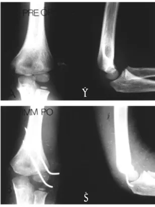
Fig 1.Case 1. 6 year old boy.
FigA. Preoperative x-ray showed Milch type II lateral condyle fracture with minimal displacement of fragment of fracture.
FigB. Emergency operation was done after 12 hours from injury. In situ percutaneous pinning with K-wires was performed.
FigC. We have got excellent reslut in view of symptoms, carrying angle and function according to criteria by Hardacre et al at one year after operation.
A
B
C
적인 견인으로 골절편의 전위가 발생할 수 있어
Finbogason 5 )등은전위가없는골절에서부목고정후
에도부가적인전위가발생한다고하였으며수상후 3주까지도전위의발생이 있을수있다고하였다 . 또 한정도의차이는있지만모든경우에서어느정도의 전위는발생하는것으로보고하고있다.
수술시내고정방법으로는봉합에의한고정, 나사 못 고정 그리고 K -강선에 의 한 고정으로 대별된다.
C r a b b e3 )는골절의내고정물로봉합에의한고정과강
선에의한고정의비교결과차이가없음을주장하면 서고정후제거술이필요치않은봉합고정술을주장 하였다. 그러나 S h a r p1 8 )는 봉합에 의한 고정은 불유 합이 증가한다고 보고하고 나사못 고정을 주장하였 으나최근에는 K -강선에의한고정이보편화된것으 로생각된다.
이상과같은근거하에최근에는경미한전위가있 는경우뿐아니라전위가없는골절에서도경피적 K - 강선고정술을시행하는추세이다.
치료의방법의결정에있어서골절의전위가중요
한 요소가 되는데 F o s t e r5 )는 비 전위 골절의 기준을 최대 2 m m로 하였으며 F l y n n6 )은 3 m m이상의 초기 전 위를보일경우 2차전위의 가능성이 높다고하였다.
이에 2 m m이내의전위를보이는경우에는경피적 K - 강선고정술을, 3mm이상의전위를보이는경우는관 혈적정복후 K -강선고정술을시행하는것이 바람직 한것으로받아들여지고있다.
한편 M i n t z e r1 4 )등은 2 m m이상의 전위가 있는 경우 라도원위상완골에부착된 골편을유지하는연골경 첩에의해관절면의일치가유지되는경우에는선택 적으로경피적 K -강선고정술로도 좋은결과를얻을 수있는것으로보고하였다.
본원에서수술적치료를 받은소아의상완골 외과 골절 6 0례중 3 0례가 경피적 K- 강선 고정술을 시행 받았으며나머지 3 0례에 대해서는 관혈적정복후 K - 강선 고정술을 시행하였으며 대부분의 경우 H a r d a c r e7 )등의기준에따른우수군이상의결과를 보 였다. 골절의추시결과골유합은모든례에서우수하 였다.
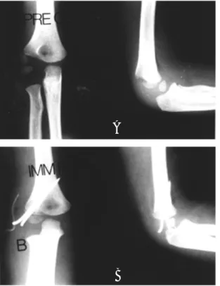
Fig 2.Case 2. 5 year old girl
FigA. Preoperative x-ray showed Milch type II lateral condyle fracture with displacement beyond 2mm of fragment of fracture but, articular congruency was maintained by cartilaginous hinge.
FigB. Emergency operation was done with closed reduction and internal fixation with K-wires.
FigC. At 15 months after operation, the result was excellent.
A
B
C
결 론
저자들은 인제대학교상계백병원정형외과교실 에서 수술적 치료를 받은 소아 상완골 외과 골절 6 0 례를통해분석한결과진정한의미의비전위골절은 드물고, 방사선촬영상골절선의증거가보이거나경 미한 전위가 보이는 골절은 경피적 내 고정술을, 2 m m이상의 전위를 보이는 골절의 경우에는 관혈적 정복후내고정술을시행하였으며, 2mm 이상의 전위 를 보이는 골절이라도 연골 경첩에 의해 관절 면의 일치성을보이는경우에는경미한 전위골절에준하 는경피적내고정술을시행하였다.
그 결과 골절의 유합 시기는 7 . 8주였으며 방사선 추시상건측의 1 3 . 6도에대해환측의운반각은 1 3 . 9도 로 의미있는 변화는 없었고 2례에서 K -강선 제거 직 후일시적인운동범위의제한을보였으나추시중완 전한운동범위로의회복을보였다.
H a r d a c r e8 )등의기준에 따라총 6 0례중 5 7례( 9 5 % )가 우수, 2례( 3 % )가 양호, 1례 ( 2 % )가 불량군에 속하는
결과를얻었다.
이상에서 전위와회전이동반된골절에 대한관혈 적정복및내고정술외에도전위가없는골절과경미 한전위를보이는골절에도 조기에적극적으로시행 하는 비 관혈적 정복 및 내고정술을 시행하는 것이 추가적인 골절의 전위와 불유합등의 합병증을 예방 할 수 있는 권장할 만한 방법으로 생각되어 문헌 고 찰과함께보고하는바이다.
R E F E R E N C E S
1) Badelon O, Bensahel H, Mazda K, Vie P L a t e r a l humeral condylar fractures in children : a report of 47 cases, J Pediatr. O r t h o p e d 8:31,1988.
2) Conner AN, Smith MG Displaced fractures of the lateral humeral condyle in children, J. Bone Joint S u r g 5 2 - B : 4 6 0 , 1 9 7 0 .
3) Crabbe W.A. Treatmewnt of fracture separation of the Capitular Epiphysis. J. Bone and Joint Surg.
Fig 3.Case 2. 5 year old girl
FigA. Preoperative x-ray showed Milch type II lateral condyle fracture with displacement beyond 2mm of fragment of fracture but, articular congruency was maintained by cartilaginous hinge.
FigB. Emergency operation was done with closed reduction and internal fixation with K-wires.
FigC. At 15 months after operation, the result was excellent.
A
B
C
5 2 : 4 6 0 - 4 6 4 , 1 9 7 0 .
4) Dallek M, Jungbluth KH H i s t o m o r p h o l o g i c a l studies on the development of the radial condyle fracture of the humerus in the growth years. J Unfall C h i r. 16:57,1990.
5) Finnbogason T, Karlsson G, Lindberg L, Mortensson W Nondisplaced and minimally displaced fractures of the lateral humeral condyle in children : a prospective radiographic investigation of fracture stability, J. Pediatr Orthoped 1 5 : 4 2 2 , 1 9 9 5 . 6) Foster DE, Sullivan JA, Gross RH Lateral humeral
condyler fractures in children. J Pediatr Orthop 5 : 1 6 - 2 2 , 1 9 8 5 .
7) Flynn JC, Richards JF, Saltzman RI P r e v e n t i o n and treatment of nonunion of slightly displaced fractures of the lateral humeral condyle in children, J . Bone Joint Surg[Am] 5 7 : 1 0 8 7 - 9 2 , 1 9 7 9 .
8) Hardacre JA,Stanley HN, Froimson AJ, Brown JE Fractures of the lateral condyle of the humerus in children, J. Bone Joint Surg[Am] 53:1083-85,1971.
9) Herring JA Lateral condyle fracture of the elbow, J Perdiatr Orthoped, 6:724-727,1986.
10) Jacob R, Fowlers J, Rang M, Kassab M Observations concerning fractures of the lateral humeral condyle in children. J Bone Joint Surg. 57- B 430, 1975.
11) Marcus NW, Agins HJ Articular cartilage sleeve fracture of the lateral humeral condyle, J. Pediatr O r t h o p e d 4 : 6 2 0 , 1 9 8 4 .
12) McIntyre W Lateral Condyle Fracture of the Humerus. In Letts RM ed. Management of Pediatric Fracture. 1st ed. Churchill Livingston Inc. 241.
1 9 9 4 .
13) Milch H Fractures and fracture-dislocations of humeral condyle, J. Trauma 4 : 5 9 2 , 1 9 6 4 .
14) Milch H Treatment of the external humeral c o n d y l e , J A M A 1 6 0 : 6 4 1 , 1 9 5 6 .
15) Mintzer CM, Waters PM, Brown DJ, Kasser JR Percutneous pinning in the treatment of displaced lateral condyle fractures, J. Pediatr Orthoped 1 4 : 4 6 2 - 4 6 5 , 1 9 9 4 .
16) Roye DP, Bini SA, Infosino A Late surgical treatment of lateral condylar fractures in children, J . Pediatr Orthoped 1 1 : 1 9 5 , 1 9 9 1 .
17) Rutherford A Fractures of the lateral humeral condyle in children J. Bone Joint Surg[Am] 6 7 : 8 5 1 - 6 , 1 9 8 5 .
18) Sharp I.K. Fracture of the lateral humeral condule in children. Acta Orthop. Belg. 31:811, 1965.
19) Silberstein JJ, Brodeur AE, Gravis ER S o m e vagaries of the lateral condyle, J. Bone Joint Surg 6 4 - 1 : 4 4 4 , 1 9 8 1 .