to carcinogens3. The initial presence of a precursor cell sub- sequently developing into cancer is well established in oral cancer4. Oral leukoplakia, submucous fibrosis, and lichen planus are major known precursor lesions. The prevalence of malignant transformation of oral lichen planus is around 0.5%
and approximately 1% for leukoplakia5. Ability to clinically predict malignant transformation is difficult and routine histo- pathological diagnosis has limited prognostic value. Despite advanced technologies for early detection of oral precancer- ous and cancerous lesions, there are limitations for its use, including that diagnosis is essentially subjective, all lesions exhibiting dysplasia do not eventually become malignant and may even regress, and carcinoma can develop from lesions in which epithelial dysplasia was not diagnosed on previous bi- opsies6,7. Therefore, it is necessary to develop other methods for predicting the malignant potential of premalignant lesions and preventive measures7.
Recently, strong evidence has suggested that the nuclear factor-κB (NF-κB) signaling pathway plays a critical role in carcinogenesis, protection from apoptosis, and chemoresis- tance in a number of cancer types, including head and neck
I. Introduction
Oral cancer is the eleventh most common cancer in the world1. Its incidence is predominantly high in northern India, other Asian countries, and in certain places in the western hemisphere. It has been reported that 90% of oral cancers in India among men were attributable to chewing and smok- ing habits. In India, the age-standardized incidence rate of oral cancer is 12.6 per 100,000 people2. Oral squamous cell carcinoma develops through a multi-step process of genetic, epigenetic, and metabolic changes resulting from exposure
Jasdeep Kaur
OMFS-IMPATH Research Group, Department of Imaging and Pathology, Faculty of Medicine, University of Leuven and Oral and Maxillofacial Surgery, University Hospitals Leuven, Herestraat 49, Leuven 3000, Belgium
TEL: +32-16-3-32452 FAX: +32-16-3-32410 E-mail: [email protected]
This is an open-access article distributed under the terms of the Creative Commons Attribution Non-Commercial License (http://creativecommons.org/licenses/by-nc/4.0/), which permits unrestricted non-commercial use, distribution, and reproduction in any medium, provided the original work is properly cited.
CC
Proinflammatory cytokine levels in oral lichen planus, oral leukoplakia, and oral submucous fibrosis
Jasdeep Kaur, Reinhilde Jacobs
OMFS-IMPATH Research Group, Department of Imaging and Pathology, Faculty of Medicine, University of Leuven and Oral and Maxillofacial Surgery, University Hospitals Leuven, Leuven, Belgium
Abstract(J Korean Assoc Oral Maxillofac Surg 2015;41:171-175)
Objectives: The objective of this study was to identify salivary and serum concentrations of interleukin (IL)-8, IL-6, and tumor necrosis factor alpha (TNF-α) in patients with oral lichen planus, oral leukoplakia, oral submucous fibrosis, and healthy controls.
Materials and Methods: Patients selected included 54 oral lichen planus (41 to 65 years), 50 oral leukoplakia (42 to 65 years), 51 oral submucous fi- brosis (41 to 65 years), and 50 healthy controls (42 to 65 years). Oral lichen planus, oral leukoplakia, and oral submucous fibrosis cases were diagnosed using histopathological analysis. Salivary and serum cytokine concentrations were measured using enzyme-linked immunoassay kits in all subjects.
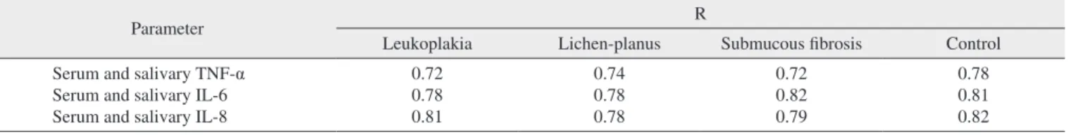
Results: The levels of serum and salivary TNF-α, IL-6, and IL-8 were statistically significantly increased in oral leukoplakia, submucous fibrosis, and lichen planus in contrast to normal healthy subjects (P<0.05). Serum and salivary correlation analysis revealed strong and highly significant correla- tions for TNF-α, IL-6, and IL-8 in all groups (r=0.72-0.82, P<0.05).
Conclusion: Salivary and serum cytokines were also elevated when analyzed in oral precancerous lesions. Thus, salivary and serum IL-8, IL-6, and TNF-α levels might act as diagnostic markers for detection of oral precancer.
Key words: Saliva, Oral lichen planus, Oral leukoplakia, Oral submucous fibrosis, Cytokines
[paper submitted 2015. 1. 4 / revised 2015. 5. 31 / accepted 2015. 6. 2]
Copyright Ⓒ 2015 The Korean Association of Oral and Maxillofacial Surgeons. All rights reserved.
tus was also ascertained. Patients and healthy subjects with matched periodontal status were selected.
All subjects had neither a smoking history nor any visible oral lesions under careful examination. Additionally, they had not received any treatments for the oral lesions within 90 days prior to the specimen collection, and had no history, symptoms, or signs of systemic infections or other diseases.
Histopathological diagnosis was made by a single oral pa- thologist. This study was approved by the ethical committee of Baba Nidhan Singh Hospital, Punjab, India and informed consents were obtained from all subjects as per the Declara- tion of Helsinki.
2. Evaluation during study
For each sample, 1.0 mL of supernatant was used for the enzyme-linked immunoassay (ELISA) cytokine assay by us- ing the human ELISA kit for TNF-α, IL-6, and IL-8 (R&D Systems Inc., Minneapolis, MN, USA), according to the manufacturer’s instructions.
3. Salivary and serum cytokine measurements
Blood and serum samples were drawn between 9:00 and 10:00 AM from all subjects. Serum was separated from blood cells by centrifugation at 1,000 g for 5 minutes. The whole unstimulated saliva was collected between 9:00 and 10:00 AM. The subjects abstained from eating and drinking for at least 2 hours prior to the sampling. All subjects were requested to swallow first, tilt their head forward at more than 45o, and then expectorate saliva (10 mL) into a sterile centrifuge tube without swallowing for 4 to 5 minutes. The saliva was then centrifuged for 25 minutes at 3,500 g, and the clarified supernatants were separated into 2.0 mL aliquots.
All samples were immediately frozen at –60oC for future use.
Prior to assays, the serum or saliva supernatants were allowed cancer, breast cancer, hepatocellular carcinoma, and gastric
cancer8-12. Recently, accumulating evidence has suggested that the NF-κB–dependent cytokine levels are elevated in saliva and tissue specimens of patients with oral premalig- nant lesions13. Furthermore, different cytokines can act as diagnostic tools for detecting oral cancerous and precan- cerous lesions and conditions. Interleukin (IL)-6 levels are significantly increased in oral cancer patients14-17. It has been evident that different cytokines are expressed by cancerous cells, the most common of which include tumor necrosis fac- tor alpha (TNF-α), IL-1, IL-6, and IL-815-17. Limited studies were conducted on roles of TNF-α, IL-6, and IL-8 in oral precancer and cancer15-17. The local and systemic nature of these responses suggests the hypothesis that cytokines with proinflammatory and proangiogenic activity are produced by precancers and cancers and could contribute to the pathogen- esis of oral malignancy. In this study, we hypothesized that salivary and serum TNF-α, IL-6, and IL-8 levels could be elevated in oral precancerous lesions and conditions. To test this hypothesis, this study was conducted to analyze the con- centrations of TNF-α, IL-6, and IL-8 in oral precancer using saliva and serum samples.
II. Materials and Methods
1. Eligibility
Consecutive patients clinically and histopathologically confirmed as having oral lichen planus lesions, oral leukopla- kia, and oral submucous fibrosis were recruited for this study from Baba Nidhan Singh Hospital, Punjab, India, based on the definition of oral cancer and precancerous lesions by the World Health Organization9. Healthy normal patients served as the control group. Demographic characteristics of patient and controls characteristics were mentioned in Table 1. The mean and standard deviation of smoking and alcoholic sta-
Table 1. Demographic characteristics of patients and controls
Variable Oral leukoplakia Oral lichen planus Oral submucous fibrosis Control
Classification
Age (yr) Smokers
(cigarettes/day) Alcoholic status (g of alcohol/day)
50 (benign hyperkeratosis: 7, mild dysplasia: 27, moderate dysplasia:
8, severe dysplasia: 8) 42-65 18±4.6 657.9±159.2
54 (benign hyperkeratosis: 6, mild dysplasia: 16, moderate dysplasia:
14, severe dysplasia: 18) 41-65 21±6.7 593.1±155.6
51 (benign hyperkeratosis: 1, mild dysplasia: 23, moderate dysplasia:
12, severe dysplasia: 15) 41-65 18±4.5 585.9±186.2
50
42-65 21±7.9 569.9±158.8 Values are presented as range or mean±standard deviation.
Jasdeep Kaur et al: Proinflammatory cytokine levels in oral lichen planus, oral leukoplakia, and oral submucous fibrosis. J Korean Assoc Oral Maxillofac Surg 2015
test. The Kruskal Wallis test was utilized for comparisons between the groups. Mann-Whitney test for comparisons performed to detect any differences between the two groups.
A P<0.05 was considered as statistically significant. All data was statistically analyzed using the SPSS version 11.5 (SPSS Inc., Chicago, IL, USA).
III. Results
The demographic characteristics of patient and controls are mentioned in Table 1. The levels of serum and salivary TNF-α, IL-6, and IL-8 were statistically significantly higher in advanced stages of precancerous lesions as compared to early stages (P<0.05).(Tables 2, 3) The levels of serum and salivary TNF-α, IL-6, and IL-8 were also statistically signifi- to thaw completely at room temperature for two hours. For
each sample, 1.0 mL of supernatant was used for the ELISA cytokine assays, using the human ELISA kit for TNF-α, IL- 6, and IL-8, according to the manufacturer’s instructions. The absorbances of the samples at 492 nm for TNF-α, IL-6, and IL-8 were measured with a spectrophotometer (Sirio; Seac, Florence, Italy). A standard curve was organized by plotting the absorbance value of the standards versus corresponding concentrations. The concentrations of the cytokines in the sample were then determined by extrapolating from the stan- dard curve. Tests were run in duplicate to check reliability.
Protein contents were expressed in pg/mL. The inter-assay coefficient of variation was 3.0% to 4.5% and the intra-assay coefficient of variation was 2.5% to 4.2%.
Data normality was evaluated by Smirnoff Kolmogorof’s
Table 3. Salivary levels of NF-κB mediators in leukoplakia, lichen, submucous fibrosis patients and control healthy
TNF-α IL-6 IL-8
Control
Benign hyperkeratosis Mild dysplasia Moderate dysplasia Severe dysplasia
Leukoplakia Lichen-planus Submucous fibrosis Leukoplakia Lichen-planus Submucous fibrosis Leukoplakia Lichen-planus Submucous fibrosis Leukoplakia Lichen-planus Submucous fibrosis
8.51±1.74 12.10±3.96 13.63±2.04
- 14.38±4.27 18.08±5.27 18.41±3.51 18.25±6.50 18.30±4.94 20.37±5.12 18.39±3.58 21.46±5.51 24.33±5.16
16.6±1.88 30.53±6.80 26.32±4.72
- 35.30±7.52 32.71±8.13 34.45±6.72 39.80±10.69 34.04±8.07 35.92±8.32 39.61±6.53 36.06±8.52 42.26±9.03
738.5±98.5 770.74±106.26 1,071.02±82.61
- 849.58±128.76 1,074.95±99.17 1,384.10±93.30 930.61±120.74 1,071.09±91.65 1,347.18±95.92 919.87±107.65 1,116.28±118.17 1,437.82±114.69 (NF-κB: nuclear factor-κB, TNF-α: tumor necrosis factor alpha, IL: interleukin)
All data except ‘Control’ are P<0.05 as compared to control.
Values are presented as mean±standard deviation.
Jasdeep Kaur et al: Proinflammatory cytokine levels in oral lichen planus, oral leukoplakia, and oral submucous fibrosis. J Korean Assoc Oral Maxillofac Surg 2015 Table 2. Serum levels of NF-κB mediators in leukoplakia, lichen, submucous fibrosis patients and healthy controls
TNF-α IL-6 IL-8
Control
Benign hyperkeratosis Mild dysplasia Moderate dysplasia Severe dysplasia
Leukoplakia Lichen-planus Submucous fibrosis Leukoplakia Lichen-planus Submucous fibrosis Leukoplakia Lichen-planus Submucous fibrosis Leukoplakia Lichen-planus Submucous fibrosis
1.31±0.40 1.18±0.30 2.19±0.88
- 2.22±0.44 3.20±0.79 4.12±0.66 3.22±0.53 3.74±0.82 5.18±0.46 3.94±0.34 4.87±0.80 6.27±0.53
5.13±2.11 11.02±3.79 12.95±1.67
- 13.38±4.16 17.10±4.78 17.76±3.63 16.15±5.18 17.01±4.05 18.90±4.91 16.07±3.63 18.79±4.02 21.70±4.04
13.45±2.71 16.77±6.03 22.16±4.54
- 19.49±6.82 23.05±7.48 26.35±5.82 20.22±6.06 21.96±6.11 24.42±6.33 20.05±7.05 23.00±5.90 28.25±6.47 (NF-κB: nuclear factor-κB, TNF-α: tumor necrosis factor alpha, IL: interleukin)
All data except ‘Control’ are P<0.05 as compared to control.
Values are presented as mean±standard deviation.
Jasdeep Kaur et al: Proinflammatory cytokine levels in oral lichen planus, oral leukoplakia, and oral submucous fibrosis. J Korean Assoc Oral Maxillofac Surg 2015
of TNF-α, IL-6, and IL-8 might be protective in action.
Smoking is a risk factor of oral precancerous and cancerous lesions2. However, while smoking and chronic alcohol usage might be risk factors for cancer, this study showed no signifi- cant difference between controls and patients. TNF-α might act as an endogenous tumor promoter as well as an inducer of tissue remodeling required for tumor growth and spread22. IL-6 signaling has also been implicated in tumorigenesis23. TNF-α, IL-6, and IL-8 were also elevated in periodontitis patients21, but in the present study, subjects with periodontitis were excluded. So, the present study shows that the eleva- tion of TNF-α, IL-6, and IL-8 is correlated with precancerous and cancerous lesions. Indeed, elevation of these cytokines in saliva might indicate the potential precancerous development into oral cancer22. Alternatively, these cytokines may be im- properly produced in precancerous lesions, which could lead to induced growth, invasion, disruption of tumor suppression, and immune status. NF-κB activation leads to the upregula- tion of anti-apoptotic genes as a cell survival mechanism, by inducing physiological stress, which triggers an inflammatory response. In addition, NF-κB induces cytokines that regulate TNF-α, IL-6, and IL-8, which leads to the recruitment of leu- kocytes to the sites of inflammation27. There was good cor- relation between salivary and serum TNF-α, IL-6, and IL-8 in all groups. Since saliva can be easily collected, measurement of these biomarkers of diseases may prove useful in early detection of oral cancer risks. The salivary analysis for oral diagnosis may prove a cost effective method for screening large populations24.
Hence, salivary levels of NF-κB mediators were signifi- cantly elevated and may have diagnostic and prognostic util- ity as useful biomarkers for detection of precancerous and cancerous lesions. This study did not take into account factors such as diet, alcoholic history, and environmental factors, so further studies are required on large samples including these variables to determine the relationship between salivary bio- markers and oral cancer, and to further clarify their mecha- nism of action. It can only be concluded that patients with cantly increased in oral leukoplakia, submucous fibrosis, and
lichen-planus in contrast to normal healthy subjects. Serum and salivary correlation analysis revealed a strong and highly significant correlation for TNF-α, IL-6, and IL-8 levels in all groups as shown in Table 4.
IV. Discussion
Serum and salivary TNF-α, IL-6, and IL-8 were higher in advanced stages as compared to early stages of precancerous lesions and conditions, data which supports previous stud- ies14,15-24. TNF-α, IL-6, and IL-8 are potent angiogenic medi- tators with significant effects on tumor growth, and are as- sociated with increased tumor vessel density and a worsened outcome15-18. Thus, these cytokines might act as surrogate biomarkers of angiogenesis and prognosis. It has been previ- ously reported that excessive cell proliferation and activation of cellular actions can be instigated by chronic inflammation, which leads to the induction of irreversible DNA damage25. TNF-α, IL-6, and IL-8 released through the inflammatory response would promote tumor growth, while tumor growth further stimulates the inflammatory response, resulting in cyclic progression26. In the present study, serum and salivary TNF-α, IL-6, and IL-8 levels were upregulated in precancer- ous lesions and conditions. This finding is in line with most previous conducted work14-17, although one study has shown that salivary TNF-α and IL-6 were downregulated18,19. This could be due to a small sample size and not using age- or gender-matched samples. Proinflammatory cytokine levels are elevated in submucous fibrosis due to immunoregula- tory activity. Increased levels of NF-κB mediators might be associated with the development of oral precancerous and cancerous lesions. In the normal cell, stimulation of cyto- kines causes growth inhibition, while in oral cancer cells, stimulation of cytokines leads to upregulation of positive cell cycle regulators including NF-κB, signal transducers, and activators of transcription and the mitogen-activated protein kinase/extracellular signal-regulated pathway16. Upregulation
Table 4. Correlations between salivary and serum biomarker estimates
Parameter R
Leukoplakia Lichen-planus Submucous fibrosis Control
Serum and salivary TNF-α Serum and salivary IL-6 Serum and salivary IL-8
0.72 0.78 0.81
0.74 0.78 0.78
0.72 0.82 0.79
0.78 0.81 0.82 (TNF-α: tumor necrosis factor alpha, IL: interleukin)
All data except ‘Control’ are P<0.05 as compared to control.
Jasdeep Kaur et al: Proinflammatory cytokine levels in oral lichen planus, oral leukoplakia, and oral submucous fibrosis. J Korean Assoc Oral Maxillofac Surg 2015
11. Aggarwal BB. Nuclear factor-kappaB: the enemy within. Cancer Cell 2004;6:203-8.
12. Shishodia S, Aggarwal BB. Nuclear factor-kappaB activation me- diates cellular transformation, proliferation, invasion angiogenesis and metastasis of cancer. Cancer Treat Res 2004;119:139-73.
13. Rhodus NL, Ho V, Miller CS, Myers S, Ondrey F. NF-kappaB de- pendent cytokine levels in saliva of patients with oral preneoplastic lesions and oral squamous cell carcinoma. Cancer Detect Prev 2005;29:42-5.
14. Aggarwal BB, Kumar A, Bharti AC. Anticancer potential of cur- cumin: preclinical and clinical studies. Anticancer Res. 2003;23:
363-98.
15. Piva MR, DE Souza LB, Martins-Filho PR, Nonaka CF, DE San- tana Santos T, DE Souza Andrade ES, et al. Role of inflammation in oral carcinogenesis (part II): CD8, FOXP3, TNF-α, TGF-β and NF-κB expression. Oncol Lett 2013;5:1909-14.
16. Chang KP, Kao HK, Wu CC, Fang KH, Chang YL, Huang YC, et al. Pretreatment interleukin-6 serum levels are associated with pa- tient survival for oral cavity squamous cell carcinoma. Otolaryngol Head Neck Surg 2013;148:786-91.
17. Chen Z, Malhotra PS, Thomas GR, Ondrey FG, Duffey DC, Smith CW, et al. Expression of proinflammatory and proangiogenic cyto- kines in patients with head and neck cancer. Clin Cancer Res 1999;
5:1369-79.
18. Cohen RF, Contrino J, Spiro JD, Mann EA, Chen LL, Kreutzer DL. Interleukin-8 expression by head and neck squamous cell car- cinoma. Arch Otolaryngol Head Neck Surg 1995;121:202-9.
19. Rhodus NL, Cheng B, Myers S, Miller L, Ho V, Ondrey F. The feasibility of monitoring NF-kappaB associated cytokines: TNF- alpha, IL-1alpha, IL-6, and IL-8 in whole saliva for the malignant transformation of oral lichen planus. Mol Carcinog 2005;44:77-82.
20. St John MA, Li Y, Zhou X, Denny P, Ho CM, Montemagno C, et al. Interleukin 6 and interleukin 8 as potential biomarkers for oral cavity and oropharyngeal squamous cell carcinoma. Arch Otolar- yngol Head Neck Surg 2004;130:929-35.
21. Brailo V, Vucićević-Boras V, Cekić-Arambasin A, Alajbeg IZ, Milenović A, Lukac J. The significance of salivary interleukin 6 and tumor necrosis factor alpha in patients with oral leukoplakia.
Oral Oncol 2006;42:370-3.
22. Frodge BD, Ebersole JL, Kryscio RJ, Thomas MV, Miller CS.
Bone remodeling biomarkers of periodontal disease in saliva. J Periodontol 2008;79:1913-9.
23. Burke F, Relf M, Negus R, Balkwill F. A cytokine profile of normal and malignant ovary. Cytokine 1996;8:578-85.
24. Hodge DR, Hurt EM, Farrar WL. The role of IL-6 and STAT3 in inflammation and cancer. Eur J Cancer 2005;41:2502-12.
25. Rai B, Kaur J, Jacobs R, Singh J. Possible action mechanism for curcumin in pre-cancerous lesions based on serum and salivary markers of oxidative stress. J Oral Sci 2010;52:251-6.
26. Coussens LM, Werb Z. Inflammation and cancer. Nature 2002;420:
860-7.
27. Scott HR, McMillan DC, Forrest LM, Brown DJ, McArdle CS, Milroy R. The systemic inflammatory response, weight loss, per- formance status and survival in patients with inoperable non-small cell lung cancer. Br J Cancer 2002;87:264-7.
oral precancerous lesions have elevated salivary and serum cytokines compared to healthy controls and that the levels of salivary cytokines increase with the severity of dysplasia.
V. Conclusion
Salivary and serum cytokines were also elevated when ana- lyzed in oral precancerous lesions. Thus, salivary and serum IL-8, IL-6, and TNF-α levels might act as diagnostic markers for detection of oral precancer.
Conflict of Interest
No potential conflict of interest relevant to this article was reported.
References
1. Petersen PE. The world oral health report 2003. Geneva: World Health Organization; 2003.
2. Petersen PE. The World Oral Health Report 2003: continuous im- provement of oral health in the 21st century--the approach of the WHO Global Oral Health Programme. Community Dent Oral Epi- demiol 2003;31(Suppl 1):3-23.
3. Lippman SM, Hong WK. Molecular markers of the risk of oral cancer. N Engl J Med 2001;344:1323-6.
4. Reibel J. Prognosis of oral pre-malignant lesions: significance of clinical, histopathological, and molecular biological characteristics.
Crit Rev Oral Biol Med 2003;14:47-62.
5. Gupta PC, Bhonsle RB, Murti PR, Daftary DK, Mehta FS, Pin- dborg JJ. An epidemiologic assessment of cancer risk in oral pre- cancerous lesions in India with special reference to nodular leuko- plakia. Cancer 1989;63:2247-52.
6. Geum DH, Roh YC, Yoon SY, Kim HG, Lee JH, Song JM, et al.
The impact factors on 5-year survival rate in patients operated with oral cancer. J Korean Assoc Oral Maxillofac Surg 2013;39:207-16.
7. Rai B, Kaur J, Jacobs R. Direct tissue fluorescence imaging in relation to tissue, serum and salivary protoporphyrin for oral pre- cancerous and cancerous lesions. Oral Oncol 2011;47(Suppl 1):S40.
8. Wang CY, Mayo MW, Baldwin AS Jr. TNF- and cancer therapy- induced apoptosis: potentiation by inhibition of NF-kappaB. Sci- ence 1996;274:784-7.
9. van der Waal I. Potentially malignant disorders of the oral and oropharyngeal mucosa; terminology, classification and present con- cepts of management. Oral Oncol 2009;45:317-23.
10. Pikarsky E, Porat RM, Stein I, Abramovitch R, Amit S, Kasem S, et al. NF-kappaB functions as a tumour promoter in inflammation- associated cancer. Nature 2004;431:461-6.