CASE REPORT
경피 내시경하 위루술 후 발생한 상장간막 동맥 손상 1예
이서희, 문희석, 박재호, 김주석, 강선형, 이엄석, 김석현, 성재규, 이병석, 정현용
충남대학교 의과대학 내과학교실 소화기내과
Percutaneous Endoscopic Gastrostomy Tube Insertion-induced Superior Mesenteric Artery Injury Treated with Angiography
Seo Hee Lee, Hee Seok Moon, Jae Ho Park, Ju Seok Kim, Sun Hyung Kang, Eaum Seok Lee, Seok Hyun Kim, Jae Kyu Sung, Byung Seok Lee and Hyun Yong Jeong
Division of Gastroenterology, Department of Internal Medicine, Chungnam National University School of Medicine, Daejeon, Korea
Percutaneous endoscopic gastrostomy (PEG) is widely used to provide nutritional support for patients with dysphagia and/or dis- turbed consciousness preventing oral ingestion, and PEG tube placement is a relatively safe and convenient non-surgical procedure performed under local anesthesia. However, the prevention of PEG-insertion-related complications is important. A 64-year-old man with recurrent pneumonia underwent tracheostomy and nasogastric tube placement for nutritional support and opted for PEG tube insertion for long-term nutrition. However, during the insertion procedure, needle puncture had to be attempted twice before success- ful PEG tube placement was achieved, and a day after the procedure his hemoglobin had fallen and he developed hypotension.
Abdominal computed tomography revealed injury to a pancreatic branch of the superior mesenteric artery (SMA) associated with bleeding, hemoperitoneum, and pancreatitis. Transarterial embolization was performed using a microcatheter to treat hemorrhage from the injured branch of the SMA, and the acute pancreatitis was treated using antibiotics and supportive care. The patient was discharged after an uneventful recovery. Clinicians should be mindful of possible pancreatic injury and bleeding after PEG tube insertion. Possible complications, such as visceral injuries or bleeding, should be considered in patients requiring multiple puncture attempts during a PEG procedure. (Korean J Gastroenterol 2018;72:308-312)
Key Words: Gastrostomy; Endoscopy; Hemorrhage; Angiography
Received April 25, 2018. Revised May 28, 2018. Accepted June 12, 2018.
CC This is an open access article distributed under the terms of the Creative Commons Attribution Non-Commercial License (http://creativecommons.org/licenses/
by-nc/4.0) which permits unrestricted non-commercial use, distribution, and reproduction in any medium, provided the original work is properly cited.
Copyright © 2018. Korean Society of Gastroenterology.
교신저자: 문희석, 35015, 대전시 중구 문화로 282, 충남대학교 의과대학 내과학교실 소화기내과
Correspondence to: Hee Seok Moon, Division of Gastroenterology, Department of Internal Medicine, Chungnam National University School of Medicine, 282 Munhwa-ro, Jung-gu, Daejeon 35015, Korea. Tel: +82-42-280-7163, Fax: +82-42-257-5753, E-mail: [email protected], ORCID: https://orcid.org/0000-0002-8806-2163 Financial support: None. Conflict of interest: None.
INTRODUCTION
Percutaneous endoscopic gastrostomy (PEG) is a procedure that is widely performed to provide nutritional support in pa- tients with dysphagia and/or disturbed consciousness. PEG was first introduced in the 1980s by Gauderer et al.,1 and has been used in patients with dysphagia secondary to stroke or brain damage and in patients in whom oral ingestion is not possible owing to facial damage, or rhinopharyngeal or
esophageal malignancies.2,3 PEG tube placement is a rela- tively safe and convenient non-surgical procedure performed under local anesthesia,4 but it is important that patients be monitored closely to prevent complications resulting from PEG tube insertion-related procedures. We report a rare case of injury to a pancreatic branch of the superior mesenteric artery (SMA) associated with bleeding after PEG tube insertion and provide a review of pertinent literature.
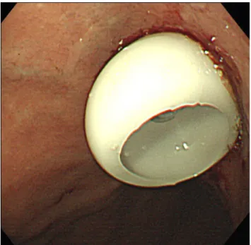
Fig. 1. The percutaneous endoscopic gastrostomy tube was inserted successfully.
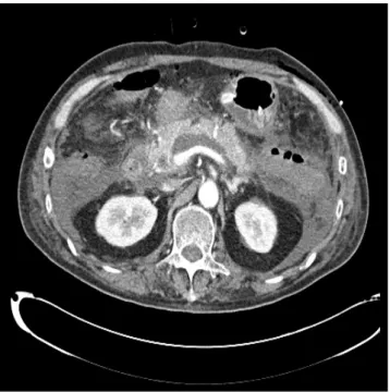
Fig. 2. (A) Abdominal CT showed contrast media leakage from a pancreatic branch of the SMA suggestive of bleeding, and (B) moderate amounts of fluid collection with high attenuation in perihepatic and perisplenic spaces and pelvic cavity suggesting hemoperitoneum. CT, computed tomography; SMA, superior mesenteric artery.
CASE REPORT
A 66-year-old man was diagnosed with herpes encephalop- athy in 1994 and reported a history of receiving medication from a Neurology Department. He had also received medi-
cation for diabetes mellitus and hypothyroidism. In addition, the patient reported a history of recurrent pneumonia and that he had undergone tracheostomy and nasogastric tube placement for nutritional support. At this presentation, he re- ported dysphagia and frequent episodes of aspiration pneu- monia, and opted for PEG tube insertion for long-term nutrition.
His vital signs were stable upon admission. Initial labo- ratory data showed: white blood cells 6,830/mm3, hemoglo- bin 12.1 g/dL, platelets 92,000/mm3, AST 18 U/L, ALT 6 U/L, BUN 15 mg/dL, and creatinine 0.61 mg/dL. The low platelet count appeared to be due to the valproate medication the patient was taking. Prior to the PEG tube insertion procedure, he had remained in a fasting state for >8 hours admission and received prophylactic antibiotics. Needle puncture was attempted twice during the PEG placement procedure, but the remainder of the procedure was completed normally (Fig. 1) without any immediate post-procedural complication. However, a day after the procedure, the patient developed hypotension, tachycardia, and dyspnea and exhibited a slight increase in abdominal circumference. In addition, his hemoglobin had dropped from 12.1 to 9.4 g/dL. Nasogastric tube washings and a rectal examination did not reveal any specific findings.
However, abdominal CT showed leakage of contrast medium
A
A BB
Fig. 3. Gelform embolization was performed with a microcatheter.
Fig. 4. Abdominal CT obtained one week after embolization showed no further bleeding but demonstrated the presence of acute pancreatitis and peripancreatic fluid collection. CT, computed tomography.
Fig. 5. Abdominal CT obtained 3 months after embolization showed reduced peripancreatic fluid collection and improved pancreatitis.
CT, computed tomography.
from a pancreatic branch of the SMA and moderately sized fluid collection of high attenuation in the perihepatic and peri- splenic spaces, and in the pelvic cavity, suggesting the pres- ence of hemoperitoneum, and the presence of acute pan- creatitis (Fig. 2). Pancreatic enzyme levels were very high, in
particular, amylase and lipase levels were 1,516 U/L and
>8,000 U/L, respectively. The pancreatitis was presumed to have been caused by injury during PEG tube insertion.
Transarterial embolization was performed to treat the hemor- rhage from the injured pancreatic branch of the SMA.
Angiography had revealed no active bleeding, and thus, the bleeding artery had not been clearly identified. Gelfoam micro- embolization was performed based on considerations of ab- dominal CT findings (Fig. 3). After embolization, vital signs stabilized without any further suspicion of bleeding.
Follow-up abdominal CT performed a week later showed peripancreatic fluid collection due to pancreatitis but no fur- ther bleeding (Fig. 4). The pancreatitis improved following sup- portive treatment (Fig. 5), and the patient was discharged after making an uneventful recovery.
DISCUSSION
PEG tube insertion is widely used to provide enteral nu- trition in patients with dysphagia or in those that have under- gone pharyngeal or esophageal surgery and in whom oral in- gestion is difficult/impossible.2,3 PEG can be performed under local anesthesia, and thus, is less invasive and more cost-ef- fective than surgery.4,5
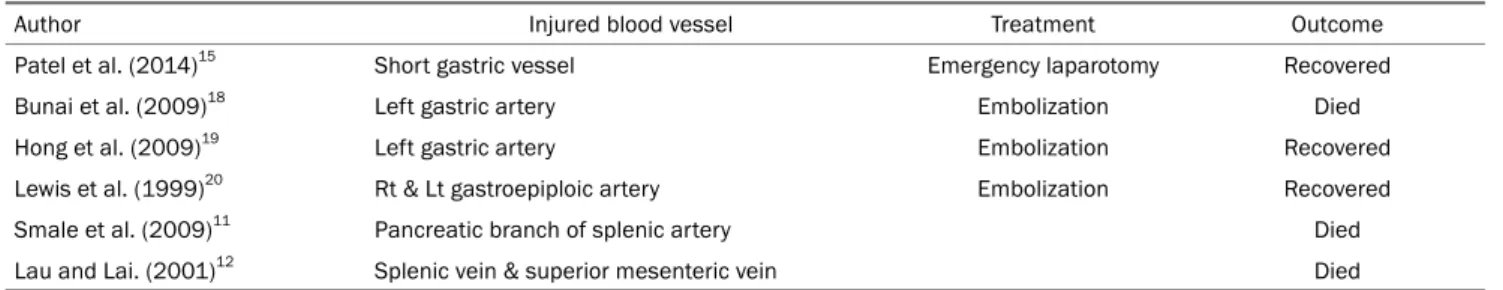
Table 1. Reported Cases of Vascular Injury during PEG Insertion
Author Injured blood vessel Treatment Outcome
Patel et al. (2014)15 Short gastric vessel Emergency laparotomy Recovered
Bunai et al. (2009)18 Left gastric artery Embolization Died
Hong et al. (2009)19 Left gastric artery Embolization Recovered
Lewis et al. (1999)20 Rt & Lt gastroepiploic artery Embolization Recovered
Smale et al. (2009)11 Pancreatic branch of splenic artery Died
Lau and Lai. (2001)12 Splenic vein & superior mesenteric vein Died
PEG, percutaneous endoscopic gastrostomy; Rt, right; Lt, left.
PEG is performed in patients diagnosed with cere- brovascular disease, dementia, or motor neurological disease, in patients with dysphagia and/or disturbed consciousness secondary to brain damage, and in patients with head and neck or esophageal cancer. It can also be used to provide enteral nutrition to patients with oral ingestion difficulties.6,7
PEG is usually a safe procedure, but complications occa- sionally occur, and although wound infection is the most com- mon complication, granuloma formation, tube dislodgement, peritonitis, visceral injuries, intestinal obstruction, aspiration pneumonia, bleeding, fistula formation, buried bumper syn- drome, and necrotizing fasciitis have all been reported.8-12 Abdominal visceral or vascular injuries are rare but serious complications, which can be fatal.
Although any abdominal organ could be injured, the colon, small intestine, liver, and spleen are at greatest risk of injury during PEG tube insertion; traumatic intestinal perforation is more common in elderly patients. Symptoms of peritoneal irrita- tion after PEG tube insertion can occur in patients with a suspected intestinal injury,13,14 and abdominal CT is warranted in such patients. Treatment of intestinal injuries often requires laparotomy. A case reported at ACG Case Reports Journal de- scribed a patient that developed a splenic laceration and a short gastric vessel injury after PEG tube insertion, who recov- ered uneventfully after emergency splenectomy.15
Intraperitoneal hemorrhage during PEG tube placement is a relatively rare adverse effect that occurs in ~1% of patients.16 Furthermore, bleeding complications after PEG tube placement are relatively rare and have been usually re- ported as case reports. Table 1 lists details of vascular in- juries that may occur during PEG tube insertion.
Amann et al.17 reported an incidence of massive bleeding secondary to arterial rupture of 0.9% (2/232) after PEG tube insertion. Bunai et al.18 described a case of left gastric artery
bleeding after a PEG placement procedure unsuccessfully treated by angiographic embolization. Hong et al.19 reported a ruptured pseudoaneurysm of the left gastric artery in a pa- tient that developed hypotension and anemia 3 days after PEG tube placement and was treated successfully by embolization. Similarly, Lewis at al.20 reported the case of a 68-year-old woman who developed bleeding after PEG tube insertion and underwent successful embolization. In our pa- tient, we performed embolization to treat bleeding from a pan- creatic branch of the SMA, which had been injured during PEG tube insertion. The above-mentioned reports and our ex- periences demonstrate embolization can serve as a good treatment option for bleeding after PEG tube insertion.
Although rare, massive bleeding after PEG tube insertion is associated with mortality. One case report described an 83-year-old man who reportedly died from intra-abdominal hemorrhage secondary to pancreatic injury after PEG tube insertion. Autopsy findings revealed bleeding from a pancre- atic branch of the splenic artery.11 Another case report de- scribed a 93-year-old woman who reportedly died from retro- peritoneal bleeding after PEG tube insertion. In this case, au- topsy findings revealed bleeding from the splenic and superior mesenteric veins. Both of these patients had undergone a cholecystectomy,12 and in both, autopsy findings revealed fib- rosis and adhesions between liver and stomach. Because of postoperative changes that occur in intraperitoneal organs, these organs appear to be more vulnerable to hemorrhage resulting from needle puncture attempted twice during PEG tube insertion.
In our case, the 64-year-old man had not undergone any abdominal surgery, but the two puncture attempts performed during PEG are shared with the previous two case reports.
In our patient, bleeding from the injured pancreatic branch of the SMA was treated by transarterial embolization and the
acute pancreatitis by using antibiotics and supportive care, and the patient made a successful and uneventful recovery.
However, our case report differs from those previously re- ported as our patient showed bleeding from an injured pan- creatic branch of the SMA, whereas previous reports describe patients that developed bleeding from an injured left gastric artery and splenic vein. In our case, the patient appeared to have been punctured from the front to the back of the stomach, resulting in damage to the pancreas situated poste- rior to the stomach.
PEG is usually safer than surgery in terms of hospital stays and costs, and thus, this endoscopic procedure is widely used by hospitals. We make the following suggestions to reduce vascular injury. First, the tip of the needle should be checked by endoscopy during the puncturing procedure. Second, the stomach must be properly inflated with air during tube insertion. Third, if possible, needle puncture should be ach- ieved on first attempt. When two or more puncture attempts are required during the procedure, patients concerned should be closely monitored for hemorrhagic complications, and in- tra-abdominal hemorrhage should be suspected in those demonstrating hypotension and anemia without melena and/or gastrointestinal hemorrhage through the gastric tube after PEG tube insertion. Transarterial embolization may pro- vide a good treatment option to manage post-procedural bleeding. We report a patient with a hemorrhagic injury to a pancreatic branch of the SMA and pancreatitis after PEG tube insertion and provide a review of the relevant literature.
REFERENCES
1. Gauderer MW, Ponsky JL, Izant RJ Jr. Gastrostomy without lapa- rotomy: a percutaneous endoscopic technique. J Pediatr Surg 1980;15:872-875.
2. Löser C, Müller MJ. Ethical guidelines for performing percuta- neous endoscopic gastrostomy (PEG catheter). Z Gastroenterol 1998;36:475-478.
3. Rabeneck L, McCullough LB, Wray NP. Ethically justified, clin- ically comprehensive guidelines for percutaneous endoscopic gastrostomy tube placement. Lancet 1997;349:496-498.
4. Löser C, Wolters S, Fölsch UR. Enteral long-term nutrition via per- cutaneous endoscopic gastrostomy (PEG) in 210 patients: a four-year prospective study. Dig Dis Sci 1998;43:2549-2557.
5. Grant JP. Comparison of percutaneous endoscopic gastrostomy with Stamm gastrostomy. Ann Surg 1988;207:598-603.
6. ASPEN Board of Directors and the Clinical Guidelines Task Force.
Guidelines for the use of parenteral and enteral nutrition in adult and pediatric patients. JPEN J Parenter Enteral Nutr 2002;26 (1 Suppl):1SA-138SA.
7. Löser C, Aschl G, Hébuterne X, et al. ESPEN guidelines on artifi- cial enteral nutrition--percutaneous endoscopic gastrostomy (PEG). Clin Nutr 2005;24:848-861.
8. Lee HJ, Choung RS, Park MS, et al. Two cases of uncommon com- plication during percutaneous endoscopic gastrostomy tube re- placement and treatment. Korean J Gastroenterol 2014;63:
120-124.
9. Schrag SP, Sharma R, Jaik NP, et al. Complications related to per- cutaneous endoscopic gastrostomy (PEG) tubes. A compre- hensive clinical review. J Gastrointestin Liver Dis 2007;16:407-418.
10. Naik RP, Joshipura VP, Patel NR, Haribhakti SP. Complications of PEG--prevention and management. Trop Gastroenterol 2009;30:
186-194.
11. Smale E, Davison AM, Smith M, Pritchard C. Fatal intra-abdominal haemorrhage following percutaneous endoscopic gastrostomy.
BMJ Case Rep 2009;2009:bcr06.2009.2044.
12. Lau G, Lai SH. Fatal retroperitoneal haemorrhage: an unusual complication of percutaneous endoscopic gastrostomy. Forensic Sci Int 2001;116:69-75.
13. Lim YJ, Yang CH. Technique, management and complications of percutaneous endoscopic gastrostomy. Korean J Gastrointest Endosc 2009;39:119-124.
14. Fang JC. Minimizing endoscopic complications in enteral access.
Gastrointest Endosc Clin N Am 2007;17:179-196, ix.
15. Patel BB, Andrade C, Doraiswamy V, Amodeo D. Splenic avulsion following PEG tube placement: a rare but serious complication.
ACG Case Rep J 2014;2:21-23.
16. Rabeneck L, Wray NP, Petersen NJ. Long-term outcomes of pa- tients receiving percutaneous endoscopic gastrostomy tubes. J Gen Intern Med 1996;11:287-293.
17. Amann W, Mischinger HJ, Berger A, et al. Percutaneous endo- scopic gastrostomy (PEG). 8 years of clinical experience in 232 patients. Surg Endosc 1997;11:741-744.
18. Bunai Y, Akaza K, Nagai A, Tsujinaka M, Jiang WX. Iatrogenic rup- ture of the left gastric artery during percutaneous endoscopic gastrostomy. Leg Med (Tokyo) 2009;11 Suppl 1:S538-S540.
19. Hong SH, Cha JM, Lee JI, et al. A case of ruptured left gastric artery pseudoaneurysm complicating Percutaneous Endoscopic Gastrostomy (PEG). Korean J Gastrointest Endosc 2009:39:
34-37.
20. Lewis MB, Lewis JH, Marshall H, Lossef SV. Massive hemorrhage complicating percutaneous endoscopic gastrostomy: treatment by means of transcatheter embolization of the right and left gas- troepiploic arteries. J Vasc Interv Radiol 1999;10:319-323.