WWW.KJOG.ORG 123
A CASE OF GROWING TERATOMA SYNDROME IN MIXED GERM CELL TUMORS OF THE OVARY
Yeri Lee, MD, Ji Yeon You, MD, Chel Hun Choi, MD, Byung Gie Kim, MD, Duk Soo Bae, MD, Je Ho Lee, MD
Department of Obstetrics and Gynecology, Samsung Medical Center, Sungkyunkwan University School of Medicine, Seoul, Korea
Growing teratoma syndrome (GTS) is an increase in tumor size containing only mature teratoma component, during or after chemotherapy for germ cell tumors. A surgical resection is important to confirm diagnosis and is considered as the most appropriate therapeutic management. GTS is rare event in association with ovarian germ cell tumors. In this report, we present a case of GTS which occurred following the treatment of mixed ovarian germ cell tumor with immature teratoma and endodermal sinus tumor. The patient was given adjuvant chemotherapy consisting of six cycles of etoposide and cisplatin. Abdomen pelvis computed tomography performed after chemotherapy demonstrated no evidence of recurrence. She is on regular follow up and remained disease-free for 8 months following second surgery.
Keywords:
Growing teratoma syndrome; Mixed germ cell tumor; Ovary
CASE REPORT
Received: 2011. 9.30. Revised: 2011.12.26. Accepted: 2012. 1. 2.
Corresponding author: Je Ho Lee, MD
Department of Obstetrics and Gynecology, Samsung Medical Center, Sungkyunkwan University School of Medicine, Irwon-ro 81, Gangnam-gu, Seoul 135-710, Korea
Tel: +82-2-3410-3519 Fax: +82-2-3410-0630 E-mail: [email protected]
Th is is an Open Access article distributed under the terms of the Creative Commons Attribution Non-Commercial License (http://creativecommons.org/licenses/
by-nc/3.0/) which permits unrestricted non-commercial use, distribution, and reproduction in any medium, provided the original work is properly cited.
Copyright © 2012. Korean Society of Obstetrics and Gynecology Korean J Obstet Gynecol 2012;55(2):123-126
http://dx.doi.org/10.5468/KJOG.2012.55.2.123 pISSN 2233-5188 · eISSN 2233-5196
Growing teratoma syndrome (GTS) is defined as an increase in tumor size in a patient with germ cell tumor during or after che- motherapy while tumor markers are normal and histology shows only mature teratoma. Logothetis et al. [1] fi rst used this term in 1982. DiSaia et al. [2], in 1977, described this phenomenon as
“chemotherapeutic retroconversion”. Later it was understood that the two terminologies were synonymous [3]. GTS appears in 1.9- 7.6% after chemotherapy in testicular non-seminomatous germ cell tumors [4]. But GTS originating from ovarian germ cell tumor is very rare [5].
In this report, we present the case of a 17-year-old GTS patients following treatment of ovarian mixed germ cell tumors.
Case Report
A 17-year-old female patient presented to gynecologic clinic in Mexico with a history of intermittent abdominal pain and abdomi- nal distension. Abdominopelvic (AP) computed tomography (CT) demonstrated 14 × 9 × 16 cm sized cystic and solid mass in the pelvic and abdominal cavity with calcifi cation. And CT demonstrat- ed ascites and omental infi ltration and peritoneal thickening and no evidence of lymph node or hematogenous metastasis (Fig. 1).
CA-125 was 159.0 U/mL and alpha fetoprotein (AFP) was 834.5
ng/mL. Exploratory laparotomy revealed a 20 cm sized right ovar- ian tumor. Right ovarian cystectomy and peritoneal tissue multiple biopsies were performed. Pathology revealed mixed germ cell tumor with grade III immature teratoma (Fig. 2) and endodermal sinus tumor (yolk sac tumor) (Fig. 3). Multiple peritoneal biopsies were positive for malignancy. She was therefore diagnosed to have mixed germ cell tumor, stage IIIc. After surgery, the patient was referred to our hospital. AP magnetic resonance imaging (MRI) and positron emission tomography/computed tomography (PET/
CT), done before chemotherapy, revealed residual cancers in the
abdominal cavity. About 5 × 5 cm sized Right ovarian mass and
WWW.KJOG.ORG 124
KJOG Vol. 55, No. 2, 2012
6x5cm sized Left ovarian mass and peritoneal carcinomatosis with metastatic lymph nodes in left paraaortic and iliac bifurcation and right common iliac area were seen. CA-125 was 75 U/mL and AFP was 149 ng/mL. Six cycles of combination chemotherapy with bleomycin, etoposide and cisplatin were given every three weeks.
Following chemotherapy, tumor markers returned to within normal limits, CA-125 was 0.5 U/mL and AFP was 1ng/mL. But the AP MRI demonstrated no interval change of residual cancer in both ovaries with peritoneal carcinomatosis (Fig. 4). She was followed
up without further treatment.
Because she was young, and following tumor markers was normal and following AP MRI, there was no interval change of both ovar- ian mass size.
The secondary laparotomy was performed 16 months following last chemotherapy because of persistent radiographic evidence of disease. The purpose of operation was diagnosis and removal
Fig. 2. Immature teratoma showing squamous epithelial component andimmature mesenchymal stromal component (H&E, ×200).
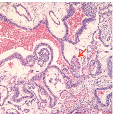
Fig. 3. Endodermal sinus tumor showing Loose reticular pattern and round papillary process with central capillary (Schiller-Duval body) (arrow, H&E, ×200).
Fig. 4. Post bleomycin, etoposide and cisplatin therapy, abdominopelvic computed tomography shows no interval change of residual cancer in both ovaries with peritoneal carcinomatosis.
Fig. 1. Abdomen pelvic computed tomography shows about 14.1 × 9.1 cm sized huge cystic and solid mass in the pelvic and abdominal cavity with fi ne calcifi cation.
141.54 mm 91.30 mm
WWW.KJOG.ORG 125
Yeri Lee, et al. Growing teratoma syndrome of the ovary
of tumor. Even more than before chemotherapy, Abdominal and pelvic examination during laparotomy revealed growing masses of both ovarian tumors. At laparotomy, 6 × 4 cm sized right ovarian tumor and 7 × 4.5 cm sized left ovarian tumor were found. In ad- dition, 10 × 6 cm sized diaphragmatic mass and 2-5 cm sized peri- toneal mass were found. And severe adhesion in abdominal and pelvic cavity, both ovary were adherent to lateral pelvic wall and uterus was adherent to rectum and posterior cul-de-sac oblitera- tion due to tumor plaques. And tumor of diaphragm was adherent to liver surface. For diagnosis, Frozen biopsies of omentum and peritoneal mass were done. The histological examination revealed the masses of biopsies to be teratoma. Total hysterectomy, bilater- al salpingo-oophorectomy, bilateral pelvic lymph node dissection, total omentectomy, appendectomy, tumorectomy and diaphrag- matic mass excisions were performed. After operation, there were 1-2 mm miliary seeding nodules on peritoneal surface. Final pa- thology revealed that tumor consisted of mature tissue including mature glial, epithelial tissue and cartilage. And tumor presented in bilateral ovary, peritoneum, mesoappendix, omentum and dia- phragm. The patient was given adjuvant chemotherapy consisting of six cycles of etoposide and cisplatin after debulking surgery. AP CT showed interval decrease in size of the calcifi ed solid nodule in the pelvic cavity and tumor markers were maintained normal range recommending regular follow-up. The patient is on regular follow up at outpatient clinic without evidence of recurrent dis- ease for eleven months.
Discussion
GTS is an extremely rare complication of malignant germ cell tu- mor, especially in females [1].
The defi nition of GTS requires three criteria [1]. 1) Clinical or ra- diological enlargement of tumors during or after chemotheraphy administered for nonseminomatous germ cell tumors (NSGCT);
2) Normalization of previously elevated tumor markers (AFP or β-human chorionic gonadotropin); and 3) Absence of any NSGCT component other than mature teratoma at histological examination of the entirely resected tumor.
Treatment depends on the age of the patient and the specific tumor type. Although surgery is the most common form of treat- ment for immature teratoma, extensively destructive surgery, including total hysterectomy, bilateral salpingo-oophorectomy, omentectomy, lymph node sampling and appendectomy is seldom considered for immature teratoma, except for those patients who
have completed their fertility. Fertility-preserving surgery (removing the affected ovary and preserving the contralateral ovary and the uterus) followed by combination chemotherapy has become the standard of care of early stages and selected advanced ovarian GCTs [6]. Since 1989, bleomycin, etoposide, and cisplatin combi- nation has become the standard chemotherapy treatment for all ovarian GCTs Three or four courses of chemotherapy have placed patients into remission with long-term survival.
In this report, we present the case of chemotherapeutic retro- conversion of residual tumor of mixed ovarian germ cell with im- mature teratoma and endodermal sinus tumor after postoperative chemotherapy. The presented case reports persistent radiographic evidence of disease in the face of normal serum tumor markers.
The normalization of AFP generally signifi es cure for endodermal sinus tumors and mixed germ cell tumors contained endodermal sinus components, particularly if the AFP remains normal over an extended period of time [7]. Decreasing of the AFP value below the normal range is recognized as an indication of remission [8]. Af- ter second laparotomy, histologically, the residual masses contained mature teratoma with no elements of immature components.
Incomplete resection of the primary tumor is considered to be one of the risk factors for GTS. Andre et al. analysis cases of 30 male patients of GTS, one patient (4%) having undergone complete resection (n = 24) had recurrent GTS, compared all but one patient (83%) in whom resection was partial (n = 6) [9]. The other risk fac- tors for GTS were histological grade 2 or 3 and clinical stage III or IV.
Despite relative benign characteristics of GTS, two types of clinical complications can be observed in GTS. First, mechanical complica- tions can occur because of the tumor growth which can compress the surrounding organs. Second, malignant transformation of GTS can occur in 3% of the cases [9]. The transformation could be into immature teratoma, sarcoma, adenocarcinoma, squamous cell carcinoma, or primitive neuroectodermal tumor. Therefore histo- logically mature teratoma may have benign appearances but with potential malignancy [10].
The treatment of GTS is surgical resection as complete as possible for several reasons. First reason is to confirm diagnosis and to exclude malignancy. Second is to relieve possible pressure mass effects from adjacent organs. Third is to prevent malignant trans- formation. Most of the patients are then in complete remission and no further treatment is indicated.
The patient was given adjuvant chemotherapy consisting of six
cycles of etoposide and cisplatin. Surgery and adjuvant platinum
based chemotherapy in GTS have been reported to give a 5-year
survival rate of 93% [11].
WWW.KJOG.ORG 126
KJOG Vol. 55, No. 2, 2012
The mechanism of the evolution from malignant germ cell tumor to mature teratomatous tissue in not fully understood. The che- motherapeutic agent used may induce somatic maturation in the malignant cell [12]. Or, the malignant elements can be destroyed by the chemotherapy. In conclusion, complete surgical resection of GTS is necessary to prevent complication and recurrence of GTS and malignant transformation. But, there is no standard approach with high dose chemotherapy after surgery from clinical experience. So, the effect of postoperative chemotherapy in GTS need further study.
Although GTS has a good prognosis, close follow-up with tumor markers and imaging studies are highly recommended, because sta- bilization of GTS can take as long as 10 years after diagnosis [13].
At the time of this report, Normalization of tumor markers and no evidence of recurrence has been observed in our patient.
References
1. Logothetis CJ, Samuels ML, Trindade A, Johnson DE. The grow- ing teratoma syndrome. Cancer 1982;50:1629-35.
2. DiSaia PJ, Saltz A, Kagan AR, Morrow CP. Chemotherapeutic retroconversion of immature teratoma of the ovary. Obstet Gynecol 1977;49:346-50.
3. Amsalem H, Nadjari M, Prus D, Hiller N, Benshushan A. Grow- ing teratoma syndrome vs chemotherapeutic retroconver- sion: case report and review of the literature. Gynecol Oncol 2004;92:357-60.
4. Maroto P, Tabernero JM, Villavicencio H, Mesía R, Marcuello E, Solé-Balcells FJ, et al. Growing teratoma syndrome: experience
of a single institution. Eur Urol 1997;32:305-9.
5. Kattan J, Droz JP, Culine S, Duvillard P, Thiellet A, Peillon C. The growing teratoma syndrome: a woman with non- seminomatous germ cell tumor of the ovary. Gynecol Oncol 1993;49:395-9.
6. Tzortzatos G, Sioutas A, Schedvins K. Successful pregnancy after treatment for ovarian malignant teratoma with growing teratoma syndrome. Fertil Steril 2009;91:936 e1-3.
7. Razzi S, Luisi S, Gabbanini M, Lazzeri L, Mazzini M, Petraglia F.
Yolk sac tumor in a young girl: a case report. Gynecol Endocri- nol 2005;20:334-5.
8. Chen CH, Yang MJ, Cheng MH, Yen MS, Lai CR, Wang PH. Fer- tility preservation with treatment of immature teratoma of the ovary. J Chin Med Assoc 2007;70:218-21.
9. André F, Fizazi K, Culine S, Droz J, Taupin P, Lhommé C, et al.
The growing teratoma syndrome: results of therapy and long- term follow-up of 33 patients. Eur J Cancer 2000;36:1389-94.
10. Sella A, el Naggar A, Ro JY, Dexeus FH, Amato RJ, Lee JS, et al.
Evidence of malignant features in histologically mature tera- toma. J Urol 1991;146:1025-8.
11. Tewari K, Cappuccini F, Disaia PJ, Berman ML, Manetta A, Kohler MF. Malignant germ cell tumors of the ovary. Obstet Gynecol 2000;95:128-33.
12. Pectasides D, Pectasides E, Kassanos D. Germ cell tumors of the ovary. Cancer Treat Rev 2008;34:427-41.
13. Hsieh TY, Cheng YM, Chang FM, Chou CY. Growing tera- toma syndrome: an Asian woman with immature teratoma of left ovary after chemotherapy. Taiwan J Obstet Gynecol 2009;48:186-9.
난소의 혼합 생식세포종양에서의 성장기형종증후군 1예
성균관대학교 의과대학 삼성서울병원 산부인과 이예리, 유지연, 최철훈, 김병기, 배덕수, 이제호
성장기형종증후군은 항암화학요법 중 또는 후 생식 세포세포종양에서 성숙기형종으로 이루어진 성장하는 종양을 의미한다. 성장기형종 증후군의 확진 및 치료를 위해 수술적 제거가 필요하며 완전한 종양의 수술적 제거는 치료를 위해 중요하다. 성장기형종증후군은 난소의 생식 세포종양에서는 매우 드물게 발생한다. 이에 미성숙 기형종과 내배엽동종양으로 이루어진 혼합 생식세포종양에 있어서 secondary cytoreduction 후 진단된 성장기형종증후군 환자에 있어서 추가 항암요법으로 etoposide와 cisplatin로 6차례 추가 항암치료 용법을 시행한 1예를 경험하였기에 문헌 고찰과 함께 보고하는 바이다.
중심단어: 성장기형종증후군, 혼합 생식세포종양, 난소