Case Report
© 2014 The Korean Ophthalmological Society
This is an Open Access article distributed under the terms of the Creative Commons Attribution Non-Commercial License (http://creativecommons.org/licenses /by-nc/3.0/) which permits unrestricted non-commercial use, distribution, and reproduction in any medium, provided the original work is properly cited.
Macular Hole Formation after Pars Plana Vitrectomy for the Treatment of Valsalva Retinopathy: A Case Report
Kook Young Kim1* , Seung-Young Yu1* , Moosang Kim2 , Hyung Woo Kwak1
1Department of Ophthalmology, Kyung Hee University Hospital, Kyung Hee University School of Medicine, Seoul, Korea
2Department of Ophthalmology, Kangwon National University School of Medicine, Chuncheon, Korea
The Valsalva maneuver involves forcible exhalation against the closed glottis and produces a sudden increase in the venous blood pressure due to an increase in intratho- racic or intraabdominal pressure. Valsalva hemorrhagic retinopathy was first described by Duane [1] in 1973 as preretinal hemorrhages observed in association with heavy lifting, vomiting, straining, or coughing. This type of reti- nopathy is caused by rupture of the superficial retinal cap- illaries due to increased intraocular venous pressure second-
ary to an abrupt increase in intrathoracic or intraabdominal pressure and results in sudden, painless loss of vision in an otherwise healthy eye. The hemorrhage typically occurs at the macula and, in the vast majority of cases, resolves spontaneously without compromising visual acuity. Gener- ally, it is an isolated and self-limited event, but even a small premacular hemorrhage of one disc diameter (DD) may take several months to clear [2].
To the best of our knowledge, this is the first reported case of complete surgical resolution of Valsalva retinopa- thy that manifested as a premacular hemorrhage with membrane followed by macular hole (MH) formation re- sulting from the first vitrectomy. We describe changes ob- served with spectral domain-optical coherence tomogra- phy (SD-OCT) following two separate 23-gauge pars plana vitrectomy procedures to treat the MH formed as a com- plication of Valsalva retinopathy.
We report a case of complete surgical resolution of Valsalva retinopathy that manifested as a premacular hemorrhage involving a membrane followed by a macular hole (MH) resulting from the first vitrectomy.
A 20-year-old female patient was referred to our hospital due to sudden vision loss in the left eye. Her best-corrected visual acuity (BCVA) in the left eye was hand motion. Fundus photographs and optical coherence tomography (OCT) revealed a premacular hemorrhage. Nine weeks later, the BCVA in the left eye had returned to 20 / 100 and the premacular hemorrhage had completely resolved, but residual sub- internal limiting membrane deposits and a preretinal membrane were present. The preretinal membrane was removed by core vitrectomy and preretinal membrane peeling, but the foveal deposits could not be excised. Two weeks after the first vitrectomy, the deposits resolved spontaneously, but a full-thickness MH was present. Six months after a second vitrectomy with fluid-gas exchange, the BCVA in the left eye had improved to 20 / 25 and OCT showed that the MH had closed. This case illustrates the possibility of MH formation following vitrectomy for Valsalva retinopathy.
Key Words: Macular hole, Valsalva retinopathy, Vitrectomy
Received: October 8, 2012 Accepted: May 28, 2013
Corresponding Author: Hyung Woo Kwak, MD, PhD. Department of Ophthalmology, Kyung Hee University Hospital, #23 Kyungheedae-ro, Dongdaemun-gu, Seoul 130-872, Korea. Tel: 82-2-958-8451, Fax: 82-2- 966-7340, E-mail: [email protected]
This paper was presented as a “Challenging Case of Symposia” at the 27th Asia-Pacific Academy of Ophthalmology meeting in April 2012.
*These authors contributed equally to this work.
Case Report
A 20-year-old female patient was referred to our hospital due to a sudden decrease in visual acuity in the left eye af- ter heavy alcohol consumption. She denied any history of trauma or sexual contact. Her medical and ocular histories were unremarkable. Previous medical records reported that the patient had a history of severe vomiting following heavy drinking. In the left eye, the best-corrected visual acuity (BCVA) was hand motion. The anterior segment was unremarkable in both eyes. Fundus examination was normal in the right eye but revealed a well-circumscribed premacular hemorrhage about 4 DDs in size beneath a transparent membrane with glistening reflexes extending over the macula in the left eye. No posterior vitreous de- tachment (PVD) was evident. Time-domain OCT (TD- OCT) revealed an intact foveal contour and two mem- branes of differing optical reflectivity, identified as the internal limiting membrane (ILM) and the posterior hy- aloid, respectively, with blood beneath the hyperreflective membrane (Fig. 1).
During a nine-week follow-up period, the premacular hemorrhage resolved spontaneously, and the BCVA in the left eye returned to 20 / 100. Yellowish residual sub-ILM deposits, however, were present, and a thick preretinal membrane was visible over the fovea (Fig. 1).
The patient requested aggressive treatment. After full explanation of the expected effects and possible complica- tions of vitrectomy, the patient provided informed consent.
A 23-gauge pars plana vitrectomy was performed using a minimal core vitrectomy technique. PVD was created us- ing triamcinolone acetonide and a vitreous cutter only around the posterior pole. The ILM was peeled off using 23-gauge microforceps after staining the ILM with indo- cyanine green (Diagnogreen; Daiichi Pharmaceutical, To- kyo, Japan). The ILM was removed from a 2- to 3-DD area centered on the fovea. The thick preretinal membrane and the ILM were removed, but the residual foveal deposits could not be extracted because they had adhered to the fo- vea. The surgeon expected the deposits to absorb sponta- neously, so fluid-air exchange was not performed. Two weeks after the first vitrectomy, the foveal deposits had re- solved, but a full thickness MH was observed. Postopera- tive SD-OCT, which was used to analyze the cross-section- al image almost every day (Fig. 2), did not reveal an MH during the first seven days. A second vitrectomy was per-
formed using standard techniques to create a total PVD and to remove the remnant peripheral vitreous with flu- id-air exchange through an extrusion cannula. The eye was flushed with a 15% perfluoropropane gas-air mixture to ensure complete exchange. After the second surgery, the patient was instructed to maintain a strict face-down posi- tion for three days in the hospital until MH closure was confirmed.
Six months after the second surgery to close the MH, the BCVA in the left eye had improved to 20 / 25. SD-OCT showed that the MH had closed with only slight scarring of the pho- toreceptor inner segment/outer segment junction (Fig. 3).
Discussion
Valsalva retinopathy develops in response to the Valsal- va maneuver. Reported causes of Valsalva retinopathy in- clude straining and physical activities, most commonly coughing, weight lifting, vomiting, aerobic exercise, sexual activity, colonoscopy procedures, and congenital retinal macroaneurysm [3-6]. A history of such activities is help- ful in establishing this diagnosis. Although in the present case, the patient did not remember the exact situation due to heavy drinking, according to the previous medical re- cords, severe vomiting could have been a possible cause of Valsalva retinopathy.
The presentation of Valsalva retinopathy varies depending on the size of the vessel involved and the location of the hem- orrhage, which can be subretinal, intraretinal and/or subhy- aloid. The exact location of premacular hemorrhage (whether sub-hyaloid or sub-ILM) has been disputed in the literature.
Hemorrhage following Valsalva retinopathy is more com- monly sub-ILM than sub-hyaloid; rarely, it may be a combi- nation of both [2]. In the present case, premacular hemorrhage manifested as a small dome-shaped, sub-ILM hemorrhage that was characterized by a glistening light reflex, fine striae on the surface of the hemorrhage, and a large dome-shaped sub-hyaloid hemorrhage located anterior and inferior to the sub-ILM hemorrhage, as determined by color fundus photo- graphs and SD-OCT (Fig. 1).
To the best of our knowledge, development of an MH af- ter vitrectomy to remove the thick preretinal membrane following spontaneous resolution of the premacular hem- orrhage has not been previously reported in the literature.
There are several possible mechanisms that could explain
Fig. 1. (A) A fundus photograph of the left eye shows a dome-shaped sub-internal limiting membrane (ILM) hemorrhage (white aster- isk) and a sub-hyaloid hemorrhage located anterior and inferotemporal to the sub-ILM hemorrhage (white cross) at initial presentation.
Note the “double ring” sign with the “inner ring” caused by the sub-ILM hemorrhage and the “outer ring” caused by the sub-hyaloid hemorrhage. (B) After four weeks, a fundus photograph showed spontaneous absorption of the sub-hyaloid hemorrhage, but the sub-ILM hemorrhage remained. (C) After nine weeks, the sub-ILM and sub-hyaloid hemorrhages were almost completely resolved, and a focal yellowish lesion was observed on the fovea. (D) The Time-domain optical coherence tomography (OCT) at the initial presentation re- vealed a premacular hemorrhage with hyperreflective ILM (white arrowhead) and a hyporeflective sub-hyaloid membrane (white arrow).
The foveal contour was intact (double arrow). (E) After four weeks, OCT showed resolution of the premacular hemorrhage with a thick preretinal membrane. (F) After nine weeks, the premacular hemorrhage was completely resolved, but a hyperreflective, thick preretinal membrane (white arrowhead) remained.
A
D E F
B C
* *
†
Fig. 2. Serial spectral domain-optical coherence tomography images before and after pars plana vitrectomy. The macular hole was not identified immediately after the first operation but was observed after resolution of the sub-internal limiting membrane deposits at two weeks after the first surgery. (A) Before 1st operation, (B) 1 day after 1st operation, (C) 4 day after 1st operation, (D) 7 day after 1st oper- ation, (E) 14 day after 1st operation, and (F) 14 day after 2nd operation.
A
C
E
B
D
F
MH formation after the initial vitrectomy.
First, the MH could be iatrogenic (caused by ILM peel- ing). Before the first procedure, a thick preretinal mem- brane and sub-ILM deposits over the foveal area were ob- served by SD-OCT. If the MH had been iatrogenic, we would have been able to observe it immediately after sur- gery using SD-OCT. However, post-surgical SD-OCT im- ages did not reveal a MH, which could be due to the sticky nature of foveal deposits (Fig. 2).
Second, the thick preretinal membrane observed after resolution of the premacular hemorrhage was considered to create a tangential tractional force over the fovea [7].
Due to this tangential force, the increased volume of the sub-ILM deposits under the thick preretinal membrane was likely to provide an additional outward expanding force into the fovea. It is possible that the thick preretinal membrane prevented formation of the MH. After the re- sidual foveal deposit was absorbed, the MH occurred with a fading holding effect of a sticky deposit, which sustained
the margin of the MH.
The prognosis of Valsalva retinopathy is generally good, and the condition in most patients resolves spontaneously over several months. In the majority of cases, conservative management is indicated with periodic observation. Thera- peutic options in Valsalva retinopathy include conservative management, vitrectomy [8], laser membranotomy [9], in- travitreal tissue plasminogen activator, and tamponade gas injection [10]. If sub-ILM and sub-hyaloid hemorrhage were present in the macular area in the presented case, we would have considered yttrium-aluminum-garnet (YAG) laser and tissue plasminogen activator as a treatment op- tion. The sub-ILM and sub-hyaloid hemorrhages, however, were absorbed completely, and only the yellowish sub-ILM deposits remained. Accordingly, we did not choose YAG laser and tissue plasminogen activator as a treatment op- tion. In the present case, the MH seemed to be a possible complication after vitrectomy to remove the thick prereti- nal membrane associated with Valsalva retinopathy.
The residual sub-ILM deposits in the fovea were not ab- sorbed spontaneously before the first vitrectomy; therefore, the patient’s visual acuity did not improve. After ILM peeling following the first vitrectomy, the surgeon waited for spontaneous absorption of the deposits. Two weeks af- ter the first procedure, the residual sub-ILM deposits had completely resolved, and the MH could be clearly diag- nosed. A second operation was then performed with stan- dard techniques to close the MH. The ILM peeling that oc- curred during the first surgery, however, likely accelerated the spontaneous absorption of the residual foveal deposits and might have contributed to the closing of the MH. De- laying the first operation may have led to a prolonged time for absorption of the deposits, thus worsening the progno- sis due to the presence of composites such as iron in the deposits or organizing scar tissue, and the MH would have continued to be masked by the deposits.
In conclusion, patients should be monitored carefully for the development of a MH after vitrectomy for Valsalva retinopathy with residual sub-ILM deposits, as described in the present case.
Conflict of Interest
No potential conflict of interest relevant to this article was reported.
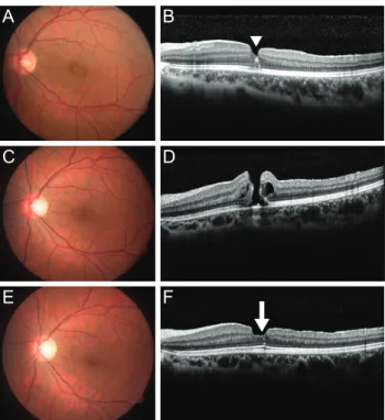
Fig. 3. (A,B) Fundus photograph and spectral domain-optical coherence tomography (SD-OCT) images after the initial pars plana vitrectomy. SD-OCT revealed hemosiderin-like deposits (arrowhead) of the fovea. (C,D) Fundus photograph and SD-OCT images before secondary pars plana vitrectomy. SD-OCT showed a full-thickness macular hole. (E,F) Six months after the second surgery, the macular hole was completely closed with minimal scarring of the photoreceptor inner/outer segment junction (ar- row), as determined by SD-OCT.
A
C
E
B
D
F
References
1. Duane TD. Valsalva hemorrhagic retinopathy. Am J Oph- thalmol 1973;75:637-42.
2. Gass JD. Stereoscopic atlas of macular diseases: diagnosis and treatment. 4th ed. St. Louis: Mosby; 1997. p. 752-4.
3. Roberts DK, MacKay KA. Microhemorrhagic maculopa- thy associated with aerobic exercise. J Am Optom Assoc 1987;58:415-8.
4. Friberg TR, Braunstein RA, Bressler NM. Sudden visual loss associated with sexual activity. Arch Ophthalmol 1995;113:738-42.
5. De Crecchio G, Pacente L, Alfieri MC, Greco GM. Valsal- va retinopathy associated with a congenital retinal mac- rovessel. Arch Ophthalmol 2000;118:146-7.
6. Choi SW, Lee SJ, Rah SH. Valsalva retinopathy associated with fiberoptic gastroenteroscopy. Can J Ophthalmol
2006;41:491-3.
7. Kwok AK, Lai TY, Chan NR. Epiretinal membrane forma- tion with internal limiting membrane wrinkling after Nd:YAG laser membranotomy in valsalva retinopathy. Am J Ophthalmol 2003;136:763-6.
8. De Maeyer K, Van Ginderdeuren R, Postelmans L, et al.
Sub-inner limiting membrane haemorrhage: causes and treatment with vitrectomy. Br J Ophthalmol 2007;91:869- 72.
9. Durukan AH, Kerimoglu H, Erdurman C, et al. Long-term results of Nd:YAG laser treatment for premacular subhy- aloid haemorrhage owing to Valsalva retinopathy. Eye (Lond) 2008;22:214-8.
10. Koh HJ, Kim SH, Lee SC, Kwon OW. Treatment of subhy- aloid haemorrhage with intravitreal tissue plasminogen ac- tivator and C3F8 gas injection. Br J Ophthalmol 2000;84:
1329-30.