© 2019 The Korean Ophthalmological Society
This is an Open Access article distributed under the terms of the Creative Commons Attribution Non-Commercial License (http://creativecommons.org/licenses /by-nc/3.0/) which permits unrestricted non-commercial use, distribution, and reproduction in any medium, provided the original work is properly cited.
Original Article
Long-term Outcomes of Macular Hole Retinal Detachment in Highly Myopic Eyes after Surgical Reattachment
Hwa Yeong Kim
1,2, Jae Jung Lee
1,2, Han Jo Kwon
1,3, Sung Who Park
1,2, Ji Eun Lee
1,21
Department of Ophthalmology, Pusan National University School of Medicine, Yangsan, Korea
2
Biomedical Research Institute, Pusan National University Hospital, Busan, Korea
3
Research Institute for Convergence of Biomedical Science and Technology, Pusan National University Yangsan Hospital, Yangsan, Korea
Purpose: To evaluate visual acuity changes over 3 years following surgical reattachment of macular hole retinal detachment (MHRD) developed in high myopia.
Methods: A retrospective analysis was performed using the medical records of patients with highly myopic eyes who underwent pars plana vitrectomy with internal limiting membrane peeling or the internal limiting mem- brane flap technique for MHRD. Changes in best-corrected visual acuity (BCVA) were measured at baseline, 6 months, 1 year, 2 years, and 3 years.
Results: Of the 22 eyes analyzed, macular hole was closed in 13 and unclosed in nine. BCVA significantly improved from 1.61 ± 0.39 logarithm of the minimum angle of resolution (logMAR) at baseline to 1.17 ± 0.43 logMAR at 6 months and 1.33 ± 0.48 logMAR at 2 years after MHRD surgery. At 3 years, BCVA significantly decreased compared with that at 6 months, and visual improvement from baseline was not significant. BCVA and proportion of vision loss ≥0.3 logMAR were not different between the closed and unclosed macular hole groups.
Conclusions: Visual improvement after surgical reattachment of MHRD in high myopia was not maintained, and favorable macular hole closure effects were not observed at 3-year follow-up.
Key Words: Degenerative myopia, Retinal detachment, Retinal perforations, Vitrectomy
Retinal detachment associated with macular hole mostly occurs in highly myopic eyes [1,2]. Although the mecha- nisms are not fully understood, it has been suggested that macular hole retinal detachment (MHRD) is caused by
tangential and anteroposterior traction due to presence of a posterior staphyloma combined with weakened retinal ad- hesion caused by retinal pigment epithelium atrophy [2,3].
These background pathologies make MHRD complicated in high myopia a surgical challenge.
Various surgical procedures including macular buckling [4,5], pars plana vitrectomy (PPV) with tamponade using gas or silicone oil [6,7], and extramacular drainage [8] have been reported to improve MHRD surgery outcomes. PPV with internal limiting membrane (ILM) peeling and gas
Received: April 19, 2019 Final revision: July 17, 2019 Accepted: August 5, 2019
Corresponding Author: Ji Eun Lee, MD, PhD. Department of Ophthal- mology, Pusan National University Hospital, 179 Gudeok-ro, Seo-gu, Bu- san 49241, Korea. Tel: 82-51-240-7957, Fax: 82-51-242-7341, E-mail: jlee@
pusan.ac.kr
tamponade has been one of the most popular surgical pro- cedures for MHRD because it effectively relieves tangen- tial macular traction by removing all the overlying fibrous membrane adjoining the macular hole (MH) [9-11]. Recent- ly, various ILM manipulations that covered the MH were reported to increase the primary success rate of MHRD surgery up to 100% [12-14]. These diverse adjunctive pro- cedures appeared to improve reattachment and MH clo- sure rates as well as functional outcomes. However, these studies examined surgical outcomes over a relatively short period of time and did not report long-term results. In ad- dition, the impact of MH closure on functional outcomes during long-term follow-up was not investigated.
As highly myopic eyes are prone to degenerative chang- es causing visual loss, the functional outcomes of MHRD surgery should be evaluated over a long period. The pur- pose of this study was to investigate long-term visual acui- ty changes after surgical reattachment of MHRD in highly myopic eyes.
Materials and Methods
The medical records of patients who underwent PPV for MHRD between November 2008 and October 2015 at Pu- san National University Hospital were retrospectively re- viewed. The study design was approved by the institutional review board of Pusan National University Hospital (1812- 020-074), and the study was conducted following the prin- ciples outlined in the Declaration of Helsinki. The in- formed consent was waived due to the retrospective nature of the study.
Patient inclusion criteria were as follows: (1) clinical pre- sentation of MHRD in a highly myopic eye, (2) MH as the primary cause of retinal detachment, (3) 3-port PPV treat- ment, and (4) reattachment maintained for ≥3 years after surgery. Eyes in which MH was not considered the primary cause of retinal detachment were excluded based on config- uration of subretinal fluid and presence of breaks other than MH. High myopia was defined as refractive error ≥-6.0 di- opters without refractive surgery or axial length ≥26.5 mm.
The cohort of 106 eyes was obtained by database search for ‘macular hole’ and ‘retinal detachment’ between No- vember 2008 and October 2015 regardless of refractive er- ror or axial length. Among them, 64 eyes were highly my- opic and underwent PPV for MHRD. Twenty-three eyes
were followed up over 3 years, and one eye was excluded due to reattachment failure. Finally, 22 eyes were included in the following analysis. Of the 22 total eyes, 20, 19, and 18 eyes were followed up at postoperative 6 months, 1 year, and 2 years, respectively. The clinical data collected from each case record were patient age; sex; preoperative best-corrected visual acuity (BCVA); postoperative BCVA at 6 months and 1, 2, and 3 years; axial length; posterior staphyloma presence; lens status; and operative variables including use of conventional ILM peeling or the inverted ILM flap technique, tamponade types, MH closure, and retinal reattachment.
All operations were conducted by experienced vitreoret- inal surgeons (JEL and SWP). Conventional 20-, 23-, or 25-gauge 3-port PPV (Constellation, Alcon, Fort Worth, TX, USA) was performed. Posterior vitreous detachment was created by vitreous aspiration using a cutter or extru- sion needle. After removing the vitreous to the periphery as much as possible, the viscous submacular fluid was gen- tly drained through the MH or extramacular sites. If there was no preexisting peripheral break, then retinotomy was performed outside of the macula to avoid chorioretinal at- rophy, and one low-barrier photocoagulation was per- formed around the retinotomy site.
The ILM was stained with indocyanine green or bril- liant blue G for ILM removal or flap technique. For the ILM flap technique, a portion of the ILM about 1-disc di- ameter in size was left at the superior margin of the MH and inverted as a single layer. The inverted flap was stabi- lized with perfluoro-n-octane (Perfluoron, Alcon) during fluid-air exchange. The vitreous was replaced by long-act- ing gas (sulfur hexafluoride [SF
6] or perfluoropropane [C
3F
8]) at a non-expanding concentration or silicone oil at the surgeon’s discretion. Patients were advised to stay in a strict face-down position for 1 to 7 days after surgery.
To confirm reattachment and MH closure, optical coher- ence tomography (OCT) examinations were performed in all patients before and after surgery using spectral-domain (Cirrus HD-OCT, Carl-Zeiss, Dublin, CA, USA) or swept- source (DRI-OCT Atlantis, Topcon, Tokyo, Japan) OCT devices. MH closure was defined as absence of a neurosen- sory defect at the fovea. Primary attachment was defined as retinal reattachment without requiring additional surgi- cal procedures except silicone oil removal. Patients were divided into two groups based on MH closure.
Visual acuity by a Snellen chart was converted to loga-
rithm of the minimum angle of resolution (logMAR) for analysis. Values are reported as the mean and standard de- viation. Wilcoxon signed-rank test was conducted to eval- uate postoperative changes for each group. Mann-Whitney U-test and Fisher’s exact test were used to compare contin- uous and categorical variables between two groups, re- spectively. A p-value <0.05 was considered significant. All analyses were performed using IBM SPSS Statistics ver.
21.0 (IBM Corp., Armonk, NY, USA).
Results
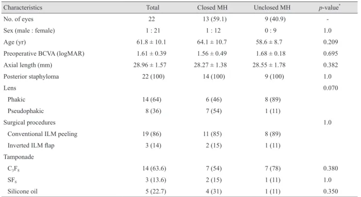
This study included 22 eyes of 22 patients with MHRD who underwent PPV. Patient characteristics are shown in Table 1. Patients comprised one male and 21 females with a mean age of 61.8 ± 10.1 years. At baseline, mean BCVA was 1.61 ± 0.39 logMAR, and mean axial length was 28.96
± 1.57 mm. All patients had staphyloma. Concurrent phacoemulsification was performed in 12/14 (85%) phakic
eyes. After primary surgery, two eyes remained phakic.
One of the phakic eyes had no significant cataract progres- sion during follow-up, whereas phacoemulsification was performed in the other eye at one year after initial MHRD surgery. PPV with conventional ILM peeling was conduct- ed in 19 eyes, and PPV with the inverted ILM flap tech- nique was performed in three eyes. The vitreous was re- placed with C
3F
8in 14 (63.6%) eyes, SF
6in three (13.6%) eyes, and silicone oil in five (22.7%) eyes.
The retina was reattached in all eyes (n = 22) with a sin- gle surgery, excluding the silicone oil removal operation.
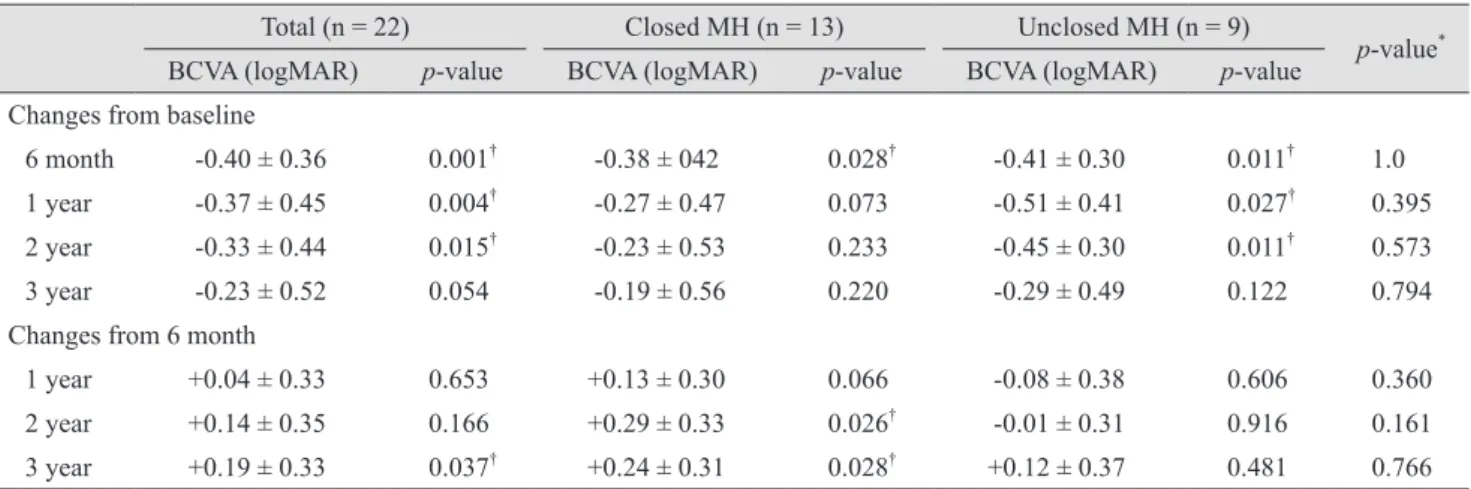
The MH was closed in 13 eyes (59.1%) and unclosed in nine eyes (40.9%). The mean BCVA significantly improved from 1.61 ± 0.39 logMAR preoperatively to 1.17 ± 0.43 log- MAR at 6 months postoperatively (p = 0.001). At 3 years postoperatively, patient visual acuity gradually decreased to 1.38 ± 0.51 logMAR, and the improvement was no lon- ger significant (p = 0.054) (Table 2). Compared with visual acuity at 6 months, that at 3 years significantly decreased (p = 0.037) (Fig. 1).
Table 1. Comparison of baseline clinical characteristics
Characteristics Total Closed MH Unclosed MH p-value
*No. of eyes 22 13 (59.1) 9 (40.9) -
Sex (male : female) 1 : 21 1 : 12 0 : 9 1.0
Age (yr) 61.8 ± 10.1 64.1 ± 10.7 58.6 ± 8.7 0.209
Preoperative BCVA (logMAR) 1.61 ± 0.39 1.56 ± 0.49 1.68 ± 0.18 0.695
Axial length (mm) 28.96 ± 1.57 28.27 ± 1.38 28.55 ± 1.78 0.382
Posterior staphyloma 22 (100) 14 (100) 9 (100) 1.0
Lens 0.070
Phakic 14 (64) 6 (46) 8 (89)
Pseudophakic 8 (36) 7 (54) 1 (11)
Surgical procedures 1.0
Conventional ILM peeling 19 (86) 11 (85) 8 (89)
Inverted ILM flap 3 (14) 2 (15) 1 (11)
Tamponade
C
3F
814 (63.6) 7 (54) 7 (78) 0.380
SF
63 (13.6) 2 (15) 1 (11) 1.0
Silicone oil 5 (22.7) 4 (31) 1 (11) 0.350
Data are expressed as number (%) of eyes or mean ± standard deviation.
MH = macular hole; BCVA = best-corrected visual acuity; logMAR = logarithm of the minimum angle of resolution; ILM = internal limiting membrane; C
3F
8= perfluoropropane; SF
6= sulfur hexafluoride.
*
Comparisons between patients with closed and unclosed MHs.
Comparison of closed and unclosed MH groups
Comparisons of the preoperative clinical characteristics between the closed and unclosed MH groups are shown in Table 1. There were no significant differences between the two groups with respect to patient sex, age, preoperative BCVA, axial length, posterior staphyloma presence, lens status, or operative technique.
There was a significant improvement of the visual acuity of the closed and unclosed MH groups at 6 months after surgery (p = 0.028 and 0.011, respectively). The mean loss of visual acuity at 3 years compared with that at 6 months after surgery was 0.24 ± 0.31 and 0.12 ± 0.37 logMAR in the closed and unclosed MH groups, respectively (Table 3).
There was no significant difference in mean visual acuity loss between the two groups (p = 0.766). The number of eyes that had decreased visual acuity by ≥0.3 logMAR at 3 years compared with 6 months after surgery was five in
Table 3. Comparison of BCVA changes after surgery
Total (n = 22) Closed MH (n = 13) Unclosed MH (n = 9)
p-value
*BCVA (logMAR) p-value BCVA (logMAR) p-value BCVA (logMAR) p-value
Changes from baseline
6 month -0.40 ± 0.36 0.001
†-0.38 ± 042 0.028
†-0.41 ± 0.30 0.011
†1.0
1 year -0.37 ± 0.45 0.004
†-0.27 ± 0.47 0.073 -0.51 ± 0.41 0.027
†0.395
2 year -0.33 ± 0.44 0.015
†-0.23 ± 0.53 0.233 -0.45 ± 0.30 0.011
†0.573
3 year -0.23 ± 0.52 0.054 -0.19 ± 0.56 0.220 -0.29 ± 0.49 0.122 0.794
Changes from 6 month
1 year +0.04 ± 0.33 0.653 +0.13 ± 0.30 0.066 -0.08 ± 0.38 0.606 0.360
2 year +0.14 ± 0.35 0.166 +0.29 ± 0.33 0.026
†-0.01 ± 0.31 0.916 0.161
3 year +0.19 ± 0.33 0.037
†+0.24 ± 0.31 0.028
†+0.12 ± 0.37 0.481 0.766
BCVA = best-corrected visual acuity; MH = macular hole; logMAR = logarithm of the minimum angle of resolution.
*
Comparisons between patients with closed and unclosed MHs;
†p < 0.05.
Table 2. Comparison of BCVA before and after surgery
Total (n = 22) Closed MH (n = 13) Unclosed MH (n = 9)
p-value
*BCVA (logMAR) No. of eyes BCVA (logMAR) No. of eyes BCVA (logMAR) No. of eyes
Baseline 1.61 ± 0.39 22 1.56 ± 0.49 13 1.68 ± 0.18 9 0.695
6 month 1.17 ± 0.43 20 1.08 ± 0.53 11 1.27 ± 0.29 9 0.456
1 year 1.24 ± 0.47 19 1.27 ± 0.52 11 1.20 ± 0.43 8 0.778
2 year 1.33 ± 0.48 18 1.39 ± 0.59 10 1.26 ± 0.32 8 0.408
3 year 1.38 ± 0.51 22 1.37 ± 0.59 13 1.39 ± 0.40 9 1.0
BCVA = best-corrected visual acuity; MH = macular hole; logMAR = logarithm of the minimum angle of resolution.
*
Comparisons between patients with closed and unclosed MHs.
lo gM A R
Mean BCVA (logMAR)
1 1.2 1.4 1.6 1.8 2
Preoperative VA 6 mon VA 1 yr VA 2 yr VA 3 yr VA
Total Unclosed MH Closed MH
Fig. 1. Changes in visual acuity (VA) after vitrectomy for macu-
lar hole (MH) retinal detachment at postoperative 6 months and 1,
2, and 3 years. BCVA = best-corrected visual acuity; logMAR =
logarithm of the minimum angle of resolution.
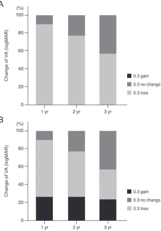
the closed (38%) and four in the unclosed (44%) MH groups (p = 1.0) (Table 4 and Fig. 1, 2A, 2B).
In one eye with an unclosed MH after primary surgery, a secondary surgery was performed to close the MH. The primary surgery was PPV combined with phacoemulsifi- cation, the ILM peeling technique, and C
3F
8tamponade.
Although the retina was reattached, the MH remained open. An additional surgery was performed 3 months after the primary surgery, and the ILM was further removed and the vitreous cavity was filled with SF
6. However, the MH remained open after the second operation. Visual acu- ity was maintained for 2 years after primary surgery.
However, at 3 years, it decreased because of expanding chorioretinal atrophy (Fig. 3A-3D).
Analysis of the long-term vision loss subgroup
Table 4 provides a summary of nine patients with BCVA loss ≥0.3 logMAR at 3 years compared with 6 months af- ter surgery. The most common cause of BCVA loss was enlargement or development of chorioretinal atrophy.
There were two cases of subretinal hemorrhage that were related to myopic choroidal neovascularization (CNV) and lacquer crack formation, respectively. One eye had vision loss from open-angle glaucoma, which developed follow- ing vitrectomy with silicone oil tamponade. Only one eye had significant vision loss that was suspected to be caused by a persistent MH, with elevated edges and macular ede- ma. In this eye, the horizontal diameter of the MH in- creased from 1,050 μm at 6 months to 1,249 μm at 3 years,
0.3 gain 0.3 no changs 0.3 loss
Change of VA (logMAR)
1 yr 2 yr 3 yr
0 40 20 60 100
A
80 (%)
0.3 gain 0.3 no changs 0.3 loss
Change of VA (logMAR)
1 yr 2 yr 3 yr
0 40 20 60 100
B
80 (%)