• 검색 결과가 없습니다.

Endovascular Stent in Traumatic Thoracic Aortic Dissection

N/A
N/A
Protected

Academic year: 2022

Share "Endovascular Stent in Traumatic Thoracic Aortic Dissection"

Copied!
4
0
0

로드 중.... (전체 텍스트 보기)

전체 글

(1)

341 Copyright © 2012 The Korean Society of Cardiology

Korean Circulation Journal

Introduction

Patients with blunt traumatic thoracic aorta injuries are increas- ingly admitted to hospital due to the increasing number of traffic accidents per day. Traumatic thoracic aortic injury is typically fatal.

The thoracic aorta wall ruptures after blunt thorax trauma and if not treated, has very poor outcome with an initial survival rate ranging from 10 to 30%. The hospital mortality rate is up to 32% during the first day, 61% within the first week and 74% after 2 weeks. More- over, according to the literature, patients surviving the acute phase without surgery had a 30% risk of late traumatic thoracic aorta an- eurysm rupture.
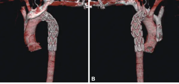
1)

Fortunately, acute and chronic traumatic lesions of the descend- ing aorta can now be treated via an endovascular approach in spe- cialized centers, with low morbidity and mortality rates.
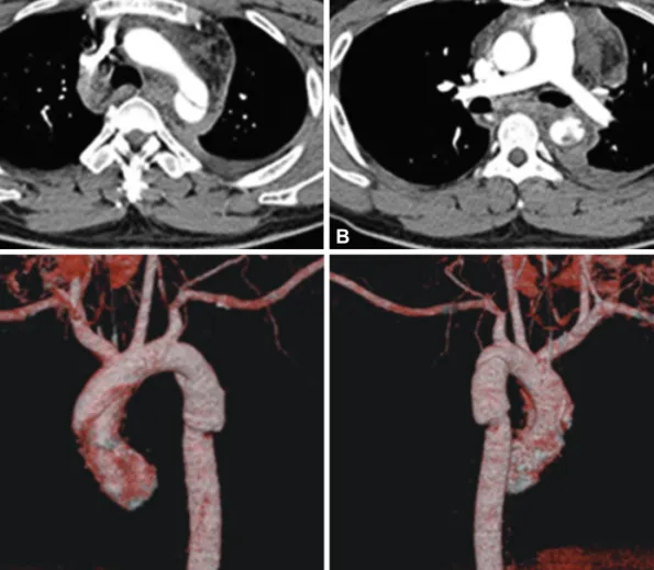
2)3)

We report two cases of endovascular stenting in traumatic thoracic aortic dis- section due to traffic accidents.

Case Report

http://dx.doi.org/10.4070/kcj.2012.42.5.341 Print ISSN 1738-5520 • On-line ISSN 1738-5555

Endovascular Stent in Traumatic Thoracic Aortic Dissection

Mi Ok Jang, MD, Ju Han Kim, MD, Sang Ki Oh, MD, Min Goo Lee, MD, Keun Ho Park, MD, Doo Sun Sim, MD, Young Joon Hong, MD, Youngkeun Ahn, MD, and Myung Ho Jeong, MD

The Heart Center of Chonnam National University Hospital, Chonnam National University Medical School, Gwangju, Korea

Traumatic thoracic aortic injury is typically fatal. However, recent improvements in pre-hospital care and diagnostic modalities have re- sulted in an increased number of patients with traumatic aortic injury arriving alive at the hospital. Also, the morbidity and mortality as- sociated with endovascular repair are significantly lower than with conventional open surgery in traumatic thoracic aorta injury. We expe- rienced two cases of successful management of traumatic thoracic aortic dissection with endovascular stents caused by traffic accidents.

(Korean Circ J 2012;42:341-344)

KEY WORDS: Aorta, thoracic; Dissection; Aortography; Multidetector computed tomography.

Received: June 9, 2011

Revision Received: August 2, 2011 Accepted: September 1, 2011

Correspondence: Myung Ho Jeong, MD, The Heart Center of Chonnam National University Hospital, Chonnam National University Medical School, 42 Jebong-ro, Dong-gu, Gwangju 501-757, Korea

Tel: 82-62-220-6243, Fax: 82-62-228-7174 E-mail: [email protected]

• The authors have no financial conflicts of interest.

This is an Open Access article distributed under the terms of the Creative Commons Attribution Non-Commercial License (http://creativecommons.

org/licenses/by-nc/3.0) which permits unrestricted non-commercial use, distribution, and reproduction in any medium, provided the original work is properly cited.

Cases

Case 1

A 28-year-old male suffered from a traffic accident while in a car.

His mental status was confused but brain CT revealed non-specific findings. He had no known history of hypertension, diabetes, hep- atitis, and pulmonary tuberculosis. He was an 8 pack-year current smoker. His family history was non-specific. On admission his vital signs were stable except for blood pressure of 100/70 mm Hg. A 12- lead electrocardiogram (EKG) showed normal sinus rhythm with 72 beat/min. The results of electrolyte panel and kidney function stud- ies were all within normal limits. The aspartate aminotransferase (AST) was 70 U/L, alanine amino-transferase (ALT) was 47 U/L, creati- nine kinase (CK) was 255 U/L and CK-MB was 28 U/L. However, se- rum level of troponin I was within normal limits. Initial 2-dimen- sional echocardiography (2DE) showed no regional wall motion ab- normality with normal left ventricular ejection fraction, but chest CT angiogram (CTA) showed traumatic aortic dissection in proximal de- scending thoracic aorta/distal aortic arch around aortic isthmus with left pleural effusion (Fig. 1). Therefore, we performed a thoracic aor- tography, which showed an aortic aneurysm distal to the subclavian artery, for which a 24×112 mm valiant thoracic stent was implanted.

Follow-up aortogram showed no evidence of leakage. Follow-up

2DE did not show evidence of turbulent flow or flow disturbance

near the entrance of the left subclavian artery. Follow-up chest CTA

showed vascular stent graft insertion state from distal aortic arch

to proximal descending thoracic aorta and resolved state of previ-

ous traumatic aortic dissection (Fig. 2). After uneventful recovery,

he was discharged on day 33 and has been followed up at the out-

(2)

342 Endovascular Stent in Traumatic Thoracic Aortic Dissection

http://dx.doi.org/10.4070/kcj.2012.42.5.341 www.e-kcj.org

patient clinic.

Case 2

A 52-year-old male suffered from a traffic accident in a car. He complained of left flank pain and had multiple left rib fracture. He had no known history of hypertension, hepatitis, or pulmonary tu- berculosis, but had been on diabetes medication for 3 years. He was a non-smoker and non-alcoholic drinker, and his family history was non-specific. On admission his blood pressure was 70/40 mm Hg. EKG showed sinus tachycardia with 138 beats/min. The results of electrolyte panel and kidney function studies were all within nor- mal limits. The AST was 219 U/L, ALT was 153 U/L, CK was 2113 U/L, CK-MB was 51.7 U/L and troponin I was 0.24 U/L. Initial chest CTA showed traumatic aortic dissection in distal aortic arch/proximal descending thoracic aorta with surrounding hematoma (Fig. 3). The patient underwent medical treatment for 3 weeks, but follow-up chest CTA showed no remarkable change of traumatic aortic dis- section and increased amount of bilateral hemothoraces. Based on this information, we performed the thoracic aortography on day 24.

Aortogram revealed dissecting aneurysm approximately 1 cm dis-

tal from the left subclavian artery ostium. Stenting was performed using a 26×112 mm valiant thoracic stent graft. Follow-up aorto- gram showed good expansion of the stent without dye leakage. Foll- ow-up chest CTA showed a graft insertion state from the distal aor- tic arch to the proximal descending thoracic aorta and a resolved st- ate of a previous traumatic aortic dissection (Fig. 4). After uneventful recovery, he was discharged on day 34 and has been followed up at the outpatient clinic.

Discussion

The main etiology of aortic injury in thoracic blunt trauma is rapid acceleration and deceleration. The trauma mechanisms described have included shear forces applied at the ligamentum arteriosum, acute compression by the diaphragm, torsion of the aorta, acute in- travascular hypertension and/or compression of the aorta between the sternum and spine (osseous pinch).

4)5)

Traumatic thoracic aortic injuries are usually located distal to the left subclavian artery. Because of the presence of intercostal arteri- es, pleura and the ligamentum arteriosum, the descending aorta is A  

C  

B  

D  

Fig. 1. Chest computed tomography angiogram shows traumatic aortic dissection in proximal descending thoracic aorta/distal aortic arch around aortic

isthmus with left pleural effusion. A: aortic arch level. B: pulmonary artery level. C: anterior view. D: posterior view.

(3)

343 Mi Ok Jang, et al.

http://dx.doi.org/10.4070/kcj.2012.42.5.341 www.e-kcj.org

Fig. 2. Follow-up chest computed tomography angiogram shows vascular stent graft insertion state from distal aortic arch to proximal descending tho- racic aorta and resolved state of previous traumatic aortic dissection. A: anterior view. B: posterior view.

A   B  

Fig. 3. Initial chest computed tomography angiogram shows traumatic aortic dissection in distal aortic arch/proximal descending thoracic aorta with sur- rounding hematoma.

Fig. 4. Follow-up chest computed tomography angiogram shows graft insertion state from distal aortic arch to proximal descending thoracic aorta and

resolved state of previous traumatic aortic dissection.

(4)

344 Endovascular Stent in Traumatic Thoracic Aortic Dissection

http://dx.doi.org/10.4070/kcj.2012.42.5.341 www.e-kcj.org

fixed more rigidly than the aortic arch and the heart during its co- urse through the vertebral sulcus. During a horizontal deceleration trauma, the descending and other parts of the aorta move at differ- ent speeds. As a result, the isthmic part of the aorta is under maxi- mum stress, and may yield total or partial rupture of the vessel.

6)

Results of a meta-analysis comparing the 30-day outcomes be- tween 278 aortic ruptures managed surgically vs. 355 managed by endovascular means showed no significant differences in injury se- verity or age between the groups.

7)

The endovascular group had significantly lower mortality (7.6% vs. 15.2%, p=0.008), paraplegia (0% vs. 5.5%, p<0.0001) and stroke (0.81% vs. 5.1%, p=0.003) com- pared to the open surgical repair cohort.

Endovascular repair is particularly attractive in managing patients whose associated injuries or comorbid conditions put them at great- er risk for open repair.

8)

Furthermore, endovascular grafts may also be used not as a definitive treatment, but in complicated cases as a bridge therapy to definitive treatment in selected patients who are not candidates for either medical treatment or operative treatment.

9)

The main advantages include shorter time procedure and lower operative risk. If the patient is not affected by other priority life-th- reatening injuries, endovascular repair should be performed at first before any other surgical treatment in order to eliminate the risk of sudden aortic rupture. Another benefit acquired from this techni- que is the absence of cardiopulmonary bypass and the low-dose systemic heparinization.

10)

Despite great achievements from endo- vascular stent grafts, several complications of endovascular stenting have remained. Although complications do not occur frequently, endoleak, stent collapse, subclavian occlusion, stroke, embolization, bronchial obstruction, implant syndrome, dissection, migration, and paralysis may develop.

4)

In our cases, procedure-related complica- tions did not develop. Blood flow to left subclavian artery was not disturbed by the endovascular stent.

More reports and follow up data about endovascular stenting in traumatic thoracic aortic injury have been presented recently. Endo-

vascular treatment for acute traumatic aortic rupture is feasible and represents a valid alternative to conventional open surgery in se- lected patients.

References

1. Finkelmeier BA, Mentzer RM Jr, Kaiser DL, Tegtmeyer CJ, Nolan SP.

Chronic traumatic thoracic aneurysm: influence of operative treatment on natural history: an analysis of reported cases, 1950-1980. J Thorac Cardiovasc Surg 1982;84:257-66.

2. Shim WH, Choi D, Yoon YS, LEE DY, Jang BC. Bifurcated stent-graft (Vanguard) for the endovascular treatment of abdominal aortic an- eurysm. Korean Circ J 1999;29:907-12.

3. Kang WC, Joung BY, Ko YG, et al. Favorable outcome of endovascular stent-graft implantation for Stanford type B aortic dissection. Korean Circ J 2003;33:457-64.

4. Karmy-Jones R, Jackson N, Long W, Simeone A. Current management of traumatic rupture of the descending thoracic aorta. Curr Cardiol Rev 2009;5:187-95.

5. Cohen AM, Crass JR, Thomas HA, Fisher RG, Jacobs DG. CT evidence for the “osseous pinch” mechanism of traumatic aortic injury. AJR Am J Roentgenol 1992;159:271-4.

6. Yilmaz O, Arbatli H, Sirin G, et al. Endovascular treatment of traumatic thoracic aortic aneurysms: report of five cases and review of the liter- ature. Ulus Travma Acil Cerrahi Derg 2010;16:575-8.

7. Tang GL, Tehrani HY, Usman A, et al. Reduced mortality, paraplegia, and stroke with stent graft repair of blunt aortic transections: a mod- ern meta-analysis. J Vasc Surg 2008;47:671-5.

8. Kasirajan K, Heffernan D, Langsfeld M. Acute thoracic aortic trauma:

a comparison of endoluminal stent grafts with open repair and non- operative management. Ann Vasc Surg 2003;17:589-95.

9. Wellons ED, Milner R, Solis M, Levitt A, Rosenthal D. Stent-graft repair of traumatic thoracic aortic disruptions. J Vasc Surg 2004;40:1095-100.

10. Ferrari E, Tozzi P, von Segesser L. Thoracic aorta emergencies: is the en-

dovascular treatment the new gold standard? Interact Cardiovasc Tho-

rac Surg 2006;5:730-4.

수치

Fig. 1. Chest computed tomography angiogram shows traumatic aortic dissection in proximal descending thoracic aorta/distal aortic arch around aortic  isthmus with left pleural effusion
Fig. 2. Follow-up chest computed tomography angiogram shows vascular stent graft insertion state from distal aortic arch to proximal descending tho- tho-racic aorta and resolved state of previous traumatic aortic dissection

참조

관련 문서

어떤 거센 태풍이 몰아친다 해도 우리는 잘사는 국민, 따뜻한 사회, 강한 나라를 향해 뚜벅뚜벅 우리의 길을 걸어갈

Pathology, Internal Medicine, Neuroscience, Psychiatry, Pediatrics, Dermatology, Surgery, Thoracic and Cardiovascular Surgery, Neurosurgery, Orthopedic Surgery,

Histogram shows the difference of pleural effusion volume depending on the experimental conditions in freshwater group... Youn Shin

Measurement of proximal contact tightness between the left first molar and second molar(dental implant) in the mandible... Clinical dental anatomy, histology,

In contrast, Chiropractic has shown better result than sports massage in the range of motion of the shoulder joint, Thoracic Scapular rhythm test, and the

 In the present study, as a preliminary study, a LACC-based Compton CT was developed to estimate the activity of the spot inside the radioactive waste drum. To

Utility of T1-and T2-weighted high-resolution vessel wall imaging for the diagnosis and follow up of isolated posterior inferior cerebellar artery dissection with

82 year-old female patient with traffic accident, (A,B,C) Initial x-rays show open fracture of left distal femur(AO 33-C3, Gustilo-Anderson II) with patella