접 수 일:2010. 4. 30.
채 택 일:2010. 6. 1.
교신저자:이귀세라 E-mail:leegsr@catholic.ac.kr
자궁 내 태아사망과 관련된 태반조직병리 분석
가톨릭대학교 의과대학 산부인과학교실
조윤성 ․ 장동규 ․ 이귀세라Analysis of placental pathological findings contributing to intrauterine fetal death
Yun Sung Jo, M.D., Dong Gyu Jang, M.D., Gui Se Ra Lee, M.D.
Department of Obstetrics and Gynecology, St. Vincent’s Hospital, The Catholic University of Korea School of Medicine, Seoul, Korea
Objective: To evaluate placental causes of fetal death intrauterine (IUFD) bases on placental pathologic findings.
Methods: Retrospective review of 123 placental pathological reports of singleton fetal deaths from 20 weeks of gestation to 41 weeks of gestation.
Results: The incidences of maternal causes, fetal causes, inflammatory causes, miscellaneous and unremarkable findings were 45.5%, 28.4%, 16.2%, 23.5%, respectively. The incidence of fetal anomaly was 8.9%. Fetal anomalies were deeply related to fetal cause (P=0.000).
Intrauterine growth restriction was significantly associated with maternal causes (P=0.038).
Conclusion: No pathological guideline regarding placental examination of intrauterine fetal death exists. In future studies, a better definition of fetal death causes and associated placental pathological findings might aid clinicians in counseling, assessing the risk of recurrence and even preventing fetal death in subsequent pregnancies.
Key Words: Fetal death, Placenta, Pathology
자궁 내 태아사망은 예측할 수 없이 갑작스럽게 일어나 부모에게 매우 고통스러운 경험을 하게 하고 의료인에게는 좌절감을 주는 사건이다. 빈도는 약 0.6%이며,1 주산기 사 망의 약 60%를 차지한다.2 지난 수십년 동안에 의료의 발 달로 특히 개발도상국에서의 태아사망 빈도가 낮아졌지만
2-4 2001년 이후 영국에서 태아사망 빈도가 의미 있게 높 아지고 있음이 발표되기도 하였다.5 이런 증가에 대한 원 인은 체계화된 통계 절차에 의하여 정확하게 집계가 이루 어졌기 때문일 수도 있지만, 위험인자의 증가 때문일 수도
있다고 해석된다.5
자궁 내 태아사망을 경험한 경우 다음 임신에서 2배에 서 10배까지 재발의 위험성이 증가하므로6 재발 가능성을 우려하게 된다. 재발의 위험성은 태아사망의 원인에 따라 서 달라지므로 자궁 내 태아사망의 원인을 규명하는 것은 다음 임신을 위해서 매우 중요하다.6 지금까지 알려진 태 아사망의 원인은 모체의 고혈압성 질환, 모체측의 다른 내 과적 질환, 태아 기형, 태반과 탯줄의 합병증, 감염, 태아 적모구증 (erythroblastosis fetalis), 원인 불명 등이다.7 원인들 중 50% 이상이 원인을 모르는 경우이므로 태아 부 검과 함께 태반에 대한 조직병리검사가 이루어진다면 원인 불명의 많은 부분이 밝혀질 수 있다.8,9 그러나, 우리나라 의 경우 사회적 통념과 태아 부검을 담당할 전문 인력의
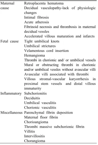
Table 1. Classification of the placental findings contributing to fetal death
Maternal cause
Retroplacenta hematoma
Decidual vasculopathy-lack of physiologic changes
Intimal fibrosis Acute atherosis
Fibrinoid necrosis and thrombosis in maternal decidual vessles
Accelerated villous maturation and infarcts Fetal cause Tight umbilical knots
Umbilical strictures Velamentous cord insertion Hemangioma
Thrombi in chorionic and/ or umbilical vessels Mural or obstructing thrombi in chorionic and/or umbilical vessles without avascular villi Avascular villi associated with thrombi Villous stromal-vascular karyorrhexis in proximal stem vessels and distal villous immaturity
Inflammatory Subchorionitis Deciduitis
Umbilical vasculitis Chorionic vasculitis
Miscellaneous Parenchymal fibrin deposition Maternal floor fibrin
Chorioangioma
Thrombi massive subchorionic fibrin Villitis
Intervillositis Chorangioma
부족으로 태아 부검의 시행에 장애가 있다. 그에 반하여 태반의 조직검사는 태아 부검에 비하여 상대적으로 쉽게 시행될 수 있으며 태아 사망을 규명하는 데 중요한 정보를 제공할 수 있다. 여러 연구에서도 태아사망의 원인이 55~
61% 정도 태반에 있는 것으로 나타났다.8,10,11
본 연구에서는 자궁 내 태아사망이 일어난 태아들의 태 반조직병리학적 결과에 따라서 태아사망의 원인을 분석하 고자 하였다.
연구 대상 및 방법
1. 연구 대상
1997년 1월부터 2007년 12월까지 가톨릭대학교 성빈센 트병원에서 자궁 내 태아사망으로 진단받고 분만한 환자를
대상으로 하였다. 이 기간에 총 253건의 태아사망이 있었 으나 이 중 20주 이하, 쌍둥이 임신 및 태반조직검사를 시 행하지 않았던 130명을 제외한 123명이 연구에 포함되었 다. 대상환자의 의무기록과 태반조직병리검사 결과에 대하 여 후향적 연구를 시행하였으며 임상시험 의료윤리위원회 (VC10R1S0042)의 승인을 받았다.
2. 태반의 조직병리검사 분류
태반조직병리검사 소견은 Lagston 등12과 Redline13에 의하여 제시된 분류에 따라 모체측 원인, 태아 원인, 염증, 그 밖의 이상 및 특이소견이 없는 경우로 5가지로 분류하 였다 (Table 1).9,11
3. 통계
모든 통계는 SAS ver. 8 (SAS institute Inc, Cary, NC, USA)을 이용하였다. 각 군 간의 비교는 Pearson’s chi- square test와 Fisher’s exact test를 이용하였으며,
P
<0.05 인 경우 통계적인 유의성이 있는 것으로 판정하였다.결 과
1. 연구대상 및 임상적 특징
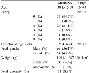
대상군 123명의 나이는 평균 30±5.38세 (16~43세)였으 며 35세 미만은 97명 (78.9%), 35세 이상은 26명 (21.1%) 이었다. 출산력이 한 번도 없는 경우는 55명 (44.7%)으로 가장 많은 비율을 차지하였다. 임신 주수는 평균 28.43±
6.74주였으며, 37주 이상의 만삭은 20명 (16.3%)이었으며 나머지 조산아 103명 중 26주 이하는 58명 (47.2%)으로 가장 많았다 (Table 2). 출생아 중 남아는 69명 (56.1%), 여아는 54명 (43.9%)으로 남아가 더 많았다. 당뇨가 있었던 환자는 11명 (8.9%), 전자간증이 있었던 환자는 14명 (11.3%) 있었으며 그 밖의 내과적 질환을 동반한 환자는 없었다.
산전 초음파와 출생 후 시진상 발견된 기형은 11명 (8.9%) 에서 있었다. 태아사망이 일어난 임신 주수를 기준으로 하 였을 때 95th percentile 이상의 거대아는 2명 (1.6%), 5th percentile 이하의 성장지연이 있었던 경우는 32명 (26%)
Table 2. Maternal and fetal general characteristics Mean±SD Range
Age 30.15±5.38 16~43
Parity 20~41
0 (%) 55 (44.7%)
1 (%) 38 (30.9%)
2 (%) 26 (21.1%)
3 (%) 2 (1.6%)
4 (%) 1 (0.8%)
8 (%) 1 (0.8%)
Gestational age (wk) 28.4±6.74 20~41 Fetal gender Male (%) 69 (56.1%) Female (%) 54 (43.9%)
Weight (g) 1,211±1,067 100~4,400
IUGR (%) 32 (26%) Macrosomia (%) 2 (1.6%)
Fetal anomaly (%) 11 (8.9%)
SD: standard deviation, IUGR: intrauterine growth restriction.
Table 3. Placental pathologic findings of 123 intrauterine fetal death
N (%)
Maternal cause 56 (45.5)
Fetal cause 35 (28.4)
Inflammatory 20 (16.2)
Miscellaneous 29 (23.5)
Unremarkable 27 (21.9)
Table 4. Placental pathologic findings of IUFD according to maternal age, parity and medical illness (preeclampsia and diabetes) Maternal age
(n) Parity
(n) Preeclampsia
(n) DM
(n)
<35 (97)
≥35
(26) P-value Primipara
(55) Multipara
(68) P-value Yes
(14) No
(109) P-value Yes (3) No
(120) P-value Materal cause 43
(44.3%) 13
(50%) 0.661 29
(52.7%) 27
(39.7%) 0.202 9
(64.2%) 47
(43.1%) 0.161 3 (100%) 53
(44.1%) 0.092 Fetal cause 26
(26.8%) 9
(34.6%) 0.467 13
(23.6%) 22
(32.3%) 0.320 5
(35.7%) 30
(27.5%) 0.538 1 (33.3%) 34
(28.3%) 1.000 Chorioamnionitis 17
(17.5%) 3
(11.5%) 0.563 11
(20%) 9
(13.2%) 0.336 3
(21.4%) 17
(15.5%) 0.669 0 (0%) 20
(16.6%) 0.584 Miscellaneous 23
(23.7%) 6
(23.0%) 1.000 11
(20%) 18
(26.4%) 0.522 2
(14.2%) 27
(24.7%) 0.516 1 (33.3%) 12
(10%) 0.557 Unremarkable 26
(26.8%) 2
(7.6%) 0.062 27
(49.0%) 1
(1.4%) 0.666 1
(7.1%) 27
(24.7%) 0.187 0 (0%) 27
(22.5%) 1.000 IUFD: intrauterine fetal death, DM: diabetes mellitus.
이었다.
2. 태반의 조직병리
태반조직병리검사상 특이할 만한 소견이 없는 경우가
27예 (21.9%)였으며 나머지 96예는 모체측 요인, 태아측 요인, 염증 소견, 기타의 4가지의 분류 중에 적어도 한 가 지 이상의 소견을 보였다. 4가지 분류 중 1가지의 소견만 보인 경우는 57예였으며 나머지 39예는 두 가지 이상의 복 합적인 소견을 보였다. 가장 흔하게 보인 소견은 모체측 요인으로 123예의 태반 중 56예 (45.5%)에서 관찰할 수 있 었으며, 두 번째로 많이 보이는 소견은 태아측 요인으로 35예 (28.4%)의 태반에서 관찰 할 수 있었다 (Table 3).
모체의 나이를 35세 미만 군과 이상 군으로 나누어서 태 반조직병리검사 소견을 비교하여 보았을 때 두 군 간에 통 계적으로 유의한 차이는 보이지 않았으며, 산과력, 당뇨 및 전자간증 유무에 따른 태반병리조직학적 차이도 보이지 않았다 (Table 4). 그러나, 모체의 나이가 35세 이상인 군, 초산모, 전자간증 및 당뇨병이 있는 군들의 태반은 50% 이 상에서 모체측 요인이 발견되었다 (Table 4).
태아의 임신 주수를 37주와 26주를 기준으로 각각 분류 하여 태반조직병리학적 소견을 비교하여 보았을 때 두 군 간 태반조직병리학적 소견에는 유의적인 차이는 없었으며 임신 37주 이상의 60%에서 모체측 요인이 발견되었다. 태 아의 성별에 따른 태반 소견에서도 각 군 간의 유의한 차 이는 보이지 않았으며 남아인 경우 50.7%에서 모체측 요 인이 발견되었다. 태아의 기형이 있었던 경우 11예에서 모 두 태반조직병리검사에서 태아측 요인으로 분류된 소견들 이 관찰되어 기형이 없었던 군보다 유의하게 태아측 요인 이 높았다 (
P<
0.0001) (Table 5).태아 체중을 1,500 g 미만과 이상으로 분류하였을 때 두 군 간의 태반 소견에는 차이가 없었으며 1,500 g 이상인 군에서는 모체측 요인이 55.5%로 나타났다. 자궁 내 성장
Table 5. Placental pathologic findings of IUFD according to gestational age, fetal gender and fetal anomaly Gestational age
(n) Gestational age
(n) Fetal gender
(n) Fetal anomaly
(n)
<26 wks
(58) ≥26 wks
(65) P-value <37 wks (103) ≥37 wks
(20) P-value Male
(69) Female
(54) P-value Yes
(11) No
(112) P-value Materal cause 25
(43.1%) 31
(47.6%) 0.171 44
(42.7%) 12
(60%) 0.220 35
(50.7%) 21
(38.8%) 0.207 5
(45.4%) 51
(45.5%) 1.000 Fetal cause 17
(29.3%) 18
(27.6%) 0.845 28
(27.1%) 7
(35%) 0.589 20
(28.9%) 15
(27.7%) 1.000 11
(100%) 24
(21.4%)<0.0001 Chorioamnionitis 10
(17.2%) 10
(15.3%) 0.811 14
(13.5%) 6
(30%) 0.094 13
(18.8%) 7
(12.9%) 0.464 0
(0%) 20
(17.8%) 0.209 Miscellaneous 17
(29.3%) 12
(18.4%) 0.202 26
(25.2%) 3
( 15%) 0.401 17
(24.6%) 12
(22.2%) 0.832 2
(18.1%) 27
(24.1%) 1.000 Unremarkable 11
(18.9%) 16
(24.6%) 0.393 25
(24.2%) 3
(15%) 0.561 11
(15.9%) 16
(29.6%) 0.131 0
(0%) 27
(24.1%) 0.453 IUFD: intrauterine fetal death.
Table 6. Placental pathologic findings according to fetal weight, IUGR and macrosomia Fetal Wt (g)
(n) IUGR
(n) Macrosomia
(n)
<1500
(87) ≥1500
(36) P-value Yes
(32) No
(91) P-value Yes
(2) No
(121) P-value Materal cause 36
(41.3%) 20
(55.5%) 0.168 20
(62.5%) 36
(39.5%) 0.038 2
(100%) 54
(44.6%) 0.205
Fetal cause 25
(28.7%) 10
(27.7%) 1.000 12
(37.5%) 23
(25.2%) 0.254 1
(50%) 34
(28.0%) 0.490 Chorioamnionitis 15
(17.2%) 5
(13.8%) 0.791 3
(9.3%) 17
(18.6%) 0.275 0
(0%) 20
(16.5%) 1.000 Miscellaneous 24
(27.5%) 5
(13.8%) 0.160 11
(34.3%) 18
(19.7%) 0.145 0
(0%) 29
(23.9%) 1.000
Unremarkable 17
(19.5%) 11
(30.5%) 0.237 3
(9.3%) 25
(27.4%) 0.049 0
(0%) 28
(23.1%) 1.000 IUGR: intrauterine growth restriction.
지연이 있었던 태아의 태반 (62.5%)에서는 모체측 요인에 의한 조직병리학적 소견이 자궁 내 성장지연이 없는 경우 (39.5%)보다 유의하게 높게 발견되었다 (
P
=0.038). 또한, 태반에 특이소견이 없는 경우는 9.3%로 성장지연이 없었 던 군의 27.4%보다 유의하게 낮았다 (P
=0.049). 거대아를 보였던 2예의 환자에서는 모두 모체측 요인이 태반에서 발 견되었으나 통계적으로 유의하지는 않았다 (Table 6).고 찰
우리나라의 자궁 내 태아사망의 빈도는 1.08~3.19%로 선진국보다 약 두 배 이상 높다.14,15 자궁 내 태아사망을 방 지하기 위해서는 자궁 내 태아사망의 원인을 밝히는 것이 중요하며 원인을 밝히기 위한 많은 검사 중 매우 중요하고 기본이 되는 것이 태아의 부검과 태반에 대한 검사이다.
태반의 검사를 포함한 태아부검이 시행된 경우 태아사망의
원인의 많은 부분을 규명할 수 있어서 원인 불명으로 분류 되는 경우는 50%에서 8~15%로 감소한다.8-11 태아의 부검 을 통한 부모의 상담을 할 경우 다음 임신에서 태아사망의 재발을 약 26~51%에서 예측할 수 있다고 하였다.16 태아 의 부검율은 아일랜드에서는 47.4%, 노르웨이에서는 62%, 영국에서는 58.1%로 높은 반면17-19 우리나라의 경우 사회 문화적 이유와 전문 인력의 부족으로 부검율은 훨씬 낮아 서 약 11% 정도이다.20
자궁 내 태아사망은 대부분 첫 임신이나 두 번째 임신에 서 일어난다고 하였고 초산부가 가장 높은 빈도를 보인다 고 하였다.19,21 본 연구에서도 첫 임신이나 두 번째 임신인 경우가 전체의 75.9%였으며 초산모가 44.7%로 가장 많았 다. 임신이 반복될수록 자궁 내 태아사망의 위험도는 감소 하는 것으로 되어 있다.22 본 연구에서 사산아 남녀비율은 1.27:1로 남아에서 높은 비율을 보였다. 대부분의 연구에 서도 남아의 비율이 높은데, 이 원인을 여아 유전자가 X
염색체에 가해지는 치명적인 손상에 대하여 보호를 받기 때문이라고 하였다.23 Hankins과 Longo24는 태아 사망 중 90% 이상은 임신 첫 20주에 발생하고 5%는 임신 20주에 서 28주 사이에 발생하며 2%는 임신 28주 이후에 발생한 다고 하였다. 본 연구에서도 29주 이전에 발생은 72건으로 약 58.5%였다.
본 연구에서 자궁 내 태아사망 중 태아의 기형빈도는 8.9%로 과거 연구보다는 낮으나,7,8 최근의 몇몇 연구와는 비슷한 빈도를 보였다.9,10 이는 과거보다 산전 검사의 질이 향상되어 태아사망에 이르기 전에 분만 또는 다른 치료를 시행하였기 때문이거나 초음파의 발달과 보급으로 임신 초 기에 진단과 임신 종결이 이루어졌기 때문으로 생각된다.
본 연구에서 태아사망의 원인을 태반조직병리학적 결과 에 따라 분류한 결과 한 가지 이상의 복합적인 소견을 보 이는 경우가 39예로 특이소견이 없는 태반을 보인 27예를 제외한 96예의 47.5%였다. 복합적 소견을 보이는 경우가 높음은 태아사망에 이르기까지 복합적인 원인이 서로 상가 효과 혹은 상승효과로 상호작용하였음을 시사한다고 하겠 다. 조직병리학적 원인 중 모체측 원인 (45.5%)이 가장 많 았으며 그 다음으로 태아측 원인 (28.4%)이 많았다. 이러 한 결과는 Kidron 등10의 연구 결과에서 모체측의 원인이 51%로 가장 많았으며 태아측 원인이 26%인 것과 유사하였 다. 태아측 원인을 제외하고 모체측 및 염증의 원인에 의 한 경우 등이 임신 시 모체의 적절한 처치에 의하여 치료 될 수 있는 요인임을 고려하면 태아사망의 반 이상이 예방 가능함을 기대할 수 있다. 태아측 원인에서 통계적으로 유 의한 차이를 보인 경우는 태아 기형이 있는 경우로 태아에 기형이 있는 경우는 태반에 반영됨을 알 수 있다. 그러나, 태반의 조직병리학적 분류에 따른 대부분의 원인들은 모체
연령, 출산력, 재태연령, 태아의 성별, 태아 출생 시 체중 이 1,500 g이 되는지 등의 요인에 대하여 통계적으로 유의 한 차이를 보이지 않았다.
자궁 내 염증은 조산과 주산기 합병증과도 관련이 있으 나 탯줄과 융모막혈관의 혈전과 염증에 의해 유발된 사이 토카인들의 자궁수축 유발에 의한 태반뒤혈종, 태아 무산 소증에 의해 유발되는 피막하 간혈종 등과 같은 일련의 일 이 태아사망을 일으키는 것으로 알려져있다.10 자궁 내 태 아사망 시 태반의 30~33%에서 염증 소견이 나타나며 이 는 정상아의 태반보다 약 2.6배 높은 것이다.13,25 염증과 관련된 자궁 내 사망은 임신 주수와 관련성이 높은데 28주 이하의 경우가 더 높은 관련성이 있는 것으로 알려져 있으 나26 본 연구에서는 유의한 차이는 없었다.
자궁 내 성장지연은 자궁 내 태아사망의 주된 위험인자 로 알려져 있으며, 자궁 내 태아사망 중 약 21~41%에서 자궁 내 성장지연이 있고, 자궁 내 성장지연인 경우 약 6.8 배 자궁 내 사망의 위험이 높아진다고 하였다.8,27 본 연구 에서는 자궁 내 성장지연이 있었던 경우는 26%였으며 태 반의 조직병리검사상 유의하게 모체측 요인이 높게 관찰되 었다. 또한, 태반조직병리검사상 특이소견이 없는 경우가 유의하게 낮았다. 이는 자궁 내 성장지연인 태아의 사망원 인이 태반과 관련이 높음을 시사하며 모체측 요인이 중요 한 역할을 하는 것으로 생각된다.
태아 부검이 어려운 현실에서 태반의 검사는 중요하다.
그러나, 아쉽게도 아직까지 자궁 내 태아사망에 시행하는 태반조직병리검사의 정형화된 지침이 없는 상태이다.28 태 아사망의 원인을 모두 설명할 수는 없었지만 향후에 체계 화된 태반조직병리학적 검사의 시행이 필요할 것으로 생각 된다.
참고문헌
1. MacDorman MF, Munson ML, Kirmeyer S. Fetal and perinatal mortality, United States, 2004. Natl Vital Stat Rep 2007; 56: 1‐19.
2. Silver RM. Fetal death. Obstet Gynecol 2007; 109:
153‐67.
3. Fretts RC. Etiology and prevention of stillbirth. Am J Obstet Gynecol 2005; 193: 1923‐35.
4. Weintraub AY, Rozen A, Sheiner E, Levy A, Press F, Wiznitzer A. Perinatal mortality: a sporadic event or a recurrent catastrophe? Arch Gynecol Obstet 2009;
279: 299‐303.
5. Smith GC, Fretts RC. Stillbirth. Lancet 2007; 370:
1715‐25.
6. Reddy UM. Prediction and prevention of recurrent stillbirth. Obstet Gynecol 2007; 110: 1151‐64.
7. Pitkin RM. Fetal death: diagnosis and management.
Am J Obstet Gynecol 1987; 157: 583‐9.
8. Horn LC, Langner A, Stiehl P, Wittekind C, Faber R.
Identification of the causes of intrauterine death during 310 consecutive autopsies. Eur J Obstet Gynecol Reprod Biol 2004; 113: 134‐8.
9. Varli IH, Petersson K, Bottinga R, Bremme K, Hofsjö A, Holm M, et al. The Stockholm classification of stillbirth. Acta Obstet Gynecol Scand 2008; 87: 1202‐12.
10. Kidron D, Bernheim J, Aviram R. Placental findings contributing to fetal death, a study of 120 stillbirths between 23 and 40 weeks gestation. Placenta 2009;
30: 700‐4.
11. Gardosi J, Kady SM, McGeown P, Francis A, Tonks A. Classification of stillbirth by relevant condition at death (ReCoDe): population based cohort study. BMJ 2005; 331: 1113‐7.
12. Langston C, Kaplan C, Macpherson T, Manci E, Peevy K, Clark B, et al. Practice guideline for examination of the placenta: developed by the Placental Pathology Practice Guideline Development Task Force of the College of American Pathologists.
Arch Pathol Lab Med 1997; 121: 449‐76.
13. Redline RW. Inflammatory responses in the placenta and umbilical cord. Semin Fetal Neonatal Med 2006;
11: 296‐301.
14. Chung WK, Kim H, Lee MJ, Lee DJ. A clinical study for fetal death in utero. Korean J Obstet Gynecol 1993; 36: 464‐72.
15. Park SH, Park JW, Song HS, Kim SD, Ahn JY. The clinical study of intrauterine fetal death. Korean J Obstet Gynecol 1994; 37: 1541‐52.
16. Hey EN, Lloyd DJ, Wigglesworth JS. Classifying perinatal death: fetal and neonatal factors. Br J Obstet Gynaecol 1986; 93: 1213‐23.
17. Thornton CM, O'Hara MD. A regional audit of perinatal and infant autopsies in Northern Ireland. Br J Obstet Gynaecol 1998; 105: 18‐23.
18. Wright C, Cameron H, Lamb W. A study of the quality of perinatal autopsy in the former northern region. The Northern Perinatal Mortality Survey Steering Group. Br J Obstet Gynaecol 1998; 105: 24‐8.
19. Holt J, Vold IN, Odland JO, Forde OH. Perinatal deaths in a Norwegian county 1986‐96 classified by the Nordic‐Baltic perinatal classification: geographical contrasts as a basis for quality assessment. Acta Obstet Gynecol Scand 2000; 79: 107‐12.
20. Park SH, Choi HM. A retrospective cohort study of maternal and perinatal risk factors on intrauterine fetal death. Korean J Obstet Gynecol 2008; 51: 965‐73.
21. Eschler G, Heidegger H, Krone HA. Stillbirth‐‐an analysis of 354 cases 1966‐88. Geburtshilfe Frauen- heilkd 1991; 51: 293‐7.
22. Kunzel W, Misselwitz B. Unexpected fetal death during pregnancy‐‐a problem of unrecognized fetal disorders during antenatal care? Eur J Obstet Gynecol Reprod Biol 2003; 110(Suppl 1): S86‐92.
23. Abramowicz M, Barnett HL. Sex ratio of infant mortality. Am J Dis Child 1970; 119: 314‐5.
24. Hankins GD, Longo M. The role of stillbirth prevention and late preterm (near‐term) births. Semin Perinatol 2006; 30: 20‐3.
25. Moyo SR, Hagerstrand I, Nystrom L, Tswana SA, Blomberg J, Bergstrom S, et al. Stillbirths and intrauterine infection, histologic chorioamnionitis and microbiological findings. Int J Gynaecol Obstet 1996;
54: 115‐23.
26. Skjoldebrand‐Sparre L, Nyman M, Broliden K, Wahren B. All cases of intrauterine fetal death should be evaluated for parvovirus B19 viral deoxyribonucleic acid. Am J Obstet Gynecol 1999; 180: 1595‐6.
27. Meyberg R, Boos R, Babajan A, Ertan AK, Schmidt W. Intrauterine growth retardation‐‐perinatal mortality and postnatal morbidity in a perinatal center. Z Geburtshilfe Neonatol 2000; 204: 218‐23.
28. Korteweg FJ, Gordijn SJ, Timmer A, Holm JP, Ravise JM, Erwich JJ. A placental cause of intra‐ uterine fetal death depends on the perinatal mortality classification system used. Placenta 2008; 29: 71‐80.
= 국문초록 =