490
Korean J Radiol 11(4), Jul/Aug 2010Clear Cell Hidradenoma of the Axilla:
a Case Report with Literature Review
Clear cell hidradenoma is an uncommon benign skin appendageal tumor that typically involves the dermal layer of the head, face, and extremities. The breast is a rare site for this lesion, with only two documented cases, which were deter- mined based on mammogram and sonogram findings. We present a case of clear cell hidradenoma of the axillary tail with radiological findings and a literature review.
here are three types of sweat glands; eccrine, apocrine, and mixed (1). A clear cell hidradenoma is a rare benign sweat gland tumor which is traditionally believed to originate from the eccrine gland (2, 3). A clear cell hidradenoma of the breast is rare. Only two cases have been reported based on mammogram and sonogram findings (3, 4). Due to the rarity of this tumor, and the common removal of it without imaging studies, typical radiological findings have not yet been well established. We present a case of clear cell hidradenoma of the axillary tail with mammography and ultrasonography in a 56 year-old woman.
CASE REPORT
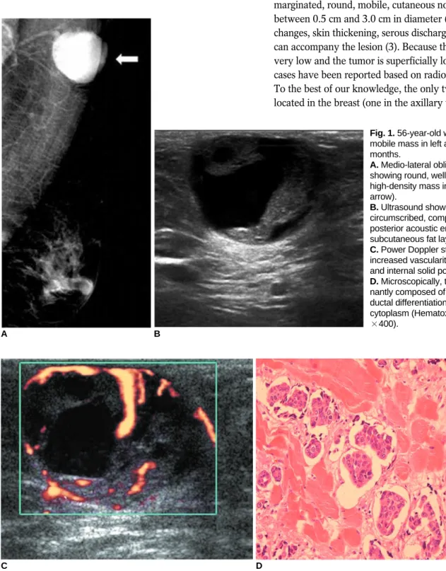
A 56 year-old woman presented with a solitary, palpable mass in the left axilla. This mass had been recognized for six months and displayed rapid growth. The patient had a personal history of ovarian cancer, but she had no remarkable family medical history. Upon physical examination, a 3-cm, soft mobile mass was identified in the axillary tail of the left breast. No nipple discharge or changes to the skin color were observed.
A left medio-lateral oblique (MLO) mammogram (Senograph DMR; GE Healthcare, Milwaukee, WI) showed a 2.7 × 2.8 cm, round, well-circumscribed, high-density mass in the left axilla (Fig. 1A). There was no combined microcalcification within or around the mass. The mass was not seen in the cranio-caudal (CC) projection. An ultrasound (iU22 unit; Philips Medical Systems, Bothell, WA) of the left breast revealed a 2.7 × 2.5 cm, oval, well-circumscribed mass with complex echogenicity in the subcutaneous layer of the left axilla (Fig. 1B). The solid portion and peripheral area of the mass showed increased vascularity following a power Doppler examination (Fig. 1C).
Radiologically, this lesion was considered to be BI-RADS (Breast Imaging Reporting and Data System) category 4a. An excisional biopsy was performed at the patient’s request due to the superficial location and palpability of the mass.
The biopsy specimen measured 3 × 2.5 × 1.5 cm. Grossly, the mass was well- encapsulated, tan-to-pink in color, and cystic. A section of the mass showed it to have solid areas with cystic cavities containing serous fluid and white-to-tan gelatinous Kyung Eun Cho, MD
1Eun Ju Son, MD
1Jeong-Ah Kim, MD
1Ji Hyun Youk, MD
1Eun-Kyung Kim, MD
1Jin Young Kwak, MD
1Joon Jeong, MD
2Index terms : Axilla mass Hidradenoma Ultrasonography
DOI:10.3348/kjr.2010.11.4.490
Korean J Radiol 2010;11:490-492 Received December 11, 2009; accepted after revision February 4, 2010.
Departments of 1Radiology and 2Surgery, Yonsei University College of Medicine, Seoul 135-720, Korea
Address reprint requests to : Eun Ju Son, MD, Department of Radiology, Yonsei University College of Medicine, Gangnam Severance Hospital, 712 Eonjuro, Gangnam-gu, 135-720 Seoul, Korea.
Tel. (822) 2019-3510 Fax. (822) 3462-5472 e-mail: [email protected]
T
material. Hematoxylin and eosin sections revealed that the tumor lobules were composed of predominantly clear cells with ductal differentiation and eosinophilic cytoplasm.
These findings were compatible with a clear cell hidrade- noma (Fig. 1D).
DISCUSSION
A clear cell hidradenoma is an uncommon benign sweat gland tumor, which is traditionally known to originate
from the eccrine gland (1, 2). This tumor occurs at any stage of life but is most common in the fourth decade.
Furthermore, it affects women 1.7 - 2 times more commonly than men (5, 6) and is usually found on the head, face, trunk, and extremities. Occurrence on the breast is considered to be rare with only two documented cases (7, 8). Typical histological locations of the tumor are the dermal layer and subcutaneous fat layer (9). The tumors are generally covered by a normal epidermis (1, 9).
The lesion is usually a single, slow-growing, well- marginated, round, mobile, cutaneous nodule ranging between 0.5 cm and 3.0 cm in diameter (2, 9). Skin color changes, skin thickening, serous discharge, or tenderness can accompany the lesion (3). Because the incidence is very low and the tumor is superficially located, very few cases have been reported based on radiological findings.
To the best of our knowledge, the only two cases that were located in the breast (one in the axillary tail and the other
Clear Cell Hidradenoma of AxillaKorean J Radiol 11(4), Jul/Aug 2010
491
Fig. 1. 56-year-old woman with soft, mobile mass in left axillary tail for six months.
A. Medio-lateral oblique mammogram showing round, well-circumscribed, high-density mass in left axilla (white arrow).
B. Ultrasound showing oval, well- circumscribed, complex mass with posterior acoustic enhancement in subcutaneous fat layer of left axillary tail.
C. Power Doppler study revealed increased vascularity in peripheral area and internal solid portion of mass.
D. Microscopically, tumor is predomi- nantly composed of clear cells with ductal differentiation and eosinophilic cytoplasm (Hematoxylin & Eosin stain,
×400).
A B
C D
Cho et al.
492
Korean J Radiol 11(4), Jul/Aug 2010in the breast parenchyma) have been diagnosed by a mammography and breast ultrasonography (3, 4). The case in the axillary tail presented as a well-circumscribed, high- density mass on mammography, just as it did in our case (3). The other case, located in the breast parenchyma, presented as a focal asymmetric density (4).
An ultrasound (1-4), performed on clear cell hidradeno- mas present as well-defined cystic masses with mural nodules or as well-defined solid tumors with
hypoechogenicity and hypervascularity. A mural nodule in a cystic lesion is also frequently highly vascular as
determined by a Doppler examination. The echogenicity of the cystic portion could be complex due to a hemorrhagic component. In addition, calcifications were reported in some cases. Common features of a clear cell hidradenoma include a well-circumscribed, lobulated, subcutaneous, cystic or solid mass with low-to-intermediate signal intensity on a T1 weighted image (WI) MRI and an
intermediate-to-high signal intensity upon a T2WI MRI and short tau inversion recovery image (2, 9). Hemorrhage and sweat gland excretion in the cystic portion may cause variable signal intensity of the fluid contents of the tumor.
The mural nodule or solid portion of the tumor could show enhancement after contrast enhancement. The differential diagnosis for a clear cell hidradenoma includes primary breast cancer and papillary neoplasm of the breast (3, 4), hemangioma, vascular leiomyoma, lymphomas, and metastases (1). Malignant transformation, or hidradenocar- cinoma, is uncommon but the differential diagnosis with a benign hidradenoma is difficult without distant metastasis or aggressive local invasion (8). Tumor remnants due to inadequate excision are a frequent cause of recurrence.
Because of this risk, an excisional biopsy with clear resection margins is necessary in order to make a final accurate diagnosis and begin the proper treatment (2, 3).
Although the radiological findings are non-specific, clear cell hidradenoma should be considered in the differential diagnosis when a superficially located, well-circumscribed, cystic mass with a variable solid portion and hypervascu- larity is encountered during a breast imaging work-up.
References
1. Jin W, Kim GY, Lew BL, Yang DM, Kim HC, Ryu JK, et al.
Sonographic findings of an eccrine spiradenoma: case report and literature review. J Ultrasound Med 2008;27:813-818
2. Mullaney PJ, Becker E, Graham B, Ghazarian D, Riddell RH, Salonen DC. Benign hidradenoma: magnetic resonance and ultrasound features of two cases. Skeletal Radiol 2007;36:1185- 1190
3. Ghai S, Bukhanov K. Eccrine acrospiroma of breast:
mammographic and ultrasound findings. Clin Radiol 2004;59:1142-1144
4. Ohi Y, Umekita Y, Rai Y, Kukita T, Sagara Y, Sagara Y, et al.
Clear cell hidradenoma of the breast: a case report with review of the literature. Breast Cancer 2007;14:307-311
5. Shaikh-Naidu N, Breitbart A. Eccrine spiradenoma of the upper extremity: case report and an algorithm for management. Eur J Plast Surg 2003;26:160-163
6. Revis P, Chyu J, Medenica M. Multiple eccrine spiradenoma:
case report and review. J Cutan Pathol 1988;15:226-229 7. El Demellawy D, Daya D, Alowami S. Clear cell hidradenoma:
an unusual vulvar tumor. Int J Gynecol Pathol 2008;27:457-460 8. Herna′ndez-Pe′rez E, Cestoni-Parducci R. Nodular hidradenoma
and hidradenocarcinoma. A 10-year review. J Am Acad Dermatol 1985;12:15-20
9. Han YD, Huan Y, Deng JL, Zhang YG, Zhang CH. MRI appear- ance of multiple eccrine spiradenoma. Br J Radiol 2007;80:E27- E29