INTRODUCTION
Lung cancer is the most commonly diagnosed cancer and the leading cause of cancer-related deaths worldwide.1 In addi- tion, there is a 40% higher mortality rate in poorer areas of the United States compared to the most affluent counties.2 Although
the underlying mechanisms of the initiation and development of lung cancer have been reported in the past decades, lung cancer prognosis and treatment still present significant chal- lenges, with high relapse and drug-resistance rates reported among lung cancer patients.3,4 Therefore, further exploration of the mechanism and development of novel therapeutic meth- ods for lung cancer treatment are required.
Over the past few decades, studies have mainly focused on the exploration of protein-coding genes, while noncoding RNA (ncRNA) was considered the result of transcription er- rors.5 However, growing evidence has revealed that ncRNAs can regulate cell proliferation, invasion, migration, and apop- tosis and a remarkable variety of other biological functions re- lated to tumors.6,7 MicroRNA (miRNA) is a class of short, sin- gle-stranded ncRNA that is <22 nucleotides in length. Recent study has shown that miRNA can modulate the expression of some key genes at both pre- and post-transcription levels. Ad- ditionally, the 3' untranslated regions (3'UTR) of some mes-
The MicroRNA hsa-let-7g Promotes Proliferation and Inhibits Apoptosis in Lung Cancer by Targeting HOXB1
Fenghe Cui*, Qian Zhou*, Kuang Xiao, and Shengwei Ma
Department of Cardiothoracic Surgery, Jingzhou Central Hospital, The Second Clinical Medical College, Yangtze University, Jingzhou, China.
Purpose: The goal of this study was to explore the effects of hsa-let-7g on cell proliferation and apoptosis, and elucidate its role in lung cancer development.
Materials and Methods: The expression levels of has-let-7g and HOXB1 in tissues and cells were measured by qRT-PCR. An inhibi- tor of hsa-let-7g or one targeting a control messenger RNA were transfected into A549 and H1944 lung cancer cells, and the effects of hsa-let-7g dysregulation on cell viability and apoptosis were analyzed using CCK-8 and apoptosis detection assays. HOXB1 was con- firmed as the target gene of hsa-let-7g, based on luciferase reporter assay results. The relationship between hsa-let-7g and HOXB1 was confirmed by co-transfection of inhibitors of hsa-let-7g and HOXB1 followed by Western blot, CCK-8, and apoptosis detection assays.
Results: We observed high expression of hsa-let-7g in lung cancer tissues compared to the corresponding normal tissues, and generally higher expression of hsa-let-7g in patients with advanced tumor classification. The results of CCK-8 and apoptosis detec- tion experiments showed that the inhibition of hsa-let-7g significantly inhibited proliferation of A549 and H1944 cells, but also pro- moted apoptosis. HOXB1 is a specific target of hsa-let-7g, and downregulation of HOXB1 in lung cancer cells reversed the suppres- sive effects caused by knocking down hsa-let-7g.
Conclusion: These data collectively suggest that the expression of hsa-let-7g inhibits lung cancer cells apoptosis and promotes proliferation by down-regulating HOXB1. The results from this study demonstrate the potential of hsa-let-7g/HOXB1 axis as a therapeutic target for the treatment of lung cancer.
Key Words: Lung cancer, miRNA, hsa-let-7g, HOXB1, proliferation, apoptosis
pISSN: 0513-5796 · eISSN: 1976-2437
Received: November 7, 2019 Revised: January 7, 2020 Accepted: January 28, 2020
Corresponding author: Shengwei Ma, PhD, Department of Cardiothoracic Sur- gery, Jingzhou Central Hospital, The Second Clinical Medical College, Yangtze Uni- versity, 1 Nanhuan Road, Jingzhou 434023, China.
Tel: 86-0761-8881888, Fax: 86-0761-8881888, E-mail: [email protected]
*Fenghe Cui and Qian Zhou contributed equally to this work.
•The authors have no potential conflicts of interest to disclose.
© Copyright: Yonsei University College of Medicine 2020
This is an Open Access article distributed under the terms of the Creative Com- mons Attribution Non-Commercial License (https://creativecommons.org/licenses/
by-nc/4.0) which permits unrestricted non-commercial use, distribution, and repro- duction in any medium, provided the original work is properly cited.
Yonsei Med J 2020 Mar;61(3):210-217 https://doi.org/10.3349/ymj.2020.61.3.210
senger RNAs (mRNAs) and the process of translation can be modulated by miRNAs.5 In the past few years, miRNA regula- tion and function have attracted growing attention due to their involvement in many regulatory functions in cancer de- velopment. Recent study has revealed substantial aberrant expression of miRNAs in lung cancer tissues compared to ad- junct normal tissues.8 In addition, several miRNAs have been reported to play vital regulatory roles in the onset and devel- opment of lung cancer. For instance, Li, et al.9 reported that miR-1254 can promote cell proliferation in lung cancer by binding to the 3'UTR of secreted frizzled related protein 1 (SFRP1) mRNA. The overexpression of stromal miR-143/145 can stimulate endothelial cell proliferation, which promotes neoangiogenesis in lung cancer development.10 By regulating the IL-6/STAT3 signaling pathway, miR-218 can inhibit lung cancer cell proliferation and invasion.11 The hsa-let-7g miRNA (also known as miR-let-7g and let-7g) is a member of the let-7 miRNA family that can exert vital biological effects in various diseases, including cancer. Hu, et al.12 reported that hsa-let-7g can exert anti-tumor effects in gastric cancer by inhibiting on- cogenesis induced by oxidative stress. Another study indicat- ed that hsa-let-7g could serve as an indicator for the prognosis of S-1-based chemotherapy.13 By binding to the 3'UTR of KRAS, hsa-let-7g can also promote the onset and progression of lung cancer in patients with chronic obstructive pulmonary disease.14 However, little is known about the role of hsa-let-7g in lung cancer cells.
The homeobox (HOX) genes encode a family of transcrip- tion factors that can bind to target genes sequence to modu- late the expression level of target genes. HOXB1 is an important member of the HOX family, and several studies have demon- strated that HOXB1 is a vital regulator in the development of many diseases, including cancer. For example, the aberrant ex- pression of HOXB1 contributes to the onset and development of glioma.15 HOXB1 is also an important factor in early verte- brate development, and HOXB1 dysregulation may result in congenital heart defects.16 Although numerous studies have re- ported crucial biological effects of HOXB1 in cancer develop- ment,17,18 there has been no analysis of the role of HOXB1 in lung cancer.
In this study, we aimed to explore the correlation and mech- anism between hsa-let-7g and lung cancer. Our analysis re- vealed significantly upregulated levels of hsa-let-7g in lung cancer tissues. The overexpression of hsa-let-7g may promote lung cancer development by directly decreasing the expres- sion of HOXB1.
MATERIALS AND METHODS
Specimen collection and processing
The sample set included 20 cases of lung cancer and 20 adja- cent normal lung tissues, which were obtained from surgical
operation and verified by postoperative pathology. The re- moved tissue was washed using DPEC water. Target tissues were cut using special scissors, transferred to refrigerated tubes, and stored in a liquid nitrogen tank. Using the WHO criteria for the classification of digestive tumors, all of the sampled patients were staged. Before collecting the samples, we obtained consent from the Ethics Committee of The Sec- ond Clinical Medical College of Yangtze University (IRB No.:
2018111029).
Cell culture
Both A549 and H1944 lung cancer cells were purchased from the Shanghai cell bank. Cells were maintained in RPMI-1640 medium, which contained 10% fetal bovine serum (Wisent, Quebec, Canada) and 100 u/mL streptomycin/penicillin at 37°C. Cells were cultivated in a humidified incubator contain- ing 5% carbon dioxide.
Plasmid construction and transfection
An inhibitor of hsa-let-7g (LV-inhibitor) and a negative control lentivirus (LV-NC) were designed as short hairpin RNA (shR- NA) sequences, and were cloned by and purchased from Ge- nepharm (Hangzhou, China). After adding 1 μg/mL polybrene (Genepharm) to each well (1×10⁵ cells/well), cells were incu- bated for 0.5 h at 37°C before the addition of 15 μL of transfec- tion regent and lentivirus vector. The transfected cells were se- lected with puromycin (2.0 μg/mL) for 14 days. Fluorescence was detected after 48 hours. Five colonies were selected, and numbered as colonies 1 to 5. After checking the effect of shR- NA transfection by qRT-PCR, colony 2 was selected for down- stream experiments.
RNA extraction and qRT-PCR
Total mRNA was extracted from A549 and H1944 lung cancer cells and tissues using TRIZOL Reagent (Invitrogen, Carlsbad, CA, USA), according to the manufacturer’s instructions. We evaluated the amount and quality of extracted RNA using a Nanodrop 2000 spectrophotometer. Using a Reverse Tran- scription Kit (Takara, Dalian, China), the extracted RNA was transcribed into complementary DNA. Subsequently, qRT- PCR was performed to measure the level of mRNA in tissues and cells through SYBR II Premix Taq (Takara), and all pro- cesses were completed according to the instructions of Prime Script RT-PCR Kit (Takara Biochemicals, Tokyo, Japan). GAP- DH was used as the internal reference. The primers utilized are listed in Table 1.
Western blot
Total protein from A549 and H1944 lung cancer cells was ex- tracted using radioimmunoprecipitation assay buffer (RIPA, Beyotime Biotechnology, Shanghai, China) and quantified by bicinchoninic acid (BCA) protein assay kit (KeyGen BioTech, Nanjing, China), according to the manufacturer’s instructions.
Subsequently, electrophoresis was performed, and the pro- tein was transferred to polyvinylidene fluoride membrane (Millipore, Billerica, MA, USA) at 300 mA for 1.2 h. The mem- branes were immersed in 5% non-fat milk for 1 hour, and then incubated with primary antibodies against HOXB1 (1:1000, Santa Cruz, Dallas, TX, USA) or GAPDH (1:1000, Santa Cruz) at 4°C overnight. After washing the membranes three times with TBS-T, appropriate secondary antibodies were added and the membranes were incubated for 1 hour at 20°C. Then, a chemiluminescence detection kit (Thermo Fisher, Waltham, MA, USA) was used to develop the blots.
Cell viability assay
Cells were transferred into a 96-well plate and cultured with a cell counting kit-8 (CCK-8) reagent (Dojindo, Kumamoto, Ja- pan). Briefly, 10 μL CCK-8 was added to each well at 24, 48, 72, 96, and 120 hours. A microplate reader (Tecan, Mechelen, Bel- gium) was then used to detect the absorbance value (OD) of each well at 450 nm. Additionally, the proliferation of A549 and H1944 lung cancer cells was detected by EdU assay. Each ex- periment was independently performed three times, and the mean values are presented.
Colony formation assay
The transfected A549 and H1944 cells were seeded into 6-well plates at a density of 1000 cells per well, and then cultured for 7 days. After formation of visible colonies, 2 mL of 4% parafor- maldehyde was added to each well and incubated for 15 min.
After washing with PBS, the wells were stained with a Giemsa stain kit for 30 min. The colonies were then counted using an ordinary optical microscope.
Apoptosis detection
Cultured cells were collected into a cell suspension through trypsinization, and the density was adjusted to 5×10⁵ cells/mL after washing twice with cold PBS. Then, 10 μL of Annexin V- FITC was added to 100-μL cell suspension in each tube, and cells were incubated in the dark for 15 min at 4°C. Flow cy- tometry analysis was performed immediately after the addi- tion of 380-μL binding buffer and 10-μL propidium iodide.
Luciferase activity assay
The wild-type (WT) or mutant (MUT) hsa-let-7g sequences encoding the binding site of HOXB1 were designed and sub- cloned into pGL3 basic vector (Promega, Madison, WI, USA).
A549 cells were seeded on 24-well plates for 24 hours. Mimics of hsa-let-7g were co-transfected with 10-μg pLUC-WT-HOXB1 or pLUC-MUT-HOXB1 using Lipofectamine 2000 reagent (Invitrogen). Luciferase activity was measured using Dual-Lucif- erase Reporter Assay System (Promega).
Statistical analysis
SPSS 19.0 (IBM Corp., Armonk, NY, USA) and GraphPad Prism 5.0 software (GraphPad Software Inc., San Diego, CA, USA) were used to perform statistical analysis. Data are presented as the mean± standard deviation. Student t-test analysis was per- formed to compare quantitative variables. Statistically signifi- cance was defined as p<0.05.
RESULTS
Overexpression of hsa-let-7g was detected in lung cancer tissues
To investigate the expression profile of hsa-let-7g in lung can-
5
4
3
2
1
0
4
3
2
1
0
1.5
1.0
0.5
0.0
Normal Tumor Stage I–II Stage III–IV Normal Tumor
Relative hsa-let-7g expression Relative hsa-let-7g expression Relative HOXB1 mRNA expression
A B C
*
*
*
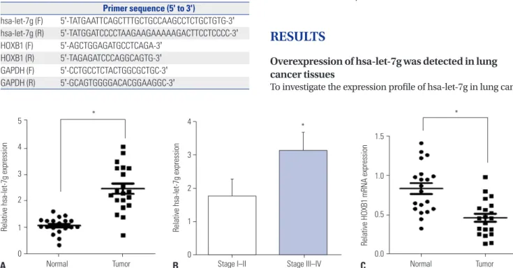
Fig. 1. hsa-let-7g levels in lung cancer. (A) The expression of hsa-let-7g in 20 paired normal-lung tumor samples. (B) Correlation of hsa-let-7g expres- sion levels at different tumor stages. (C) HOXB1 expression in 20 paired normal-tumor lung samples. *p<0.05. HOXB1, homeobox B1, mRNA: messenger RNA.
Table 1. Primer Sequences for qRT-PCR
Primer sequence (5' to 3')
hsa-let-7g (F) 5'-TATGAATTCAGCTTTGCTGCCAAGCCTCTGCTGTG-3' hsa-let-7g (R) 5'-TATGGATCCCCTAAGAAGAAAAAGACTTCCTCCCC-3' HOXB1 (F) 5'-AGCTGGAGATGCCTCAGA-3'
HOXB1 (R) 5'-TAGAGATCCCAGGCAGTG-3' GAPDH (F) 5'-CCTGCCTCTACTGGCGCTGC-3' GAPDH (R) 5'-GCAGTGGGGACACGGAAGGC-3'
cer tissues and adjacent normal tissues, we extracted mRNA from the 20 pairs of tissues samples and detected the expres- sion level of hsa-let-7g by qRT-PCR. We observed significantly increased hsa-let-7g expression in lung cancer tissues com- pared to the adjacent normal tissues (Fig. 1A). The pathologi- cal features of the 20 lung cancer patients were then compared to hsa-let-7g expression level, and we found significantly in- creased expression level of hsa-let-7g in patients with higher tumor classification (Fig. 1B). Additionally, the expression level of HOXB1 was measured, and significantly downregulated HOXB1 levels were observed in cancer tissues (Fig. 1C). All of these results were statistically significant.
Silencing of hsa-let-7g expression resulted in decreased proliferation and increased apoptosis in both A549 and H1944 cells
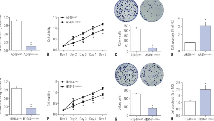
To further investigate the regulatory mechanism of hsa-let-7g in lung cancer cells, a lentivirus-mediated inhibitor of hsa-let- 7g (LV-inhibitor) was tested in A549 and H1944 lung cancer cells. Transfection efficiency was measured by qRT-PCR, and significantly decreased hsa-let-7g expression level was ob- served after transfection with the hsa-let-7g LV-inhibitor (Fig.
2A and E). CCK-8 assay and colony formation assay showed suppression of cell proliferation in the hsa-let-7g LV-inhibitor group in both A549 and H1944 cells compared to non-trans-
fected cells (Fig. 2B, C, F, and G). Moreover, cell apoptosis was significantly promoted in the hsa-let-7g LV-inhibitor group (Fig.
2D and H). These results suggested that hsa-let-7g promotes lung cancer progression.
hsa-let-7g targets HOXB1
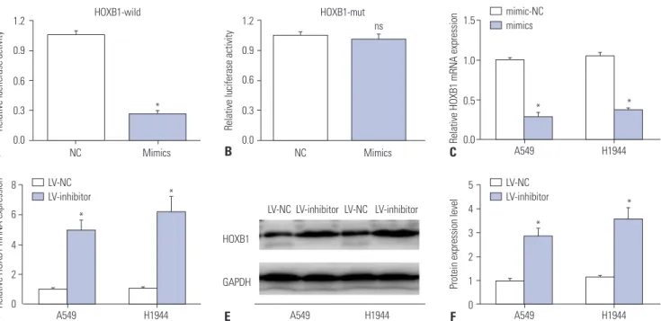
A recent study reported that has-let-7g can promote the de- velopment of osteosarcoma by targeting HOXB1.19 We pre- dicted a similar mechanism to exist in lung cancer. We used the TargetScan website (http://www.targetscan.org) and con- firmed HOXB1 as a direct target of hsa-let-7g, with the target binding site in the 987-994 nt region of 3' UTR in HOXB1 mRNA.
The binding was re-confirmed by reporter assay using lucifer- ase (Fig. 3A and B). We further analyzed the correlation be- tween HOXB1 and hsa-let-7g by measuring the expression level of HOXB1 mRNA by qRT-PCR, and measuring the protein ex- pression level by Western Blot in A549 and H1944 cells trans- fected with the hsa-let-7g LV-inhibitor or a negative control.
The results revealed increased expression levels of HOXB1 mRNA and protein after silencing hsa-let-7g (Fig. 3C-F).
hsa-let-7g regulates A549 and H1944 cells viability and apoptosis by regulating HOXB1
To further investigate whether hsa-let-7g regulates the develop- ment of lung cancer by targeting HOXB1, we next performed a
Fig. 2. Promotion of lung cancer by hsa-let-7g. (A) Expression of hsa-let-7g in A549 cells with a negative control lentivirus (LV-NC) or hsa-let-7g LV-in- hibitor. (B) Proliferation in A549 cells after inhibition of has-let-7g expression. (C) Cell apoptosis in A549 cells after inhibition of has-let-7g expression.
(D) Expression of hsa-let-7g in A549 cells transfected with LV-NC or hsa-let-7g LV-inhibitor. (E) Expression of hsa-let-7g in H1944 cells with a negative control lentivirus (LV-NC) or hsa-let-7g LV-inhibitor. (F) Proliferation in H1944 cells after inhibition of has-let-7g expression. (G) Cell apoptosis in H1944 cells after inhibition of has-let-7g expression. (H) Expression of hsa-let-7g in H1944 cells transfected with LV-NC or hsa-let-7g LV-inhibitor. *p<0.05.
1.2 0.9 0.6 0.3 0.0 1.2 0.9 0.6 0.3 0.0
2.5 2.0 1.5 1.0 0.5 0.0 4 3 2 1 0
1.5
1.0
0.5
0.0 1.5
1.0
0.5
0.0
Day 1 Day 2 Day 3 Day 4 Day 5 Day 1 Day 2 Day 3 Day 4 Day 5
H1944LV-NC A549LV-NC
H1944LV-inhibitor
A549LV-inhibitor
H1944LV-NC H1944LV-inhibitor
A549LV-NC A549LV-inhibitor
Relative hsa-let-7g expressionRelative hsa-let-7g expression Cell apoptosis (% of NC)Cell apoptosis (% of NC)
Cell viabilityCell viability
E A
H D
G C
F B
*
*
300 200 100
0 H1944LV-NCH1944LV-inhibitor
Colony cells *
250 200 150 100 50
0 A549LV-NC A549LV-inhibitor
Colony cells *
H1944LV-NC A549LV-NC
H1944LV-inhibitor
A549LV-inhibitor
*
*
Fig. 4. Reversion in A549 cells. (A, B) The expression level of HOXB1 was significantly downregulated in A549 cells after co-transfection with LV-sh- HOXB1 and has-let-7g inhibitor, compared to cells transfected with has-let-7g inhibitor or negative congtrol lentivirus (LV-NC). (C) Proliferation in- creased in A549 cells after co-transfection with LV-sh-HOXB1 and LV-inhibitor, compared to LV-inhibitor. (D) Apoptosis decreased in A549 cells after co-transfection with LV-sh-HOXB1 and LV-inhibitor, compared to LV-inhibitor alone. *p<0.05. HOXB1, homeobox B1.
1.2 0.9
0.6 0.3 0.0
1.2 0.9 0.6 0.3 0.0
HOXB1-wild HOXB1-mut
Mimics Mimics
ns
NC NC
Relative luciferase activity Relative luciferase activity
A B
E 8
6 4 2 0
5 4 3 2 1 0 1.5
1.0
0.5
0.0
A549 H1944 A549 H1944
A549 H1944
A549 H1944 HOXB1
GAPDH
Relative HOXB1 mRNA expression Protein expression levelRelative HOXB1 mRNA expression
D F
C
*
* *
*
* *
*
LV-NC LV-inhibitor
LV-NC LV-inhibitor mimic-NC mimics
LV-NC LV-inhibitor LV-NC LV-inhibitor
Fig. 3. Modulation of HOXB1 by hsa-let-7g. (A, B) Direct targeting of hsa-let-7g on HOXB1 was confirmed by luciferase activity assay. (C, D) Expres- sion level of HOXB1 mRNA in A549 and H1944 cells post-transfection with hsa-let-7g mimics or mimic-NC and LV-inhibitor or negative congtrol lentivi- rus (LV-NC). (E, F) HOXB1 protein expression level in A549 and H1944 cells transfected with LV-NC or LV-inhibitor. *p<0.05. HOXB1, homeobox B1, mRNA: messenger RNA.
1.5
1.0
0.5
0.0
1.5
1.0
0.5
0.0 1.2
0.9
0.6
0.3
0.0
Day 1 Day 2 Day 3 Day 4 Day 5 LV-NC LV-NC
A549 A549
LV-inhibitor+
LV-sh-HOXB1 LV-inhibitor+
LV-sh-HOXB1
Cell viability Cell apoptosis (% of NC)
Protein expression level
C A
D B
LV-inhibitor
LV-inhibitor+
LV-sh-HOXB1
HOXB1
GAPDH
A549
*
*
*
A549LV-inhibitor
A549LV-inhibitor+LV-sh-HOXB1
co-transfection assay. The increased HOXB1 protein expression level induced by transfection with the hsa-let-7g LV-inhibitor was significantly reversed by co-transfection of hsa-let-7g LV- inhibitor and an inhibitor targeting HOXB1, LV-sh-HOXB1, in both A549 and H1944 cells (Fig. 4A, 4B, 5A, and 5B). This sug- gested that hsa-let-7g modulates HOXB1 expression level in lung cancer cells. Additionally, CCK-8 and apoptosis assays showed increased cell viability and decreased apoptosis after co-transfection, which differed from cells that were transfect- ed with only the hsa-let-7g LV-inhibitor (Fig. 4C, 4D, 5C, and 5D). All of these results suggested that hsa-let-7g may pro- mote proliferation and inhibit apoptosis in lung cancer by modulation of HOXB1 expression level.
DISCUSSION
In this study, we investigated a potential role of hsa-let-7g in lung cancer. We found significantly increased levels of hsa-let- 7g in lung cancer tissues and downregulated expression of HOXB1. The overexpression of hsa-let-7g may promote lung
cancer development by directly decreasing the expression level of HOXB1. HOXB1 is a vital regulator in cancer develop- ment,17,18 and this is the first study to suggest a role of HOXB1 in lung cancer.
Previous studies have shown key roles of miRNAs in both physiological and pathological processes.20,21 In particular, miR- NAs can modulate the initiation and development of lung can- cer. Xu, et al.22 reported that increased expression of miR-4326 was positively associated with lung cancer cell proliferation.
Analysis of clinical data of 154 non-small-cell lung cancer pa- tients and 63 benign lung disease patients by Jiang, et al.23 re- vealed that miR-26b can act as a diagnostic and prognostic biomarker for lung cancer. Another miRNA, miR-1284, can inhibit cell proliferation, induce apoptosis, and exert anti-tu- mor effects in lung cancer.24 In this study, we observed that hsa- let-7g miRNA exerted oncogenic influence in A549 and H1944 lung cancer cells.
The hsa-let-7g is a vital member of the miRNA let-7 family, and several studies have reported vital roles of hsa-let-7g in the onset, progression, and migration of various cancers. For instance, has-let-7g can decrease the expression level of onco- Fig. 5. Reversion in H1944 cells. (A, B) The expression level of HOXB1 was significantly downregulated in H1944 cells after co-transfection with LV-sh- HOXB1 and has-let-7g inhibitor, compared to cells transfected with has-let-7g inhibitor or negative congtrol lentivirus (LV-NC). (C) Proliferation in- creased in H1944 cells after co-transfection with LV-sh-HOXB1 and LV-inhibitor, compared to LV-inhibitor alone. (D) Apoptosis decreased in H1944 cells after co-transfection with LV-sh-HOXB1 and LV-inhibitor, compared to LV-inhibitor alone. *p< 0.05. HOXB1, homeobox B1.
1.5
1.0
0.5
0.0
1.5
1.0
0.5
0.0 1.2
0.9
0.6
0.3
0.0
Day 1 Day 2 Day 3 Day 4 Day 5 LV-NC LV-NC
H1944 H1944
LV-inhibitor+
LV-sh-HOXB1 LV-inhibitor+
LV-sh-HOXB1
Cell viability Cell apoptosis (% of NC)
Protein expression level
C A
D B
LV-inhibitor
LV-inhibitor+
LV-sh-HOXB1
HOXB1
GAPDH
H1944
*
*
*
H1944LV-inhibitor
H1944LV-inhibitor+LV-sh-HOXB1
gene Myc, increase tumor suppressor gene p16 expression, and inhibit hepatocellular carcinoma cells proliferation.25 Ad- ditionally, by targeting HOXB1, hsa-let-7g can activate the NF- kB pathway that contributes to the initiation of osteosarcoma.19 Also, hsa-let-7g plays vital roles in breast cancer migration by negatively regulating FOXC2.26
Previous studies motivated us to explore the role of hsa-let- 7g in lung cancer cells, and we predicted that miRNA could be a potential therapeutic target. In our study, we used qRT-PCR to measure the expression profile of has-let-7g in 20 paired samples, and found a higher expression level in lung cancer tissues compared to that in normal tissues. In addition, we found a positive association between tumor classification and hsa-let-7g expression level by analyzing pathological data of the patients. An inhibitor of hsa-let-7g was used to detect bio- logical function in A549 and H1944 lung cancer cells. CCK-8 and apoptosis assays revealed that silencing hsa-let-7g de- creased cell viability and induced apoptosis, suggesting that the overexpression of hsa-let-7g in lung cancer patients may similarly inhibit apoptosis and promote tumor initiation. Act- ing as a sponge of mRNA is the primary mechanism by which miRNAs exert regulatory effects,27 and TargetScan analysis identified potential binding sites between has-let-7g miRNA and HOXB1 3' UTR, and luciferase activity assay confirmed the direct targeting of HOXB1 by has-let-7g. Zhou, et al.19 previ- ously pointed out that has-let-7g could exert oncogenic effects in osteosarcoma by binding the 3' UTR of HOXB1. We pre- dicted that hsa-let-7g may regulate the initiation and progres- sion of lung cancer by targeting HOXB1. The inhibition of has- let-7g resulted in increased expression levels of HOXB1 mRNA and proteins. Co-transfection of has-let-7g LV-inhibitor and LV-sh-HOXB1 confirmed that has-let-7g promoted lung cancer development by targeting HOXB1, since decreasing HOXB1 could reverse the effect.
HOXB1 is an anti-tumor gene that belongs to the HOX fam- ily, and is located in chromosome 17q21.32. Growing evi- dence has indicated that the deregulation of HOXB1 makes vital contributions to the progression of various diseases, in- cluding cancers.28,29 HOXB1 can exert tumor suppressive ef- fects by inhibiting the expression of survival oncogenic genes.30 Han, et al.15 reported that low expression level of HOXB1 con- tributed to cell invasion and proliferation, and inhibited apop- tosis in glioma.
In this study, upregulated HOXB1 was observed in A549 and H1944 lung cancer cells after transfection with hsa-let-7g LV-inhibitor. Additionally, the knockdown of HOXB1 contrib- uted to cell viability by inhibiting apoptosis, which was the opposite of the effect observed when hsa-let-7g was inhibited.
AUTHOR CONTRIBUTIONS
Conceptualization: Shengwei Ma. Data curation: Qian Zhou and Fenghe Cui. Formal analysis: Fenghe Cui. Funding acquisition: Shengwei Ma.
Investigation: Kuang Xiao. Methodology: Fenghe Cui. Project admin-
istration: Shengwei Ma. Resources: Kuang Xiao. Software: Kuang Xiao. Supervision: Shengwei Ma. Validation: Qian Zhou. Visualiza- tion: Qian Zhou. Writing—original draft: Fenghe Cui. Writing—re- view & editing: Shengwei Ma. Approval of final manuscript: All au- thors.
ORCID iDs
Fenghe Cui https://orcid.org/0000-0002-3250-1171 Qian Zhou https://orcid.org/0000-0002-8901-6185 Kuang Xiao https://orcid.org/0000-0001-5678-1019 Shengwei Ma https://orcid.org/0000-0001-9422-5427
REFERENCES
1. Bray F, Ferlay J, Soerjomataram I, Siegel RL, Torre LA, Jemal A.
Global cancer statistics 2018: GLOBOCAN estimates of incidence and mortality worldwide for 36 cancers in 185 countries. CA Can- cer J Clin 2018;68:394-424.
2. Siegel RL, Miller KD, Jemal A. Cancer statistics, 2019. CA Cancer J Clin 2019;69:7-34.
3. Woodard GA, Jones KD, Jablons DM. Lung cancer staging and prognosis. Cancer Treat Res 2016;170:47-75.
4. Nasim F, Sabath BF, Eapen GA. Lung cancer. Med Clin North Am 2019;103:463-73.
5. Cech TR, Steitz JA. The noncoding RNA revolution-trashing old rules to forge new ones. Cell 2014;157:77-94.
6. Hu X, Feng Y, Zhang D, Zhao SD, Hu Z, Greshock J, et al. A func- tional genomic approach identifies FAL1 as an oncogenic long noncoding RNA that associates with BMI1 and represses p21 ex- pression in cancer. Cancer Cell 2014;26:344-57.
7. Martens-Uzunova ES, Böttcher R, Croce CM, Jenster G, Visakorpi T, Calin GA. Long noncoding RNA in prostate, bladder, and kid- ney cancer. Eur Urol 2014;65:1140-51.
8. Del Vescovo V, Denti MA. microRNA and lung cancer. Adv Exp Med Biol 2015;889:153-77.
9. Li H, Yang T, Shang D, Sun Z. miR-1254 promotes lung cancer cell proliferation by targeting SFRP1. Biomed Pharmacother 2017;
92:913-8.
10. Dimitrova N, Gocheva V, Bhutkar A, Resnick R, Jong RM, Miller KM, et al. Stromal expression of miR-143/145 promotes neoangio- genesis in lung cancer development. Cancer Discov 2016;6:188- 201.
11. Yang Y, Ding L, Hu Q, Xia J, Sun J, Wang X, et al. MicroRNA-218 functions as a tumor suppressor in lung cancer by targeting IL-6/
STAT3 and negatively correlates with poor prognosis. Mol Cancer 2017;16:141.
12. Hu H, Zhao X, Jin Z, Hou M. Hsa-let-7g miRNA regulates the anti- tumor effects of gastric cancer cells under oxidative stress through the expression of DDR genes. J Toxicol Sci 2015;40:329-38.
13. Nakajima G, Hayashi K, Xi Y, Kudo K, Uchida K, Takasaki K, et al.
Non-coding microRNAs hsa-let-7g and hsa-miR-181b are associ- ated with chemoresponse to S-1 in colon cancer. Cancer Genom- ics Proteomics 2006;3:317-24.
14. Hu H, Zhang L, Teng G, Wu Y, Chen Y. A variant in 3'-untranslated region of KRAS compromises its interaction with hsa-let-7g and contributes to the development of lung cancer in patients with COPD. Int J Chron Obstruct Pulmon Dis 2015;10:1641-9.
15. Han L, Liu D, Li Z, Tian N, Han Z, Wang G, et al. HOXB1 is a tumor suppressor gene regulated by miR-3175 in glioma. PLoS One 2015;
10:e0142387.
16. Roux M, Laforest B, Eudes N, Bertrand N, Stefanovic S, Zaffran S.
Hoxa1 and Hoxb1 are required for pharyngeal arch artery devel- opment. Mech Dev 2017;143:1-8.
17. Morgan R, Boxall A, Harrington KJ, Simpson GR, Gillett C, Mi- chael A, et al. Targeting the HOX/PBX dimer in breast cancer.
Breast Cancer Res Treat 2012;136:389-98.
18. Guazzi S, Pintonello ML, Viganò A, Boncinelli E. Regulatory inter- actions between the human HOXB1, HOXB2, and HOXB3 pro- teins and the upstream sequence of the Otx2 gene in embryonal carcinoma cells. J Biol Chem 1998;273:11092-9.
19. Zhou JL, Deng S, Fang HS, Yu G, Peng H. Hsa-let-7g promotes os- teosarcoma by reducing HOXB1 to activate NF-kB pathway.
Biomed Pharmacother 2019;109:2335-41.
20. Tafrihi M, Hasheminasab E. MiRNAs: biology, biogenesis, their web-based tools, and databases. Microrna 2019;8:4-27.
21. Rupaimoole R, Slack FJ. MicroRNA therapeutics: towards a new era for the management of cancer and other diseases. Nat Rev Drug Discov 2017;16:203-22.
22. Xu G, Zhang Z, Zhang L, Chen Y, Li N, Lv Y, et al. miR-4326 pro- motes lung cancer cell proliferation through targeting tumor sup- pressor APC2. Mol Cell Biochem 2018;443:151-7.
23. Jiang LP, Zhu ZT, He CY. Expression of miRNA-26b in the diagno- sis and prognosis of patients with non-small-cell lung cancer. Fu- ture Oncol 2016;12:1105-15.
24. Li J, Jin H, Yu H, Wang B, Tang J. miRNA 1284 inhibits cell growth
and induces apoptosis of lung cancer cells. Mol Med Rep 2017;16:
3049-54.
25. Lan FF, Wang H, Chen YC, Chan CY, Ng SS, Li K, et al. Hsa-let-7g inhibits proliferation of hepatocellular carcinoma cells by down- regulation of c-Myc and upregulation of p16 (INK4A). Int J Cancer 2011;128:319-31.
26. Wang L, Li M, Zhou Y, Zhao Y. MicroRNA let-7g directly targets forkhead Box C2 (FOXC2) to modulate bone metastasis in breast cancer. Open Med (Wars) 2017;12:157-62.
27. Salmena L, Poliseno L, Tay Y, Kats L, Pandolfi PP. A ceRNA hy- pothesis: the Rosetta Stone of a hidden RNA language? Cell 2011;
146:353-8.
28. Arenkiel BR, Tvrdik P, Gaufo GO, Capecchi MR. Hoxb1 functions in both motoneurons and in tissues of the periphery to establish and maintain the proper neuronal circuitry. Genes Dev 2004;18:1539- 52.
29. Roux M, Laforest B, Capecchi M, Bertrand N, Zaffran S. Hoxb1 regulates proliferation and differentiation of second heart field progenitors in pharyngeal mesoderm and genetically interacts with Hoxa1 during cardiac outflow tract development. Dev Biol 2015;406:247-58.
30. López R, Garrido E, Piña P, Hidalgo A, Lazos M, Ochoa R, et al.
HOXB homeobox gene expression in cervical carcinoma. Int J Gynecol Cancer 2006;16:329-35.