양측성 완전 구순구개열 환자에서 전상악골에 대한 처치
이의룡, 정필훈*
서울대학교 치의학대학원 구강악안면외과, 치학연구소, BK21 Korea
ABSTRACT
Management of Premaxilla in Patient with Bilateral Complete Cleft Lip and Palate
Ui-Lyong Lee, Pill-Hoon Choung*
Department of Oral and Maxillofacial Surgery, Dental Research Institute, BK21 Korea, Seoul National University
전방으로 심하게 돌출된 전상악골로 인하여 구순성형술 및 비성형술의 결과가 악화 될 수 있다. 따 라서 변위된 전상악골의 이상적인 위치로 재위치 시키려는 다양한 노력이 시도되어 왔다. 에디오피아 와 같은 개발도상국에서는 어른이 되어서도 수술을 받지 못하는 구순구개열 환자가 많이 있다. 성인이 될 때까지 수술받지 못한 양측성 완전 구순구개열 환자에서는 근육, 골, 피부, 점막의 연속성이 없어서 전상악골이 심하게 전방으로 혹은 하방, 좌우측으로 변위된 경우가 대부분이다. 이 경우 구순성형술이 거의 불가능하며, 시도된다 할지라도 돌출된 전상악골 때문에 양쪽 구륜근을 봉합하여 주기가 대단히 어렵다. 따라서 이상적인 결과를 얻기 위해서는 구순성형술 전 혹은 동시에 전상악골의 재위치 술식이 필수적이다. 저자는 한국국제협력단에서 국제협력의사로 선발되어 에디오피아에서 30 개월간 근무하였 다. 그 동안 다양한 양측성 완전 구순구개열 환자에서 전상악골의 재위치 술식을 경험하였다. 저자가 경험한 전상악골의 재위치 술식(전상악골의 재위치와 골이식술 동시 시행, 전상악골의 재위치와 구개열 성형술 동시시행)에 대하여 문헌고찰과 함께 보고하고자 한다.
Key words : Premaxilla, Bilateral complete cleft lip and palate
I. INTRODUCTION
The Korea International Cooperation Agency (KOICA) was founded as a government agency to maximize the effectiveness of Korea's grant aid programs for developing countries by implementing the government's grant aid and technical cooperation
programs. The author was dispatched by KOICA to Ethiopia as voluntary international cooperative doctor program from June, 2006 to Nov, 2008.
During that time the author had a lot of chances to operate various types of cleft lip and palate victims. In Ethiopia, it is very difficult for cleft lip and palate patients who live in rural area to
get medical services at a proper time such as cheiloplasty, palatoplasty and speech therapy and so on. Because there were only 11 plastic surgeons and 4 maxillofacial surgeons (including author) all over Ethiopia in 2007. Consequently, the author encounter many adult patients who was older than 20 years of age with unoperated bilateral cleft lip and palate. These patients always had a protrusive premaxilla with intensified overjet and overbite as a result of horizontal and vertical displacement of the premaxilla. Primary cheiloplasty in adult patients with complete bilateral cleft lip and palate is quite challenging because of premaxillary anterior projection as a result of the absence of the constrictor action of labial musculature. The most noticeable feature in adult patients with unoperated bilateral complete cleft lip and palate was the extremely prominent and twisted premaxilla. In this study, 2 cases of management of premaxilla are presented; 1.
premaxillary repositioning with alveolar bone graft, 2. premaxillary repositioning with concomitant palatoplasty.
1. Case 1
(surgical repositioning of the premaxilla with bone graft)
The patient was a 23-year-old man in whom bilateral complete cleft lip and complete palatal closure had been achieved elsewhere (Figure 1).
His present complaint was the prominent premaxilla. Physical examination was within normal limits, and growth of the maxillofacial
Figure 1. A 23-year-old man with prominent premaxilla.
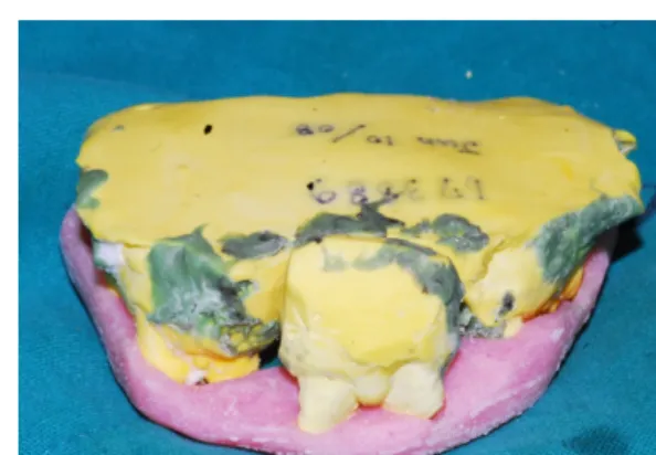
Figure 2. Model surgery and surgical template fabrication.
region seemed to be relatively normal except for the prominent maxilla. The occlusion of the dental arches posteiorly was satisfactory. The premaxilla had dropped down to obscure the mandibular teeth. Before operation, upper and lower arch impressions were taken and plaster models were mounted on articulator. Premaxilla repositioning was carried out in the model to fabricate the surgical splint with self‐curing orthodontic resin (Figure 2). Premaxilla should
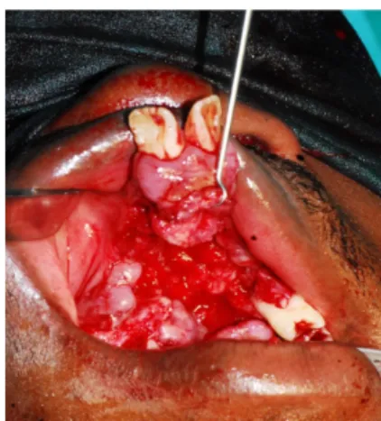
Figure 3. Premxilla was sectioned and Figure 4. Bone from iliac crest was packed into bony gap.
mobilized.
Figure 5. Surgical template was fixed to maxilla using Figure 6. After removal of surgical template.
wire.
be repositioned without premature contact with lower anterior teeth.
After induction of general anesthesia and orotracheal intubation, A circular incision was made along the lateral segments and extended into the region of the alveolar fistula and then relaxing incision was performed toward the buccal fold. Premaxilla was sectioned using reciprocating saw and surgical template in place,
the premaxilla was repositioned (Figure 3). Bone was packed into the region of the alveolus and palate (Figure 4), and a buccal-layer closure of the fistula was performed. Bone for the graft was harvested from the iliac crest. The surgical template was fixed to maxilla using wire (Figure 5). After 3 months of operation, the surgical template was removed and premaxillary mobility was assessed (Figure 6).
A
C B
Figure 7. Unoperated BCLP 20- Figure 8. Simultaneous palatal closure and premaxillary repositioning year-old woman.
2. Case 2
(Simultaneous palatoplasty and premaxillary repositioning)
Simultaneous palatoplasty and premaxillary setback was carried out in 20-year-old woman (Figure 7). The two‐flap palatoplasty for palatal closure was undertaken (Figure 8. B). Using no.
15 blade, full thickness relaxing incisions were made through the mucoperiosteum down to the bone of the maxilla. Subperiosteal undermining was then performed anterolaterally, working toward the midline, where the bony margin of the cleft was identified. Mucoperiosteal flaps were elevated on both tha palatal shelves. The nasal mucosa was separated from the palatal shelves, and vomer flaps were raised. The nasal layer is then sutured up to the junction of the hard and soft palate, intravelar veloplasty was
carried out. Vomer was fractured 5mm posterior to vomero-premaxillary suture using reciprocating saw (Figure 8. A). The superior aspect of premaxilla was separated from the nasal septum. And then the osteomized premaxilla was freely movable. With the surgical splint in place, the premaxilla was repositioned (Figure 8. C). A surgical template, fabricated preoperatively after model surgery, was assessed to ensure that bony interferences did not hinder the designed position of the premaxilla. Most of the cases, the fractured premaxilla moved backward and upward. To maximize the bony contact bone grinding or cutting was never carried out and both fractured ends were overlapped. 3 months after operation, the surgical template was removed (Figure 9).
Figure 9. After removal of surgical template
II. RESULTS
There was no severe complication, such as avascular necrosis of premaxilla or wound dehiscence at repaired palate. The movement of premaxilla was quite complicated. There were backward movement and vertical movement. No premaxilla mobility was observed. In one patient (case 1), after removal of surgical template, premature contact between upper central incisor on repositioned premaxilla and lower incisor was observed. For that reason, incisor edge of lower incisor was grinded using diamond burr.
III. DISCUSSION
Latham reported premaxillary protrusion in BCLP can be observed as early as 10 weeks of gestation1). Unrestricted growth of vomero-
premaxillary suture and anterior nasal septum accompanied with absence of bony and soft tissue connection, and constrictor action of labial musculature are considered to cause the anterior premaxillary projection2, 3). Accodingly, in many European centers, synchronous surgical repositioning of premaxilla and lip repair had been performed. However, early surgical setback of the premaxilla is now an abandoned procedure because of severe maxillary retrusion4). Secondary alveolar bone grafting with concomitant repositioning of the premaxilla in BCLP has been carried out with good result5-8). Narayaman et al. mentioned that in a developing nation, great number of cleft victims reside in rural area and have to travel long distance to get surgery, accordingly these patients like come for fewer surgeries with maximum benefits9). Consequently, we performed simultaneous palatal closure and premaxillary reposition. A rectangular sliding push back of the premaxilla after excising of the vomer using 0.8 mm Kirscher wire was introduced by Cronin10). In synchronous palatal closure and premaxillary setback, Kirschner wire fixation was performed to hold the premaxilla in a new position9). Silva Filho et al.
showed in adult patients with unoperated complete bilateral cleft lip and palate, the premaxilla, in both its basal (SN.ANS) and its dentoalveolar parts (SNA), is exteremely prominent11). Movement of premaxilla was complicated with horizontal, vertical, and lateral shifting and cutting surface of both
bony segment were not contacted. These facts hindered routine use of Kirscher wire. The author could not obtain the K-wire in Ethiopia, accordingly wiring was used for fixing osteomized premaxilla to vomer instead of K-wire. In many studies, surgical template has been used successfully for immobilizing the premaxillary segment with bone graft9, 12-15).We used surgical template succeddfully. But there were few articles about management of prominent premaxilla in adult patients with unoperated BCLP. After removal of surgical template (3 monts after operation), occlusion was carefully checked using articulation paper.
Heavy contact between upper central incisor in premaxilla and lower incisor may produce detrimental force to repositioned premaxilla.
Tight contact points on teeth were grinded with rotary instrument. To prevent premature contact, surgical simulation of repositioning of premaxilla on model should preceed operation.
Maintenance of the blood supply to the premaxilla is a matter of paramount importance in this procedure. The main blood supply to the premaxilla is through the posterior septal branch of the sphenopalatine artery and anterior septal branch from anterior ethmoidal artery16). The author did not perform cheiloplasty and open rhinoplasty during these procedure not to compromise blood supply to premaxilla. When osteotomy and surgical repositioning of premaxilla is carried out, gentle handling of premaxillary segment and minimum incision on vemerine mucosa is
necessary. There was no severe complication, such as avascular necrosis of premaxilla or wound dehiscence at repaired palate.
IV. REFERENCES
1. Latham RA. Development and structure of the premaxillary deformity in bilateral cleft lip and palate. Br J Plast Surg 1973 Jan;26(1):1-11.
2. Latham RA, Deaton TG, Calabrese CT. A question of the role of the vomer in the growth of the premaxillary segment. Cleft Palate J 1975;12:351-5.
3. Eppley BL, Sclaroff A, Delfino JJ. Secondary management of the premaxilla in bilateral cleft lip and palate patients. J Oral Maxillofac Surg 1986;44(12):987-98.
4. Friede H, Pruzansky S. Long‐term effects of premaxillary setback on facial skeletal profile in complete bilateral cleft lip and palate. Cleft Palate J 1985;22(2):97-105.
5. Freihofer HP, van Damme PA, Kuijpers‐
Jagtman AM. Early secondary osteotomy‐
stabilization of the premaxilla in bilateral clefts. J Craniomaxillofac Surg 1991;19(1) :2-6.
6. Hayward JR. Management of the premax- illa in bilateral clefts. J Oral Maxillofac Surg 1983;41(8):518-24.
7. Aburezq H, Daskalogiannakis J, Forrest C.
Management of the prominent premaxilla in bilateral cleft lip and palate. Cleft
교신 저자
정필훈, 서울대학교 치의학대학원 구강악안면외과학교실 서울시 종로구 연건동 275-1 우편번호 110-768/
Tel : 02-2072-3477 / E-mail : [email protected] Palate Craniofac J 2006;43(1):92-5.
8. Heidbuchel KL, Kuijpers-Jagtman AM, Freihofer HP. An orthodontic and cephalo- metric study on the results of the com- bined surgical‐orthodontic approach of the protruded premaxilla in bilateral clefts. J Craniomaxillofac Surg 1993;21(2):60-6.
9. Narayanan RK, Hussain SA, Murukesan S, Murthy J. Synchronous palatal closure and premaxillary setback in older children with bilateral complete cleft of lip and palate.
Plast Reconstr Surg 2006;117(2):527-31.
10. Cronin TD. Surgery of the double cleft lip and protruding premaxilla. Plast Reconstr Surg (1946) 1957;19(5):389-400.
11. da Silva Filho OG, Carvalho Lauris RC, Capeloz z a Filho L, Semb G. Craniofacial morphology in adult patients with un- operated complete bilateral cleft lip and palate. Cleft Palate Craniofac J 1998
;35(2):111-9.
12. Geraedts CT, Borstlap WA, Groenewoud JM, Borstlap‐Engels VM, Stoelinga PJ.
Long-term evaluation of bilateral cleft lip and palate patients after early secondary closure and premaxilla repositioning. Int J Oral Maxillofac Surg 2007;36(9):788-96.
13. Iino M, Sasaki T, Kochi S, Fukuda M, Takahashi T, Yamaguchi T. Surgical re- positioning of the premaxilla in combina- tion with two-stage alveolar bone grafting in bilateral cleft lip and palate. Cleft Palate Craniofac J 1998;35(4):304-9.
14. Carlini JL, Biron C, Gomes KU, Da Silva RM. Surgical repositioning of the premax- illa with bone graft in 50 bilateral cleft lip and palate patients. J Oral Maxillofac Surg 2009;67(4):760-6.
15. Scott JK, Webb RM, Flood TR. Premaxillary osteotomy and guided tissue regeneration in secondary bone grafting in children with bilateral cleft lip and palate. Cleft Palate Craniofac J 2007;44(5):469-75.
16. Mixter RC, Stroncek G, Doyle J, Carson L.
Endonasal premaxillary osteotomy. Plast Reconstr Surg 1996;97(7):1494-6.