• 검색 결과가 없습니다.

Multidisciplinary Treatment Approach in a Secondary Cleft Lip and Palate Patient for Functional Occlusal Rehabilitation

N/A
N/A
Protected

Academic year: 2021

Share "Multidisciplinary Treatment Approach in a Secondary Cleft Lip and Palate Patient for Functional Occlusal Rehabilitation"

Copied!
10
0
0

로드 중.... (전체 텍스트 보기)

전체 글

(1)

성인 구순구개열 환자에서 Multidisciplinary 치료로 기능성 교합을 형성한 증례

이지나*

이지나 치과의원

ABSTRACT

Multidisciplinary Treatment Approach in a Secondary Cleft Lip and Palate Patient for Functional Occlusal Rehabilitation

Jina-Lee Linton*

Dr. Linton's Orthodontic Clinic

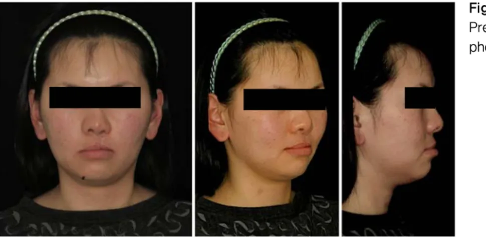
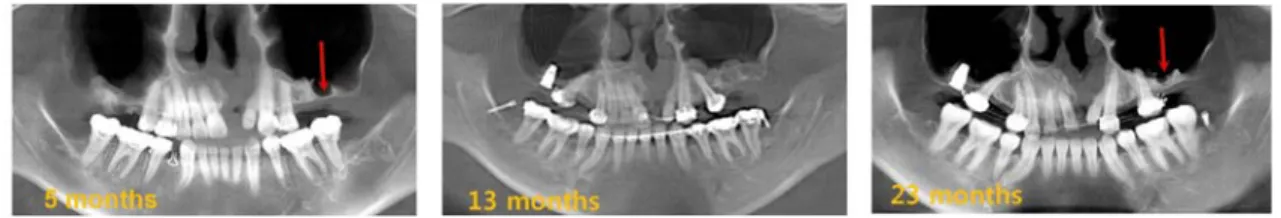
A 20 year-old cleft lip and palate patient came for occlusal rehabilitation, but the constricted maxilla and early loss of posterior teeth called for an unusual treatment modalities. Distraction osteogenesis in the edentulous areas followed by artificial bone graft, dental implant along with orthodontic tooth movement were planed. Multidisciplinary treatment enabled both esthetic and functional oral rehabilitation of this patient.

Key words

: multidisciplinary treatment, functional occlusal rehabilitation, distraction osteogenesis

Ⅰ. 서론

20세의 편측성 구순구개열인 이 여환은 코와 입 술의 이차성형수술 직후인 2007년 1월에 본원에 내원하였다. 수술 직후여서 상담만 하고 돌아갔다.

일년이 지난 2008년에 환자가 다시 내원하여 치료 받기를 원했다. 구순구개열 환자들은 흔히 겉으로

Ⅱ. 증례보고

이차성형 전의 기록이 없어 비교할 수는 없지만 nasal base의 비대칭이 수술 전에 심했을 것으로 추측된다. 정면 사진에서 코의 비대칭이 있으나 하 안면은 비교적 대칭을 이루고 있다(Figure 1).

측면 안모 사진에서 zygomatic arch에서

(2)

Figure 2.

Pre-treatment vertical proportion of the face.

하악의 고경상실과 그에 따른 mandibular over closure 때문이었다.

안모의 수직고경의 비율은 trichion에서 soft tissue menton 까지의 길이를 3등분 했을 때, 하 안모의 길이인 subnasale에서 soft tissue menton 까지의 길이가 중 안모의 길이인 glabella에서 subnasale 까지의 길이보다 조금 짧았다

4

(Figure 2). 측두방사선사진에서 구치부의 고경이 낮아지면 서 하악골은 autorotation close되었고 상하 전치 부의 수직피개가 증가했음을 짐작할 수 있었다.

상악 우측에는 중절치에서 제2소구치가, 좌측에 는 견치와 제1소구치만이 남아있었다(Figure 3).

치조골 파열은 그대로 남아있었고, 경구개는 수술

상흔으로 좁고 낮았다. 하악에는 제3대구치를 제외

한 모든 치아가 남아있었다. 하악 전치는 반대교합

으로 정출되어 있었고 하악 제2대구치는 대합치의

소실로 상악 연조직까지 정출되어 약 5mm 깊이의

curve of Spee가 관찰되었다. 상악치열의 중심선

은 하악치열의 중심선 보다 파열된 왼쪽으로 약

2mm 편이 되어있었다. 반대교합이 전치부에서 구

치부까지 관찰된다. 오른쪽 견치는 앵글씨의 제1

급 관계이고 왼쪽 견치는 제3급 관계를 보이고 있

었다.

(3)

Figure 3. Pre-treatment intraoral photographs.

파노라믹 방사선사진에서 작고 짧은 ramus와 condyle이 관찰된다. 상악 우측 제2대구치의 잔존 치근이 주위 상악동의 pneumatization으로 인한 치조골 소실과 함께 관찰되고 있다(Figure 4). 정중 선 부위에 파열된 치조골이 넓게 자리잡고 있고, 좌 측의 중절치가 결손되어있으며 측절치 위치에는 치 근의 길이가 짧은 peg lateral가 관찰된다. 좌측 제2 소구치 결손부위에는 치조골이 아직 남아있으나, 대 구치가 있었던 후방으로는 상악동의 pneumatiza-

tion이 진행되어 있었다. 하악 3대구치가 수평매복 되어있으면서 제2대구치의 치근을 흡수시켰는데, 왼쪽 제2대구치의 원심쪽 치근은 흡수되면서 주위 에 방어기전으로 secondary dentin을 만들었다.

하악의 좌우 제1소구치를 발거하고 고정성 교정 장치를 이용해서 하악 전치를 후방견인 하여 배열 하였다. 하악 우측 견치는 제2급 관계로, 좌측견치 는 제1급 관계로 견인하여 전치부 반대교합을 교정 하고자 하였다.

하악치열 교정을 시작한지 10개월 후 상악 우측

제2대구치 잔존치근을 발거하고 임플란트를 식립

(4)

Figure 5. Intraoral photographs of 5 months after orthodontic treatment.

Figure 6. Periapical x-rays shows progressive increase of interdental spaces on the right and the left premolar areas.

Figure 7.

Intraoral

photographs

shows treatment

progress from

the pre-treatment

to 10 months and

20 months of

orthodontic tooth

movement.

(5)

Figure 8. Panograms before artificial bone graft, immediately after, and 10 months later are shown.

Corticotomy 이후 3개월까지의 치아 이동은 신 속하며 bodily movement를 하였다. 3개월 이후에 는 후방치아의 치관이 치관보다 빨리 움직임으로 인해 기울어짐이 심하였다. 치근의 uprighting이 쉽지 않았는데 그 이유 중 하나는 치근이 이동해야 할 방향에 상악동에 인접해 있어 치아이동을 허용 할 골조직이 없다는 점이다(Figure 6).

하악 교정치료후 10개월 경과 되었을 때 전치부 의 반대교합이 절단면 교합의 상태로 되었다 (Figure 7). 20개월이 경과하였을 때는 상악 전치

가 하악전치를 피개하게 되었다. 우측 상하 견치는 앵글씨 제1급 관계에서 제2급 관계로 변하였고, 상 악 제1소구치와 제2소구치 간에 간격이 벌어졌다.

좌측 견치들은 제3급 관계에서 제1급 관계로 변하 였고, 상악 견치와 제1소구치 사이에 간격이 벌어 져있다.

치아가 상실됐던 시간이 오래 경과된 악 죄측

제1대구치 위위에는 치조골 흡수로 상악동 거상술

로 인공골 이식이 필요했다(Figure 8). 인공골 이

식 10개월 경과 후(23개월 째) 방사선 사진에서 골

(6)

Figure 10. Intraoral photographs of pre-treatment, after orthodontic treatment, and after restorations are shown. The premolars that were moved distally to create space for implants are marked with blue dots.

이식 전보다 두터운 치밀골이 형성되었음이 관찰 된다.

상악 최후방 소구치를 후방으로 이동하는 과정 중에 전치부에서는 다음의 치아이동이 일어났다:

1) 상악의 중심은 우측 중절치와 우측 측절치 사이 로 옮겨졌다. 2) 상악 좌측의 구개파열 부위의 치 간 간격이 좁아졌다(Figure 9).

구치부의 임플란트 치관이 하악의 자연치와 교 합 되게 제작되었다(Figure 10). 구개봉합 수술의 후유증으로 온 상악궁 협착은 구치부 반대교합을 가져왔다. 상악의 확장이 불가능했으므로 교정치료 가 끝난 시점에도 반대교합은 크게 개선되지 않았 다. 이 점은 보철물을 수복하는 과정에서 반대교합 을 좀 더 개선해 줄 수 있었다.

치료 후 전면사진을 보면 상순의 상하 폭경이 치 료 전 보다 두 배 증가 되었고, 입술 중앙보다 처졌

던 구각이 올라간 모습을 보인다(Figure 11). 또 하 악골의 윤곽이 또렷해졌다.

교정치료를 통해 하악전치가 하순의 stomium 보다 5mm 상방으로 정출된 상태에서, 치료 후에 는 하악전치의 절단면이 stomium 높이와 같도록 압하된 것을 볼수 있다(Figure 12). 구치부의 curve of Spee가 평면으로 바뀌었고, 무치악이었 던 상악 구치부위가 보철치료를 통해 하악의 구치 들과 교합되고 있다.

치료 후(붉은 선) 하악 과두가 치료 전(검은 선)

하악과두에 비해 전방으로 2.5mm 위치이동 하고

상방으로는 1.5mm 이동하였다. 치료 전 하악 전치

가 edge-to-edge로 물림을 피하기 위해 전상방으

로 과피개 되고, 과두는 후하방으로 변이되었던 것

으로 여겨진다. 치료 후 상악 교합평면의 전방부가

하방으로 6도 경사도가 증가하였고, Gnathion이

(7)

Figure 11. Facial photographs before and after treatment.

Figure 12. Cephalograms before and after treatment, and superimposition tracings.

1mm 전방으로, 4mm 하방으로 이동하여 하안면 고경이 증가되었다. 그러나 bony chin이 전방으로 이동했음에도 불구하고 하안면 근육의 긴장이 완화 로 인해 soft tissue chin이 치료 전 보다 후퇴되어

어 볼 수 없지만, 전치부 반대교합이 치료된 교정

치료가 끝난 후의 사진에서도 상악전치가 상순아래

로 보이지 않았다(Figure 14). 보철치료로 상악전

치의 길이가 정상으로 회복되자 웃는 입술 아래로

(8)

Figure 14. Smiling facial photographs before and after orthodontic treatment, and after restorative treatment.

한 골격구조로 인해 치료가 거의 불가능한 경우를 가끔 보게 된다. 본 증례는 통상적인 방법으로는 구치부 교합 재건이 불가능한데, 교정치료로 부정 교합을 교정하고, distraction osteogenesis로 치 조골을 형성한 후 dental implant와 치아수복을 통해 심미적 기능적 회복이 가능하였다.

참고문헌

1. Arnett GW and Bergman RT. Facial keys to orthodontic diagnosis and treatment planning. Part I, Am J Orthod Dentofacial Orthop 1993;103:299-312.

2. Spradley FL, Jacobs JD, Crowe DP.

(9)

Assessment of the anterioposterior soft‐tissue contour of the lower facial third in the ideal young adult. Am J Orthod 1981;79:316‐25.

3. Arnett GW, Jelic JS, Kim J, Cummings DR, Beress A, Worley CM Jr, Chung B, Bergman R. Soft tissue cephalometric analysis: diagnosis and treatment planning of dentofacial deformity. Am J Orthod Dentofacial Orthop 1999;116:239‐53.

4. Scheideman GB, Bell WH, Lefan HL,

Finn RA, Reisch JS. Cephalometric analysis of dentofacial normal. Am J Orthod 1980;78:404‐20.

5. Yen SL, Yamashita DD, Gross J, Meara JG, Yamazaki K, Kim TH, Reinisch J.

Combining orthodontic tooth move-

ment with distraction osteogenesis to

close cleft spaces and improve maxil-

lary arch form in cleft lip and palate

patients. Am J Orthod Dentofacial

Orthop 2005;127:224‐232.

(10)

수치

Figure 2.  Pre-treatment  vertical proportion  of the face. 하악의  고경상실과  그에  따른  mandibular  over closure 때문이었다
Figure 3. Pre-treatment intraoral photographs. 파노라믹  방사선사진에서  작고  짧은  ramus와  condyle이 관찰된다
Figure 7.  Intraoral  photographs  shows treatment  progress from  the pre-treatment to 10 months and 20 months of  orthodontic tooth movement.
Figure 8. Panograms before artificial bone graft, immediately after, and 10 months later are shown
+4

참조

관련 문서

In a prospective 2-year study by Kaandorp et al with 154 patients (adults and children), 21% of cases resulted in poor patient outcome (death or severe functional deterioration),

occlusal

Basic aspects of AUTOSAR architecture and methodology Safety mechanisms supported by AUTOSAR.. Technical safety concepts supported by AUTOSAR Relationship to ISO

Occlusal contact patterns of digitalized casts articulated with six interocclusal records were compared to the occlusal contact of accurately articulated

An Articulator is a device that can imitate inferior half of face and reproduce mandibular movement, so it can use in diagnosis, decision of treatment plan and education

At present, the caterpillar-type Fe electrode electrolysis system is known for its high phosphorous removal rate in sewage treatment, and in this study,

Relationship between Medical Service Satisfaction and Customer's Loyalty for Secondary Hospital in Rural Areas.. 2012 년

지진동이 OBE 수준보다 작고 주요 설비에서 지진으로 인한 손상이 발생 되지 않은 것으로 확인된 경우 계속 운전9. 지진동이 OBE 수준을 초과하거나 주요 설비에서