Prognosis following dental implant treatment under general anesthesia in patients with special needs
Il-hyung Kim
1, Tae Seong Kuk
1, Sang Yoon Park
1, Yong-suk Choi
1, Hyun Jeong Kim
2, and Kwang-Suk Seo
21Department of Oral and Maxillofacial Surgery, Seoul National University, School of Dentistry, Seoul, Korea
2Department of Dental Anesthesiology, Seoul National University, School of Dentistry, Seoul, Korea
Background: This study retrospectively investigated outcomes following dental implantation in patients with special needs who required general anesthesia to enable treatment.
Method: Patients underwent implant treatment under general anesthesia at the Clinic for the Disabled in Seoul National University Dental Hospital between January 2004 and June 2017. The study analyzed medical records and radiographs. Implant survival rates were calculated by applying criteria for success or failure.
Results: Of 19 patients in the study, 8 were males and 11 were females, with a mean age of 32.9 years. The patients included 11 with mental retardation, 3 with autism, 2 with cerebral palsy, 2 with schizophrenia, and 1 with a brain disorder; 2 patients also had seizure disorders. All were incapable of oral self-care due to serious cognitive impairment and could not cooperate with normal dental treatment. A total of 27 rounds of general anesthesia and 1 round of intravenous sedation were performed for implant surgery. Implant placement was performed in 3 patients whose prosthesis records could not be found, while 3 other patients had less than 1 year of follow-up after prosthetic treatment. When the criteria for implant success or failure were applied in 13 remaining patients, 3 implant failures occurred in 59 total treatments. The cumulative survival rate of implants over an average of 43.3 months (15-116 months) was 94.9%.
Conclusion: For patients with severe cognitive impairment who are incapable of oral self-care, implant treatment under general anesthesia showed a favorable prognosis.
Keywords: Dental Implants; Disabled Persons; General Anesthesia; Prognosis
This is an Open Access article distributed under the terms of the Creative Commons Attribution Non-Commercial License (http://creativecommons.org/licenses/by-nc/4.0/) which permits unrestricted non-commercial use, distribution, and reproduction in any medium, provided the original work is properly cited.
Received: August 12, 2017•Revised: September 11, 2017•Accepted: September 14, 2017
Corresponding Author:Kwang-Suk Seo, Department of Dental Anesthesiology, Seoul National University Dental Hospital, 101 Daehak-ro, Jongno-gu, Seoul, Korea Tel: +82-2-2072-0622 Fax: +82-2-766-9427 E-mail: [email protected]
Copyrightⓒ 2017 Journal of Dental Anesthesia and Pain Medicine
INTRODUCTION
Patients with poor dental treatment compliance due to mental disability are known to have poor oral hygiene [1]. Studies have reported that patients with intellectual disability have a higher prevalence and greater severity of periodontal diseases than the general population [2].
These patients require comprehensive dental treatment, but do not appear to be receiving proper dental treatment at the proper time. This is supported by various studies
reporting that patients with mental disabilities have more decayed teeth than the general population, but fewer filled teeth and more missing teeth [3-5]. Consequently, prosthetic treatments are needed for recovery of missing teeth in patients with mental disabilities. However, they have greater difficulty with use of a removable prosthesis than the general population. Acclimation to a removable prosthesis is more difficult than for a fixed prosthesis, and the wear and removal maneuvers that patients must perform may be challenging. Therefore, use of fixed partial dentures after implant placement may be re-
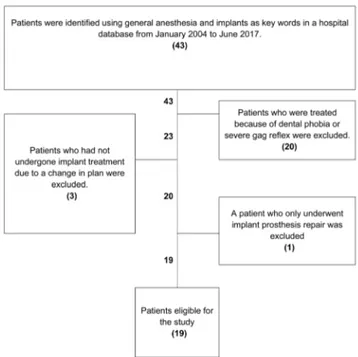
Fig. 1. Flow chart of patient selection.
1) An individual, unattached implant is mobile when tested clinically.
2) A radiograph demonstrates certain evidence of peri-implant radiolucency.
3) Individual implant performance is characterized by signs and symptoms such as pain, infection, neuropathy, paresthesia, or violation of the mandibular canal.
4) A marginal bone resorption of >1.5 mm occurs in the first year after implantation.
The occurrence of any of the above is considered an implant failure.
Table 1. Criteria for the assessment of implant survival rate derived by Albrektsson and Sennerby [7] and Albrektsson and Isidor [8]
commended as a method for providing high quality dental care for these patients [6].
However, performing implant treatments on patients with poor compliance may require several rounds of intravenous sedation or general anesthesia, which also results in higher cost. Moreover, it is difficult to recommend implant treatment for cases in which it is difficult to predict the life of the implant due to poor self-care.
Our hospital operates the Clinic for the Disabled and is equipped to perform dental treatment under general anesthesia. Implant treatment has been performed in several patients with special care needs at the request of their guardians. The present study analyzed the outcomes and success rates of dental implant treatment under general anesthesia in these patients. The findings will be used to establish future treatment plans and provide high-quality care for patients with special needs.
METHODS
The present study was conducted after approval from the Institutional Review Board of Seoul National University School of Dentistry (IRB No. S-D20170037).
The study included outpatients who underwent implant treatment under general anesthesia at the Clinic for the Disabled in Seoul National University Dental Hospital between January 2004 and June 2017.
The Seoul National University Dental Hospital database was searched for general anesthesia and implant treatment-related order codes for the study period, and a total of 43 patients were identified. Of these, 20 who required general anesthesia for dental phobia or a severe
gag reflex were excluded since they were capable of oral self-care. Four patients who did not proceed with planned implant treatment under general anesthesia for various reasons were also excluded. Ultimately, a total of 19 patients were identified and their implant treatment details were investigated and analyzed by accessing medical records and radiographs (Fig. 1).
Items investigated included age, sex, type of disability, presence of oral parafunctional habits, method of anesthe- sia used for implant surgery and prosthetic treatment, location of installed implant fixture, diameter and length of the implant fixture, surgical procedure used for implant placement, types of prostheses, surgery- or prosthesis- related complications, and subjective patient symptoms.
The extent of marginal bone resorption was evaluated by a single oral and maxillofacial surgeon using periapical and panoramic radiographs. Radiographic imaging was
Patient
No. Gender Age
(y) Disability Cause of
edentulism
Implant Surgery Prosthetic Treatment
Follow-up
(Month) Implant
Location Prosthesis Anesthetic
methods
Number of G/A or
IV/S
Anesthetic methods
Number of G/A or
IV/S
1 M 39 Autism Periodontitis
Dental Caries G/A1 2 - - 56.9
#15 SC3
#16
#25 FB4
#27
#36 SC
#37
#45 SC
#46
2 F 22 Mental Retardation
Dental Caries G/A 1 - - 119.8
#31
SC
#41
#42
Dental Caries G/A 1
- -
112.9 #15 SC
0.8 #36*
112.9 #37 SC
Periodontitis IV/S2 1 108.3 #36 SC
3 M 31 Mental Retardation Maxillary
Sinusitis Periodontitis
G/A 2 G/A 1 27.6
#24
SC
#25**
#26**
#46 SC
#47
4 M 31 Autism Dental Caries G/A 1 - - 31.4 #46 SC
#47
5 F 27 Mental Retardation Dental Caries G/A 1 - - 78.0 #26 SC
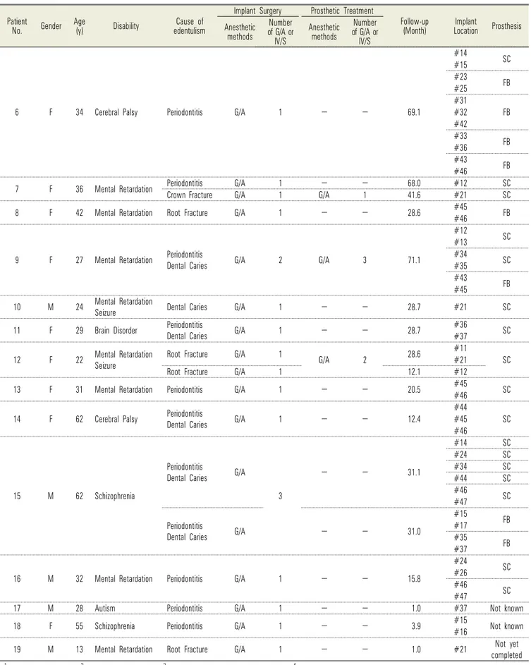
(Continued to the next page) Table 2. Details of patient distribution and treatment
performed immediately after implant placement, after implant abutment connection, after loading, and during regular visits. The interval between regular visits was set as 3-4 months during the first year following prosthesis loading if there were no specific issues, and 6-12 months thereafter.
The success criteria reported by Albrektsson and Isidor [7] and the failure criteria reported by Albrektsson and Sennerby [8] were used to obtain the survival rate of implants. The implant was considered a failure if any one of 4 criteria used in this study was met (Table 1).
Cumulative survival rates were calculated.
RESULTS
Of 19 patients in this study, 8 were males and 11 were females, with a mean age of 32.9 yrs. The patients included 11 with mental retardation, 3 with autism, 2 with cerebral palsy, 2 with schizophrenia, and 1 with a brain
disorder; 2 patients also had seizure disorders. A total of 27 rounds of general anesthesia and 1 round of intravenous sedation were performed until all implant surgeries were completed, while an additional 7 rounds of general anesthesia were performed for subsequent prosthetic treatment on 4 patients with poor compliance.
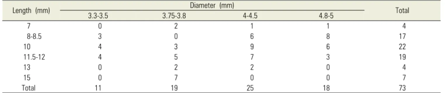
The causative dental diseases that necessitated implant treatment were mostly periodontitis and dental caries, while tooth fracture and maxillary sinusitis also contributed to the need for treatment. A total of 73 implants were placed in 19 patients, while prosthetic treatments were performed using a single implant- supported crown or an implant-supported fixed bridge (Table 2). The 73 implants had a wide range of diameters and lengths; from 3.3 mm to 5.0 mm and from 7.0 mm to 15 mm, respectively (Table 3).
With respect to surgical procedures used for implant placement, a conventional method was used most commonly in 28 cases, followed in order by a 1-step procedure in 12 cases, a conventional method with guided
Patient
No. Gender Age
(y) Disability Cause of
edentulism
Implant Surgery Prosthetic Treatment
Follow-up
(Month) Implant
Location Prosthesis Anesthetic
methods
Number of G/A or
IV/S
Anesthetic methods
Number of G/A or
IV/S
6 F 34 Cerebral Palsy Periodontitis G/A 1 - - 69.1
#14 SC
#15
#23 FB
#25
#31
FB
#32
#42
#33 FB
#36
#43 FB
#46
7 F 36 Mental Retardation Periodontitis G/A 1 - - 68.0 #12 SC
Crown Fracture G/A 1 G/A 1 41.6 #21 SC
8 F 42 Mental Retardation Root Fracture G/A 1 - - 28.6 #45#46 FB
9 F 27 Mental Retardation Periodontitis
Dental Caries G/A 2 G/A 3 71.1
#12 SC
#13
#34 SC
#35
#43 FB
#45
10 M 24 Mental Retardation
Seizure Dental Caries G/A 1 - - 28.7 #21 SC
11 F 29 Brain Disorder Periodontitis
Dental Caries G/A 1 - - 28.7 #36#37 SC
12 F 22 Mental Retardation
Seizure Root Fracture G/A 1 G/A 2 28.6 #11
SC
#21
Root Fracture G/A 1 12.1 #12
13 F 31 Mental Retardation Periodontitis G/A 1 - - 20.5 #45
#46 SC
14 F 62 Cerebral Palsy Periodontitis
Dental Caries G/A 1 - - 12.4
#44
SC
#45
#46
15 M 62 Schizophrenia
Periodontitis
Dental Caries G/A
3
- - 31.1
#14 SC
#24 SC
#34 SC
#44 SC
#46 SC
#47
Periodontitis
Dental Caries G/A - - 31.0
#15 FB
#17
#35 FB
#37
16 M 32 Mental Retardation Periodontitis G/A 1 - - 15.8
#24 SC
#26
#46 SC
#47
17 M 28 Autism Periodontitis G/A 1 - - 1.0 #37 Not known
18 F 55 Schizophrenia Periodontitis G/A 1 - - 3.9 #15#16 Not known
19 M 13 Mental Retardation Root Fracture G/A 1 - - 1.0 #21 Not yet
completed
1General Anesthesia, 2Intravenous Sedation, 3Single implant-supported crown(s), 4Implant-supported fixed bridge.
*The implant was spontaneously lost within the osseointegration phase and placed again later, **The implants were classified as failed by criteria, but still in use.
Table 2. Continued
Length (mm) Diameter (mm)
Total
3.3-3.5 3.75-3.8 4-4.5 4.8-5
7 0 2 1 1 4
8-8.5 3 0 6 8 17
10 4 3 9 6 22
11.5-12 4 5 7 3 19
13 0 2 2 0 4
15 0 7 0 0 7
Total 11 19 25 18 73
Table 3. Distribution of implants according to length and diameter
Time No. of implants No. of failed Time not passed CSR (%)
Placement to Loading 59 1 0 98.3
Loading to 1 year 58 2* 0 94.9
1 to 2 year 56 0 17 94.9
2 to 3 year 39 0 5 94.9
Longer than 3 year 34 0 34 94.9
*More than 1.5 mm of marginal bone loss was observed within 1 year after the loading.
CSR: Cumulative survival rate
Table 4. Cumulative survival rate of implants
bone regeneration (GBR) in 11, immediate placement with GBR in 7, a 1-step procedure with GBR in 5, immediate placement with a 1-step procedure in 4, a conventional method with sinus lift in 3, a 1-step proced- ure with sinus lift in 2, and immediate placement in 1.
The cumulative survival rate was evaluated for 59 implants in 13 patients who participated in at least 1 year of follow-up after completion of implant prosthetic treatment. Among the 19 patients, 3 had not passed the 1-year mark since their prosthetic treatment, while 3 did not undergo prosthetic treatment following implant surgery or did not undergo prosthetic treatment at our hospital, which made it impossible to evaluate the prosthesis. The average time required from implant placement to completion of prosthetic treatment in these patients was 7.7 months (3-27 months), while the average follow-up period following completion of prosthetic treatment was 43.3 months (15-116 months). In the pre-prosthodontic treatment phase, 1 implant fixture was considered a failed case for not achieving osseointegra- tion, resulting in a survival rate of 98.3%. In the post-prosthodontic treatment phase, 2 implants showed failure associated with marginal bone resorption of ≥1.5 mm within 1 year from prosthesis loading, resulting in a cumulative survival rate of 94.9% (Table 4).
Oral parafunctional habits such as involuntary mandi- bular movement and muscular hyperfunction were found in 4 patients, but these did not lead to implant failure.
Although there were some minor prosthodontic com- plications, including screw loosening and discomfort from poorly-fitting prostheses, there were no major complications associated with the implant procedure.
DISCUSSION
Patients with mental disabilities face many difficulties in relation to implant treatment. Treatment of patients with poor compliance requires the use of general anesthesia or intravenous sedation, which is only possible if a proper facility, equipment, and personnel are available. Moreover, patients who have difficulty with self-care are known to have poor oral hygiene; since little information pertaining to prognosis following implant treatment is available, it is difficult for dentists to choose an implant procedure as part of the treatment. Further- more, for patients with serious medical conditions, lack of knowledge about patient characteristics and fear of possible medical emergencies can limit the treatments provided by dentists [9]. For these reasons, implant
treatment has been excluded in most cases involving patients with mental disability.
Generally, poor oral hygiene with an O’Leary plaque index of ≥20% is a contraindication for implant treatment. However, The O’Leary plaque indices in patients with intellectual disabilities are reported at levels ranging from 60.2% to 100%. Such facts would have made dentists negatively consider implant treatment for them [10-12].
Nevertheless, several reports have been published after completing dental implant treatment for patients with mental disabilities. In 1995, Rogers et al. [13] reported a case in which positive results were obtained by placing 4 implants in the mandible of a patient with cerebral palsy under general anesthesia and completing prosthodontic treatment via implant overdenture. In 2000, Heckmann et al. [14] placed 2, 3, and 4 implants in the mandibles of 3 patients with Parkinson’s disease and achieved remarkable improvement in chewing function through fabrication of implant-supported prostheses, while also achieving reduction in gastrointestinal symptoms. Lustig et al. [15] published a case report in 2002 in which placement of 3 implants and prosthetic restoration were successfully completed on a Down’s syndrome patient.
According to a 2003 study by Lopez et al. [16], placement of 67 implants in 18 patients with cerebral palsy, head injuries, pyknodysostosis, Down’s syndrome, Rieger syndrome, and dementia resulted in only 4 implant failures in 3 patients during the osseointegration stage, with no other recorded failure upon completion of fixed prosthesis oral rehabilitation.
Implant treatment has continued to evolve, broadening the range of indications for its use. Studies have reported no differences in implant treatment outcomes between medically compromised and healthy patients, and even patients aged 79 years or older can undergo implant treatment if medically stable [17-20]. There are virtually no absolute medical contraindications for implant treatment, although individualized medical management is required for conditions that may increase the risk of treatment failure or complications throughout all stages
of implant treatment [21]. There have also been advances in implant surgery techniques. Since the introduction of the 2-stage surgical procedure by Branemark et al. [22], use of a 1-stage surgical procedure that skips the second- stage surgery for implant exposure is also known to produce excellent outcomes [23,24]. Even in cases where immediate placement is performed without a post- extraction healing period, a similar level of treatment outcomes was seen, as compared to cases with delayed placement [25-27].
In 2008, Isaksson et al. [28] reported that treatment using implant-supported prostheses in 35 patients incapable of oral self-care was performed without any cases of fixture mobility or prosthesis fracture among 229 implants, while there were only 15 cases of intraoral fixture exposure, 1 of purulent exudate, and 2 of peri-implant gingival hyperplasia. In 2009, Kim et al. [29]
found that there were no significant associations between psychiatric diseases and implant complication and failure rates. Based on these study results, it is assumed that treatment using implants is feasible for mentally disabled patients with poor oral hygiene.
The 19 patients in the present study had mental disabilities that compromised compliance with treatment, but appropriate implant fixture installation was possible under general anesthesia. In 1 patient, implant placement was performed with 2 rounds of general anesthesia, but a third implant surgery was performed under intravenous sedation. This was based on good treatment compliance observed in the postoperative outpatient examination room. Moreover, 15 of 19 patients were able to undergo implant prosthetic treatment without general anesthesia.
Accordingly, the surgeon should decide the need for general anesthesia by identifying the level of treatment compliance following the implant placement procedure.
The number of times that general anesthesia was performed was reduced by performing immediate place- ment, in which all procedures from extraction to implant placement was performed under a single round of general anesthesia, or by using a 1 step-procedure during implant placement [27]. To accomplish this, it was necessary to
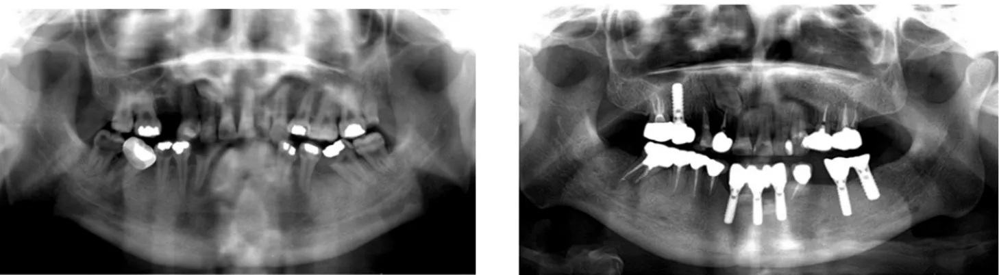
Fig. 2. A panoramic view of a patient under follow-up for the longest period after completion of implant treatment. When comparing images before (left) and after (right) completion of treatment, there was no loss of marginal bone or radiolucent lesion around the implants.
evaluate bone quality via computed tomography (CT) when establishing a preoperative treatment plan. How- ever, it is often difficult to acquire radiographic images in patients who lack treatment compliance. Successful CT imaging using deep sedation with propofol, with consulta- tion from a dental anesthesiologist, has been reported [30,31]. A more precise preoperative plan can reduce the number of times that general anesthesia is performed.
Among 13 patients in whom the failure criteria were applied, 3 of 59 implants failed. Of these, 1 implant was removed 3 weeks after placement, as osseointegration could not be achieved. However, reinstallation 4 months later was successful; the patient has used that implant and others for a prolonged period without any specific problems (Fig. 2). The other 2 cases of failure occurred in the left maxillary second premolar and first molar in a patient with a history of treatment for left maxillary sinusitis. These were considered implant failures based on marginal bone loss of approximately 2.0 mm at these sites in the first year after loading; however, the implants remain in use 2 years since completion of the procedure, without specific problems such as implant mobility or complaint of discomfort. In the present study, the cumulative survival rate of implants was found to be 98.4% and 94.9% during pre- and post-prosthodontic treatment phases, respectively. These results exceeded the cumulative survival rates of 91.4% and 80.5% during pre- and post-prosthodontic treatment phases reported in 2005 by Ekfeldt et al., after placement of 35 implants in 14 patients with neurological impairments [32]. The cumula-
tive survival rates found in the present study were similar to those from studies in the general population. A study by Romeo et al. in 2004 reported a cumulative survival rate of 95.6-96.1% for single tooth implants and implant-supported fixed partial dentures in a 7-year survey in the general population [33]. High implant success rates similar to those in healthy patients were achieved in the present study because the patients had no other known risk factors for implant failure, such as osteoporosis or a smoking habit; moreover, the surgeon was able perform procedures in a stable environment since almost all implants were performed under general anesthesia. Therefore, even in patients with impediments to general dental treatment due to lack of compliance, prognoses similar to those in the general population may be expected if implant placement and implant-supported prosthetic treatments are performed under general anesthesia.
One patient who had been visiting the hospital for the longest period after completion of treatment (9 years and 8 months) was confirmed to have no discomfort associated with the implant. Comparing this patient's panoramic radiographs taken at the first visit and the most recent visit, any radiologic problems could be found, and this indicates good oral rehabilitation had been achieved (Fig 2). Accordingly, a favorable prognosis can be guaranteed for patients with poor compliance if appropriate oral rehabilitation is administered through implant-supported dental prostheses and continued care is provided through regularly scheduled visits.
AUTHOR ORCIDs
Il-hyung Kim: https://orcid.org/0000-0003-1386-6391 Tae Seong Kuk: https://orcid.org/0000-0001-9804-7162 Sang Yoon Park: https://orcid.org/0000-0001-7624-3373 Yong-suk Choi: https://orcid.org/0000-0001-8618-4100 Hyun Jeong Kim: https://orcid.org/0000-0002-9265-7549 Kwang-Suk Seo: https://orcid.org/0000-0001-5906-0639
CONFLICTS OF INTEREST: The authors have no conflicts of interest to declare.
REFERENCES
1. Gabre P, Martinsson T, Gahnberg L. Longitudinal study of dental caries, tooth mortality and interproximal bone loss in adults with intellectual disability. Eur J Oral Sci 2001; 109: 20-6.
2. Anders PL, Davis EL. Oral health of patients with intellectual disabilities: A systematic review. Spec Care Dentist 2010; 30: 110-7.
3. Cheng RH, Leung WK, Corbet EF, King NM. Oral health status of adults with down syndrome in hong kong. Spec Care Dentist 2007; 27: 134-8.
4. Cumella S, Ransford N, Lyons J, Burnham H. Needs for oral care among people with intellectual disability not in contact with community dental services. J Intellect Disabil Res 2000; 44: 45-52.
5. Tiller S, Wilson KI, Gallagher JE. Oral health status and dental service use of adults with learning disabilities living in residential institutions and in the community. Com- munity Dent Health 2001; 18: 167-71.
6. Moon SY, Kim SG. Implants in psychiatric patients. J Korean Assoc Disabil Oral Health 2007; 3: 1-5.
7. Albrektsson T, Isidor F. Consensus report of session iv.
In: Lang NP, Karring T, editors. Proceedings of the 1st european workshop on periodontology. London: Quintessence;
1994. p. 165-9.
8. Albrektsson T, Sennerby L. State of the art in oral implants.
J Clin Periodontol 1991; 18: 474-81.
9. Seo KS, Jang KT, Kim HJ, Yum KW. The status of
comprehensive dental treatment and type of disabilities of the patients treated under outpatient general anesthesia at the clinic for the disabled in seoul national university dental hospital. J Korean Dent Soc Anesthesiol 2006; 6:
82.
10. Kim CS. Research on oral status of hearing impaired youth by using qlf-d. J Korea Contents Assoc 2013; 13: 305-11.
11. Romero-Perez MJ, Mang-de la Rosa Mdel R, Lopez-Jimenez J, Fernandez-Feijoo J, Cutando-Soriano A. Implants in disabled patients: A review and update. Med Oral Patol Oral Cir Bucal 2014; 19: e478-82.
12. Binkley CJ, Johnson KW, Abadi M, Thompson K, Shamblen SR, Young L, et al. Improving the oral health of residents with intellectual and developmental disabilities:
An oral health strategy and pilot study. Eval Program Plann 2014; 47: 54-63.
13. Rogers JO. Implant-stabilized complete mandibular denture for a patient with cerebral palsy. Dent Update 1995; 22:
23-6.
14. Heckmann SM, Heckmann JG, Weber HP. Clinical out- comes of three parkinson's disease patients treated with mandibular implant overdentures. Clin Oral Implants Res 2000; 11: 566-71.
15. Lustig JP, Yanko R, Zilberman U. Use of dental implants in patients with down syndrome: A case report. Spec Care Dentist 2002; 22: 201-4.
16. Lopez-Jimenez J, Romero-Dominguez A, Gimenez-Prats MJ. Implants in handicapped patients. Med Oral 2003;
8: 288-93.
17. Smith RA, Berger R, Dodson TB. Risk factors associated with dental implants in healthy and medically compromised patients. Int J Oral Maxillofac Implants 1992; 7: 367-72.
18. Anner R, Grossmann Y, Anner Y, Levin L. Smoking, diabetes mellitus, periodontitis, and supportive periodontal treatment as factors associated with dental implant survival:
A long-term retrospective evaluation of patients followed for up to 10 years. Implant Dent 2010; 19: 57-64.
19. Engfors I, Ortorp A, Jemt T. Fixed implant-supported prostheses in elderly patients: A 5-year retrospective study of 133 edentulous patients older than 79 years. Clin Implant Dent Relat Res 2004; 6: 190-8.
20. Grant BT, Kraut RA. Dental implants in geriatric patients:
A retrospective study of 47 cases. Implant Dent 2007;
16: 362-8.
21. Diz P, Scully C, Sanz M. Dental implants in the medically compromised patient. J Dent 2013; 41: 195-206.
22. Branemark PI, Hansson BO, Adell R, Breine U, Lindstrom J, Hallen O, et al. Osseointegrated implants in the treatment of the edentulous jaw. Experience from a 10-year period.
Scand J Plast Reconstr Surg Suppl 1977; 16: 1-132.
23. Friberg B, Henningsson C, Jemt T. Rehabilitation of edentulous mandibles by means of turned branemark system implants after one-stage surgery: A 1-year retrospective study of 152 patients. Clin Implant Dent Relat Res 2005; 7: 1-9.
24. Carr AB, Choi YG, Eckert SE, Desjardins RP.
Retrospective cohort study of the clinical performance of 1-stage dental implants. Int J Oral Maxillofac Implants 2003; 18: 399-405.
25. Rosenquist B, Grenthe B. Immediate placement of implants into extraction sockets: Implant survival. Int J Oral Maxillofac Implants 1996; 11: 205-9.
26. Evian CI, Emling R, Rosenberg ES, Waasdorp JA, Halpern W, Shah S, et al. Retrospective analysis of implant survival and the influence of periodontal disease and immediate placement on long-term results. Int J Oral Maxillofac Implants 2004; 19: 393-8.
27. Nam H, Sung KW, Kim MG, Lee K, Kwon D, Chi SI, et al. Immediate implant placement for schizophrenic
patient with outpatient general anesthesia. J Dent Anesth Pain Med 2015; 15: 147-51.
28. Isaksson R, Becktor JP, Brown A, Laurizohn C, Isaksson S. Oral health and oral implant status in edentulous patients with implant-supported dental prostheses who are receiving long-term nursing care. Gerodontology 2009; 26:
245-9.
29. Kim JW, Kim YK. Clinical study about the implant treatment in the patients with systemic disease. Implantology 2009;
13: 64-75.
30. Seo KS, Lee JH, Shin TJ, Yi YE, Kim HJ, Yum KW, et al. Intravenous sedation of cerebral palsy patient for dental implant ct taking-a case report. J Korean Assoc Disabil Oral Health 2008; 4: 21-5.
31. Hong YJ, Dan JB, Kim MJ, Kim HJ, Seo KS. Prognosis after treatment with multiple dental implants under general anesthesia and sedation in a cerebral palsy patient with mental retardation: A case report. J Dent Anesth Pain Med 2017; 17: 149-55.
32. Ekfeldt A. Early experience of implant-supported prostheses in patients with neurologic disabilities. Int J Prosthodont 2005; 18: 132-8.
33. Romeo E, Lops D, Margutti E, Ghisolfi M, Chiapasco M, Vogel G. Long-term survival and success of oral implants in the treatment of full and partial arches: A 7-year prospective study with the iti dental implant system.
Int J Oral Maxillofac Implants 2004; 19: 247-59.
![Bid Invitation [Security Guard]](data:image/gif;base64,R0lGODlhAQABAIAAAP///wAAACH5BAEAAAAALAAAAAABAAEAAAICRAEAOw==)