Journal of Bacteriology and Virology 2013. Vol. 43, No. 4 p.297 – 306 http://dx.doi.org/10.4167/jbv.2013.43.4.297
Cytoplasmic Translocation of p53 by Human Cytomegalovirus Infection
Yejin Kwon1, Jiyeon Kim1,2, Jung Heon Kim1 and Eung Soo Hwang1,3*
1Department of Microbiology and Immunology and 2BK21 Plus Biomedical Science Project, 3Seoul National University College of Medicine, Seoul; Institute of Endemic Diseases, Seoul National University Medical Research
Center, Seoul, Korea
p53 is a well-known multi-functional transcription regulator and is critical in the induction of apoptosis in response to various stresses. Human cytomegalovirus (HCMV) infection induced the accumulation of p53, which was partly relocalized in cytoplasm, but no apparent cell death in human fibroblasts. p53 in HCMV-infected cells was mainly mono-ubiquitinated, which might be resulted from the decreased expression of MDM2 in the course of HCMV infection.
Ubiquitinated p53 was also phosphorylated at serine 20. CRM1 increased in the cytoplasm of HCMV-infected cells. It was found that p53 and its mutant in nuclear export sequences were localized in the cytoplasm of cells when co-expressed with CRM1. Collectively, our data suggest that HCMV infection modifies p53 into a stable and exportable form and accumulates it in the cytoplasm, but does not result in apoptotic death of cells.
Key Words: Human cytomegalovirus, p53, MDM2, Nuclear exportation, CRM1
INTRODUCTION
p53 responds to various stimuli to regulate target genes that induce cell cycle arrest, apoptosis, senescence, DNA repair, or metabolic changes in cells (1). In normal cells, p53 is maintained inactive and low in level through MDM2, which ubiquitinates p53 to promote its degradation.
Transcriptional regulation of p53 occurs in the nucleus, but other biological activities of p53 such as the induction of apoptosis via mitochondrial pathway and the inhibition of autophagy were also reported to take place in the cytoplasm in transcription-independent manner (2). Many posttrans-
lational modifications modulate the biological activity and the cellular localization of p53 (3~5).
The functions of p53 are impaired by viral proteins of some small DNA viruses, for example, SV40 T antigen (6, 7), adenovirus E1B-55 kDa (8), and human papillomavirus E6 (9). They all contribute in the transformation of the virus- infected cells. It was also reported that human cytomegalo- virus (HCMV), one of large DNA viruses, induced the accumulation of p53 in the infected cells and altered cellular regulation and functions (10, 11). HCMV has been related to cell cycle disturbance in some diseases. Several HCMV gene products are responsible for transforming cells. HCMV IE2-86 protein can reduce transcriptional activity of p53 by
297
Original Article
Received: November 1, 2013/ Revised: November 8, 2013/ Accepted: November 14, 2013
*Corresponding author: Eung Soo Hwang. Department of Microbiology and Immunology, Seoul National University College of Medicine 28 Yeongeon dong, Jongro gu, Seoul 110-799, Korea.
Phone: +82-2-740-8307, Fax: +82-2-743-0881, e-mail: hesss@snu.ac.kr
**This study was partially supported by a grant of the Korea Healthcare technology R&D Project, MHWFA, Republic of Korea (A080460) and a grant 04-2012-0480 (2012-1275) from Seoul National University Hospital, Republic of Korea. We thank Jisung Kim for his excellent technical support.
○CCThis is an Open Access article distributed under the terms of the Creative Commons Attribution Non-Commercial License (http://creativecommons.org/license/by-nc/3.0/).
the direct interaction with it and result in a cellular pro- liferation, which might involve in the restenosis of coronary artery after surgery (12). Our previous studies show HCMV IE1-72 and UL44 also inhibit p53-dependent transcription in different ways (13, 14). Besides transcriptional regulation p53 facilitates the transportation of one of HCMV matrix proteins from nucleus to cytoplasm and induces the production of viruses (15). The sequestration of p53 was reported in the cytoplasm of endothelial cells after HCMV infection (16). Although the interactions of p53 with some HCMV proteins such as HCMV major capsid protein (MCP, UL86), UL25, pp65 (UL83), and UL44 were reported by our previous work (14), the mechanism of p53 exportation to the cytoplasm are not fully elucidated. This study was devised to explore the mechanisms of exportation of p53 in HCMV-infected cells.
MATERIALS AND METHODS Cells and virus culture
HEL 299 (ATCC, CCL-137), U373MG (ATCC, HTB-17), and H1299 (ATCC, CRL-5803) cells were cultured in Dulbecco's modified Eagle's Media (DMEM, Invitrogen, Carlsbad, CA, USA) supplemented with 10% fetal bovine serum (FBS, Invitrogen) and penicillin-streptomycin.
Synchronized cells were adsorbed with 4 multiplicity of infection (m.o.i.) of HCMV Towne (ATCC VR-977) for 90 min. After washing with media, cells were cultured with DMEM supplemented with 2% FBS and incubated for the indicated time. Virus titer was measured by a conventional plaque assay.
Antibodies and chemicals
For primary mouse monoclonal antibodies, anti-p53 (DO-1, Santa Cruz, Santa Cruz, CA, USA), anti-MDM2 (SMP14, Santa Cruz), anti-CRM1 (clone 17, BD Bioscience, San Jose, CA, USA), and anti-GAPDH (R&D systems, Minneapolis, MN, USA) were used. For primary rabbit polyclonal antibodies, anti-annexin V (Abcam, Cambridge, UK), anti-ubiquitin (FL-76, Santa Cruz) and anti-phospho- p53 serine 20 (R&D systems) were used. For secondary
antibodies, FITC- (Merck, Darnstadt, Germany) or Alexa 568- (Invitrogen) conjugated anti-mouse IgG and horse radish peroxidase-conjugated anti-mouse IgG and anti-rabbit IgG (Promega, Madison, WI) were used. For immuno- precipitation, agarose-conjugated anti-p53 (FL393, Santa Cruz) and anti-ubiquitin (FL-76, Santa Cruz) were used.
Concentration of Nutlin-3 (Merck), MG132, and doxorubicin in this experiment was 5 μM, 30 μM, and 500 ng/ml, respectively. Staurosporine (STS) with 50 nM was used as a positive control inducer of apoptosis in HEL299 (17). All the chemicals were purchased from Sigma Chemical (St.
Louise, MO, USA) unless otherwise stated.
Microarray
HEL 299 cells were infected with 4 m.o.i. of HCMV.
The infected cells were harvested at 0, 12, 24, 48, 72, and 96 h postinfection (h.p.i.). Total RNA was extracted from infected cells using Trizol (Invitrogen, Carlsbad, CA, USA) at each time point. Total RNA (2 μg) was reverse transcribed with a Chemiluminescent RT-IVT Labeling kit (Applied Biosystems, Foster City, CA, USA) and hybridized to the Human Genome Survey Microarray (Applied Biosystems).
For each time point, data were initially normalized, and differential gene expression at each time point was calculated by comparing the differences of values between expression levels at each time point and the level in mock-infected HEL 299 cells at 0 h.p.i.
Plasmids and transfection
pEGFP-N1 was purchased from Invitrogen. GFP-p53 (NES-) and Flag-hCRM1 were purchased from Addgene (Cambridge, MA, USA). pEGFP-p53 was constructed from pEGFP-N1 and pCMV-p53 (Clontech, Mountain View, CA, USA), and pEGFP-p53NLS(-) was constructed by site- directed mutagenesis kit (Agilent Life Science Technologies, Santa Clara, CA, USA) and verified by DNA sequencing.
Cells were transfected with Lipofectamine (Invitrogen) according to the manufacturer's manual.
Immunofluorescent staining
Cells were washed twice with phosphate buffered saline
(PBS), fixed with 10% paraformaldehyde for 10 min at room temperature, and permeabilized with 0.2% Triton X-100 for 5 min. After washed with PBS containing 0.05%
Tween 20, cells were treated successively with the primary antibody and FITC- or Alexa 568-conjugated secondary antibody at 4℃. Fluorescent signal was observed under confocal laser scanning microscope (LSM5 Pascal, Carl Zeiss, Oberkochen, Germany).
Cell fractionation
For the fractionation of nuclear and cytoplasmic com- ponents, HCMV-infected cells were harvested and lysed with cell lysis buffer (10 mM HEPES, 1.5 mM MgCl2, 10 mM KCl, 0.5 mM DTT, 0.2 mM EDTA, 0.2 mM EGTA, 0.2 mM PMSF) on ice for 10 min. After centrifugation at 13,000 rpm for 10 min the supernatant was collected as the cytoplasmic fraction, and the pellet was further treated with nuclear extract buffer (20 mM HEPES, 1.5 mM MgCl2, 420 mM NaCl, 0.5 mM DTT, 0.2 mM EDTA, 0.2 mM EGTA, 0.2 mM PMSF, 25% glycerol) on ice for 10 min.
After centrifugation at 13,000 rpm for 10 min the super- natant was collected as the nuclear fraction. For the whole cell preparation, the extraction buffer (1% Nonidet P-40, 0.05% deoxycholate, 140 mM NaCl, 20 mM Tris HCl, pH 7.5) with proteinase inhibitor cocktail (Roche Molecular Diagnostics, Indianapolis, IN, USA) was used. The amount of protein was measured with BCA kit (Thermo Scientific, Marietta, OH, USA) according to manufacturer's manual.
Immunoprecipitation
Each cell extract sample was reacted with agarose- conjugated antibody at 4℃ for 60 min with constant mixing.
After washed three times with buffer (50 mM Tris-Cl, pH 7.5, 150 mM NaCl, 1% NP-40, 0.5% deoxycholic acid, 0.1% sodium dodecyl sulfate) containing phosphatase inhibitor and proteinase inhibitor cocktail (Roche), the final agarose pellet was prepared for a further analysis.
Western blot
Each sample containing the equal amount of proteins was mixed with Laemmli SDS sample buffer (50 mM Tris,
pH 6.8, 2% SDS, 10% glycerol, 50 mM dithiothreitol, 0.5%
bromophenol blue) containing 1.25% mercaptoethanol, and was boiled for 5 min. Proteins in cleared samples were separated on 10% SDS-polyacrylamide gel electrophoresis and transferred to PVDF membranes. The blots were probed with the appropriate primary antibody and peroxidase- conjugated secondary antibody, and were visualized by enhanced chemiluminescence (Amersham) according to the manufacturer's protocol.
Apoptosis assay
HEL299 were adsorbed with 4 m.o.i. of HCMV for 90 min. Thereafter the infected cells were harvested at 0, 1 and 2 days postinfection (d.p.i). Approximately 5 × 10⁵ cells were trypsinized and fixed with BD Fixation/
Permeabilization solution at 4℃ for 15 min. Cells were washed twice with FACS buffer containing 0.5% BSA and 0.1% NaN in PBS, and treated with anti-annexin V antibody in BD Perm/WashTM Buffer at 4℃ for 30 min. Cells were spun down and washed twice with FACS buffer, and stained with FICT-conjugated goat anti-rabbit IgG (Invitrogen) at 4℃ 30 min. FITC-positive cells were measured with FACSCanto II (BD). Data of cell apoptosis were analyzed with FACS DIVA (BD).
RESULTS
p53 accumulation after HCMV infection without the induction of apoptosis
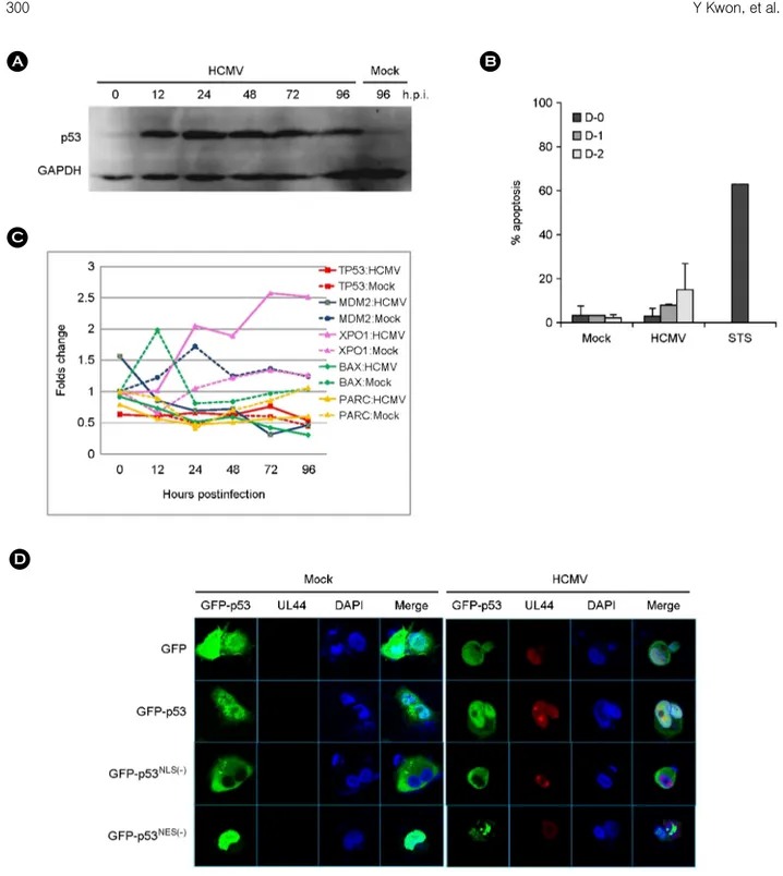
HCMV infection induced the accumulation of p53 in HEL 299 cells during the course of infection as shown in Fig. 1A. p53 was accumulated in HCMV-infected HEL 299 cells at an early phase, 12 to 24 h.p.i., and maintained in a later phase of infection. Whether increased p53 after HCMV infection involved the induction of apoptosis was assayed with annexin V staining. Apoptosis assay showed 3.3+3.0%, 8.1+0.6%, and 14.7+12.1% on day 0, 1 and 2, respectively, in HCMV-infected HEL 299 while 68.0% in staurosporine- treated cells (Fig. 1B).
Microarray screening data showed that mRNA levels of p53 decreased and was maintained at a low level in HCMV-
Figure 1. Characteristics of p53 induced by HCMV infection. (A) Western blot analysis with anti-p53 (DO-1) antibody for the detection of p53 accumulation in HEL 299 infected with HCMV during the course of infection. (B) Apoptosis in HCMV-infected human fibroblasts determined by annexin V staining during the course of infection. Experiments were performed twice independently, and results were expressed mean + standard deviation. Staurosporine (STS) was treated in fibroblast for a positive control of apoptosis. (C) mRNA expression pattern of TP53, MDM2, XPO1, BAX and PARC, in HEL 299 cells infected with HCMV. mRNA levels of each gene were detected by microarray assay as described in Materials and Methods. Values shown are the differences between those in HCMV- or mock-infected HEL 299 cells at the indicated time and in mock-infected HEL 299 cells at 0 h.p.i. (D) Subcellular localization of transfected GFP-p53, p53NLS(-) and p53NES(-) in U373MG after HCMV infection at 48 h.p.i. HCMV UL44 was stained with monoclonal anti-UL44 antibody and Alexa 568-conjugated anti-mouse IgG as a marker for HCMV infection.
A
C
D
B
infected HEL 299 cells as compared with mock-infected ones during the course of infection except the early phase of infection. At all the time during infection mRNA level of MDM2 decreased up to 23% level in HCMV-infected HEL 299 cells as compared with one in mock infection at 72 h.p.i. mRNA of PARC and BAX was downregulated during the whole time of HCMV infection, and mRNA of XPO1 (CRM1/exportin1) increased to 2.05 folds at 24 h.p.i., and up to 2.57 folds at 72 h.p.i. (Fig. 1C).
Subcellular localization of GFP-p53, GFP-p53NLS(-) and GFP-p53NES(-) was analyzed in U373MG infected with HCMV at 48 h.p.i. (Fig. 1D). Concomitant detection of UL44, which is present mainly in nucleus, was used as a marker of HCMV infection. GFP-p53 was localized dominantly in nucleus, but also in cytoplasm, and GFP- p53NLS(-) was present only in cytoplasm. GFP-p53NES(-) was observed only in nucleus in mock-infected cells, but present also in cytoplasm after HCMV infection. These results indicated that a part of p53 shifted into cytoplasm after HCMV infection.
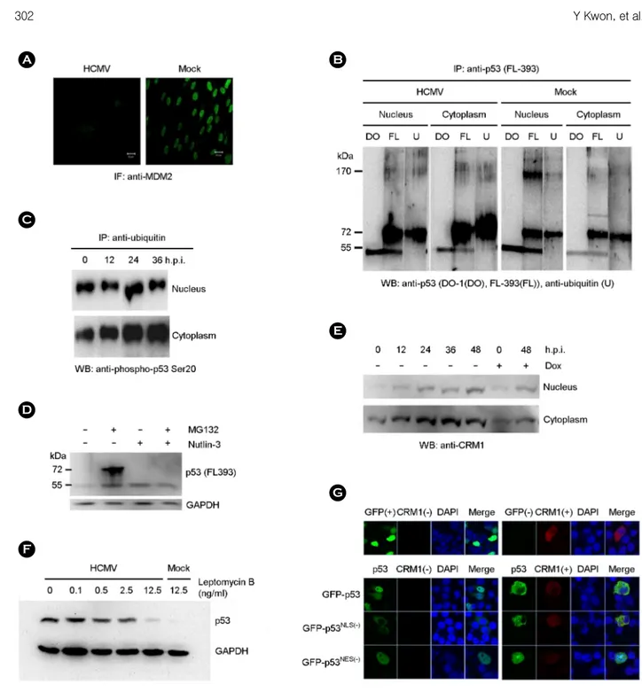
Nuclear exportation of p53 by HCMV infection MDM2 was hardly detected in HCMV-infected cells at 48 h.p.i. by immunofluorescent staining (Fig. 2A). Each nuclear and cytoplasmic fraction of HCMV-infected HEL 299 was immunoprecipitated with anti-p53 full length peptide (FL-393) antibody and immunoblotted with anti- p53 (DO-1), anti-p53 (FL-393) and anti-ubiquitin antibody, respectively. Anti-p53 (DO-1) antibody reacted only to peptides with 53 kDa in all the fraction of HCMV- or mock-infected cells. Anti-p53 (FL-393) antibody reacted to molecules with 53, 60~75, and 170 kDa in both fraction of HCMV-infected cells while it did with molecules with 53, 72, and 170 kDa in the nuclear fraction and with 53, 72, and 95 kDa in the cytoplasmic fraction of mock-infected cells (Fig. 2B). Especially molecules with 60~75 kDa were predominantly reacted with anti-p53 (FL-393) antibody in case of HCMV infection as compared with mock infection.
Reactive patterns of anti-ubiquitin antibody were similar to those of anti-p53 (FL-393) antibody except the molecule with 53 kDa and it suggested that the major portion of p53
with 60~75 kDa might be ligated mainly with one or two ubiquitins after HCMV infection.
Immunoprecipitation and western blot with the specific antibodies revealed that ubiquitinated p53 was also phos- phorylated at serine 20 in p53 (Fig. 2C). In spite of the decreased expression of MDM2 after HCMV infection, p53 could be still ubiquitinated and accumulated with the treat- ment of proteasome inhibitor, MG132, and ubiquitination of p53 was inhibited by MDM2 antagonist, Nutlin-3 (Fig.
2D). These results suggested MDM2 could work in mono- ubiquitination but not in poly-ubiquitination of p53 in HCMV-infected cells.
CRM1 increased in both nuclear and cytoplasmic fractions of HCMV-infected HEL 299 with time progression, but more prominent in the cytoplasmic fraction (Fig. 2E).
Leptomycin B (LMB), a specific inhibitor of CRM1- mediated nuclear export of proteins (18), inhibited the accumulation of p53 in the cytoplasm of HCMV-infected HEL 299 in a dose-dependent manner (Fig. 2F). Subcellular localization of transfected GFP-p53, GFP-p53NLS(-) and GFP-p53NES(-) in p53-defective H1299 transfected along with CRM1 was analyzed. P53 was localized predominantly in nucleus when expressed alone, but in cytoplasm when co-expressed with CRM1. Localization of p53NLS(-) was not changed with the addition of CRM1. p53NES(-) alone was present only in nucleus, but in both nucleus and cytoplasm after co-expressed with CRM1 (Fig. 2G).
DISCUSSION
The accumulation of p53 by HCMV infection coincides with other previous reports (10, 19). Cytoplasmic seques- tration of p53 after HCMV infection was also reported in endothelial cells (16, 20, 21). mRNA of p53 decreased and maintained at a low level after HCMV infection (Fig. 1C).
These results suggest that p53 might be stabilized and not involved in apoptosis after HCMV infection. This possibility is supported by other works of p53 with the prolongation of half-life in human fibroblasts and human endothelial cells after HCMV infection (16, 22).
p53 is not an indispensable protein in HCMV replication,
Figure 2. Nuclear export of p53. (A) MDM2 was stained with anti-MDM2 antibody and FITC-conjugated anti-mouse IgG in HEL 299 infected with HCMV at 48 h.p.i. (B) Ubiquitination of p53 was determined by immunoprecipitation with anti-p53 (FL-393) and western blot with anti-p53 (DO-1; lane DO), anti-p53 (FL-393; lane FL), and anti-ubiquitin (lane U) antibody, respectively, in the nuclear and cytoplasmic fraction of HCMV- and mock-infected HEL 299 at 48 h.p.i. (C) The presence of phospho-p53 serine 20 among the ubiquitinated proteins was determined by immunoprecipitation with agarose-conjugated anti-ubiquitin antibody and western blot with anti-phospho-p53 serine 20 antibody in the nuclear and cytoplasmic fraction of HCMV-infected HEL 299 during the course of infection. (D) The effect of proteasome inhibitor, MG132, and MDM2 antagonist, Nutlin-3, on the accumulation of p53 was determined by western blot with anti-p53 (FL-393) antibody in the nuclear fraction of HCMV-infected HEL 299 at 48 h.p.i. (E) CRM1 expression was determined by western blot with anti-CRM1 antibody in the nuclear and cytoplasmic fraction of HCMV-infected HEL 299 at 48 h.p.i. Doxorubicin (Dox) was treated as a control expression of CRM1. (F) The effect of a nuclear export inhibitor, leptomycin B, on the nuclear exportation of p53 was determined by western blot with anti-53 antibody in the cytoplasmic fraction of HCMV-infected HEL 299 at 48 h.p.i. (G) Subcellular localization of transfected GFP-p53, GFP-p53NLS(-) and GFP-p53NES(-) in H1299 transfected along with CRM1. CRM1 was stained with the monoclonal antibody and Alexa568-conjugated anti-mouse IgG.
A
C
B
D
F
E
G
but it may affect the viral replication or other functions through its interactions with the identified HCMV proteins.
p53 facilitates the transportation of pp65 from nucleus to cytoplasm and enhances the production of viruses (15).
HCMV MCP, UL25, pp65, and UL44, which are co- precipitated with p53 (14), are components of virions and dense body (23). It suggests that although p53 is not a component of virion (23), it might react with HCMV proteins and be protected from attacks by outside factors, or be changed into a stable form by the posttranslational modification such as phosphorylation at serine15 or serine20 (24, 25). p53 may play an important role in recruitment of proteins involving in virion assembly, but the exact mechanism should be elucidated in the future studies.
p53 is maintained at low levels in normal condition with a very short half-life mainly through the regulation of p53 and MDM2 circuit (26). MDM2 has an intrinsic E3 ubiquitin ligase activity, and poly-ubiquitinates p53 and degrades it in proteasome (27). p53 might be degraded less in HCMV- infected cells than in mock-infected ones. mRNA level of MDM2 decreased in HCMV-infected HEL 299 during infection (Fig. 1C) and MDM2 was hardly detected in HCMV-infected cells at 48 h.p.i. by immunofluorescent staining (Fig. 2A). It was reported that MDM2 was seques- tered in the cytoplasm of HCMV-infected fibroblasts, and MDM2 decreased with time progression after 24 h.p.i. (22).
Other report described the proteasome-independent degra- dation of MDM2 by specific interactions between IE2-86 and MDM2 (28). To find out the role of MDM2 in ubiquitination of p53 in HCMV-infected HEL 299 cells, cells were treated with the combination of proteosome in- hibitor, MG132, and MDM2 antagonist, Nutlin-3. In spite of the decreased expression of MDM2 after HCMV infection, p53 could be still ubiquitinated and accumulated with the treatment of proteosome inhibitor, and ubiquitination of p53 was inhibited by MDM2 antagonist (Fig. 2D). These results suggested MDM2 could work in mono-ubiquitination but not in poly-ubiquitination of p53 in HCMV-infected cells.
Especially molecules with 60~75 kDa were predom- inantly reacted with anti-p53 (FL-393) antibody in case of
HCMV infection as compared with mock infection. Mole- cules with 60~75 kDa might be p53 which was ligated with one or two ubiquitins. Reactive patterns of anti-ubiquitin antibody were similar to those of anti-p53 (FL-393) antibody except the molecule with 53 kDa and suggested that the major portion of p53 might be mainly mono-ubiquitinated after HCMV infection. MDM2 is responsible for mono- or poly-ubiquitination of p53 depending on the ratio of MDM2 and p53. Mono-ubiquitinated p53 is favorable for nuclear export while poly-ubiquitinated p53 is easily degraded (29).
There was a possibility of masking an epitope recognized by anti-p53 (DO-1) antibody among molecules with 60~75 and 170 kDa, which were immunoprecipitated with anti-p53 (FL-393) antibody. The epitope recognized with anti-p53 (DO-1) antibody may be modified in the conformation by the phosphorylation of serine 20 on p53 (30, 31), and/or the ubiquitination of lysine 24 on p53 because anti-p53 (DO-1) antibody recognizes amino acid 20~25 epitope on p53 (32). The possibility of N-terminal ubiquitination in p53 is very low because it was reported that lysine 24 itself is not a ubiquitin acceptor site and is not necessary for the binding of MDM2 or the ubiquitination of p53 (33).
Immunoprecipitation and western blot with the specific antibodies revealed that ubiquitinated p53 was also phos- phorylated at serine 20 in p53 (Fig. 2C). It was also reported that the poly-ubiquitination of p53 could be inhibited when p53 is phosphorylated at serine 15 or 20 (24, 25). Therefore mono-ubiquitinated p53 after HCMV infection was also phosphorylated on serine 20, which resulted in unrecognition by anti-p53 (DO-1) antibody and establishing its stable form.
These results prompted us to draw the conclusion that anti- p53 (DO-1) antibody is not suitable to probe whole p53 located in the cytoplasm after HCMV infection.
It can be considered the involvement of the cytoplasmic anchor of p53, PARC (34), or the nuclear exporter, XPO1 (CRM1/exportin1) (35), in the cytoplasmic localization of p53 in HCMV-infected fibroblasts. The nuclear exportation of p53 is more feasible mechanism of the cytoplasmic translocation of p53 than the cytoplasmic parking in HCMV- infected HEL299 because that the gene of CRM1 was upregulated and CRM1 protein was exported to the cyto-
plasm with p53 in HCMV-infected cells, and Leptomycin B inhibited cytoplasmic accumulation of p53 (Fig. 1C, Fig.
2E and Fig. 2F). These mechanisms of translocation were verified by the experiment of co-expression of CRM1 with p53 nuclear localizing signal (NLS) and nuclear export signal (NES) mutants (Fig. 2G). p53 contains NES in N terminal and C terminal (36, 37). CRM1, an export receptor for leucine-rich nuclear export signals (38), mediates nuclear export of p53 through not only C-terminal NES but N-terminal NES (35). Because the last 30 amino acids from C-terminal of p53 involve p53 binding to F-actin (39), C-terminal ubiquitination of p53 contributes to the alteration of p53 binding to actin and the nuclear export of p53 through N-terminal NES (35). These results mean that CRM1 increased and was exported to the cytoplasm with p53 in HCMV-infected cells.
Although the effect of p53 in the cytoplasm on the cellular functions is not fully understood in HCMV- infected cells, it may create a favorable environment for the survival of HCMV in early phase of infection. p53 facilitates the transportation of HCMV matrix protein, pp65, from nucleus to cytoplasm and the production of viruses (15).
mRNA of Bax decreased (13) and the cytoplasmic p53 was not localized into mitochondria (data not shown), which suggested that p53 in the cytoplasm of HCMV-infected cells at 48 h.p.i. was not involved in apoptosis (Fig. 1B). In other aspects, cytoplasmic p53 may affect the accurate synthesis of DNA by reverse transcriptase of retrovirus (40).
The functional roles of p53 in the cytoplasm of HCMV- infected cells should be elucidated further in future studies.
Conclusively, HCMV infection induces mono- ubiquitinated and serine20 phosphorylated p53 and facili- tates exportation of it to the cytoplasm with CRM1, resulting in the cytoplasmic localization and the stabilization of p53 without induction of apoptosis in human cells.
REFERENCES
1) Moll UM, Schramm LM. p53--an acrobat in tumori- genesis. Crit Rev Oral Biol Med 1998;9:23-37.
2) Green DR, Kroemer G. Cytoplasmic functions of the
tumour suppressor p53. Nature 2009;458:1127-30.
3) Brooks CL, Li M, Gu W. Monoubiquitination: the signal for p53 nuclear export? Cell Cycle 2004;3:436-8.
4) Carter S, Bischof O, Dejean A, Vousden KH. C-terminal modifications regulate MDM2 dissociation and nuclear export of p53. Nat Cell Biol 2007;9:428-35.
5) Halaby MJ, Yang DQ. p53 translational control: a new facet of p53 regulation and its implication for tumori- genesis and cancer therapeutics. Gene 2007;395:1-7.
6) Linzer DI, Levine AJ. Characterization of a 54 K dalton cellular SV40 tumor antigen present in SV40- transformed cells and uninfected embryonal carcinoma cells. Cell 1979;17:43-52.
7) Lane DP, Crawford LV. T antigen is bound to a host protein in SV40-transformed cells. Nature 1979;278:
261-3.
8) Sarnow P, Ho YS, Williams J, Levine AJ. Adenovirus E1b-58kd tumor antigen and SV40 large tumor antigen are physically associated with the same 54 kd cellular protein in transformed cells. Cell 1982;28:387-94.
9) Scheffner M, Werness BA, Huibregtse JM, Levine AJ, Howley PM. The E6 oncoprotein encoded by human papillomavirus types 16 and 18 promotes the degra- dation of p53. Cell 1990;63:1129-36.
10) Muganda P, Mendoza O, Hernandez J, Qian Q. Human cytomegalovirus elevates levels of the cellular protein p53 in infected fibroblasts. J Virol 1994;68:8028-34.
11) Castillo JP, Yurochko AD, Kowalik TF. Role of human cytomegalovirus immediate-early proteins in cell growth control. J Virol 2000;74:8028-37.
12)Speir E, Modali R, Huang ES, Leon MB, Shawl F, Finkel T, et al. Potential role of human cytomegalo- virus and p53 interaction in coronary restenosis. Science 1994;265:391-4.
13)Hwang ES, Zhang Z, Cai H, Huang DY, Huong SM, Cha CY, et al. Human cytomegalovirus IE1-72 protein interacts with p53 and inhibits p53-dependent trans- activation by a mechanism different from that of IE2-86 protein. J Virol 2009;83:12388-98.
14) Kwon Y, Kim MN, Young Choi E, Heon Kim J, Hwang ES, Cha CY. Inhibition of p53 transcriptional activity by human cytomegalovirus UL44. Microbiol Immunol 2012;56:324-31.
15)Casavant NC, Luo MH, Rosenke K, Winegardner T, Zurawska A, Fortunato EA. Potential role for p53 in the permissive life cycle of human cytomegalovirus. J Virol 2006;80:8390-401.
16)Utama B, Shen YH, Mitchell BM, Makagiansar IT, Gan Y, Muthuswamy R, et al. Mechanisms for human cytomegalovirus-induced cytoplasmic p53 sequestration in endothelial cells. J Cell Sci 2006;119:2457-67.
17)Shao H, Yi XM, Wells A. Epidermal growth factor protects fibroblasts from apoptosis via PI3 kinase and Rac signaling pathways. Wound Repair Regen 2008;
16:551-8.
18) Kudo N, Wolff B, Sekimoto T, Schreiner EP, Yoneda Y, Yanagida M, et al. Leptomycin B inhibition of signal- mediated nuclear export by direct binding to CRM1.
Exp Cell Res 1998;242:540-7.
19) Jault FM, Jault JM, Ruchti F, Fortunato EA, Clark C, Corbeil J, et al. Cytomegalovirus infection induces high levels of cyclins, phosphorylated Rb, and p53, leading to cell cycle arrest. J Virol 1995;69:6697-704.
20) Kovacs A, Weber ML, Burns LJ, Jacob HS, Vercellotti GM. Cytoplasmic sequestration of p53 in cytomegalovirus-infected human endothelial cells. Am J Pathol 1996;149:1531-9.
21) Wang J, Belcher JD, Marker PH, Wilcken DE, Vercellotti GM, Wang XL. Cytomegalovirus inhibits p53 nuclear localization signal function. J Mol Med 2001;78:642-7.
22) Chen Z, Knutson E, Wang S, Martinez LA, Albrecht T.
Stabilization of p53 in human cytomegalovirus-initiated cells is associated with sequestration of HDM2 and decreased p53 ubiquitination. J Biol Chem 2007;282:
29284-95.
23)Varnum SM, Streblow DN, Monroe ME, Smith P, Auberry KJ, Pasa-Tolic L, et al. Identification of proteins in human cytomegalovirus (HCMV) particles:
the HCMV proteome. J Virol 2004;78:10960-6.
24) Shieh SY, Ikeda M, Taya Y, Prives C. DNA damage- induced phosphorylation of p53 alleviates inhibition by MDM2. Cell 1997;91:325-34.
25) Chehab NH, Malikzay A, Stavridi ES, Halazonetis TD.
Phosphorylation of Ser-20 mediates stabilization of human p53 in response to DNA damage. Proc Natl Acad Sci U S A 1999;96:13777-82.
26)Momand J, Wu HH, Dasgupta G. MDM2--master
regulator of the p53 tumor suppressor protein. Gene 2000;242:15-29.
27) Fang S, Jensen JP, Ludwig RL, Vousden KH, Weissman AM. Mdm2 is a RING finger-dependent ubiquitin protein ligase for itself and p53. J Biol Chem 2000;275:
8945-51.
28) Zhang Z, Evers DL, McCarville JF, Dantonel JC, Huong SM, Huang ES. Evidence that the human cytomegalo- virus IE2-86 protein binds mdm2 and facilitates mdm2 degradation. J Virol 2006;80:3833-43.
29) Li M, Brooks CL, Wu-Baer F, Chen D, Baer R, Gu W.
Mono- versus polyubiquitination: differential control of p53 fate by Mdm2. Science 2003;302:1972-5.
30)Bond JA, Webley K, Wyllie FS, Jones CJ, Craig A, Hupp T, et al. p53-Dependent growth arrest and altered p53- immunoreactivity following metabolic labelling with 32P ortho-phosphate in human fibroblasts. Oncogene 1999;18:3788-92.
31)Craig AL, Burch L, Vojtesek B, Mikutowska J, Thompson A, Hupp TR. Novel phosphorylation sites of human tumour suppressor protein p53 at Ser20 and Thr18 that disrupt the binding of mdm2 (mouse double minute 2) protein are modified in human cancers.
Biochem J 1999;342:133-41.
32) Stephen CW, Helminen P, Lane DP. Characterisation of epitopes on human p53 using phage-displayed peptide libraries: insights into antibody-peptide interactions. J Mol Biol 1995;248:58-78.
33) Chan WM, Mak MC, Fung TK, Lau A, Siu WY, Poon RY. Ubiquitination of p53 at multiple sites in the DNA- binding domain. Mol Cancer Res 2006;4:15-25.
34) Kastan MB, Zambetti GP. Parc-ing p53 in the cytoplasm.
Cell 2003;112:1-2.
35) Lohrum MA, Woods DB, Ludwig RL, Bálint E, Vousden KH. C-terminal ubiquitination of p53 contributes to nuclear export. Mol Cell Biol 2001;21:8521-32.
36) Stommel JM, Marchenko ND, Jimenez GS, Moll UM, Hope TJ, Wahl GM. A leucine-rich nuclear export signal in the p53 tetramerization domain: regulation of subcellular localization and p53 activity by NES masking.
EMBO J 1999;18:1660-72.
37) Zhang Y, Xiong Y. A p53 amino-terminal nuclear export signal inhibited by DNA damage-induced phosphory- lation. Science 2001;292:1910-5.
38) Fornerod M, Ohno M, Yoshida M, Mattaj IW. CRM1 is an export receptor for leucine-rich nuclear export signals. Cell 1997;90:1051-60.
39)Okorokov AL, Rubbi CP, Metcalfe S, Milner J. The interaction of p53 with the nuclear matrix is mediated by F-actin and modulated by DNA damage. Oncogene
2002;21:356-67.
40) Bakhanashvili M, Novitsky E, Lilling G, Rahav G. P53 in cytoplasm may enhance the accuracy of DNA synthesis by human immunodeficiency virus type 1 reverse transcriptase. Oncogene 2004;23:6890-9.