http://dx.doi.org/10.4014/kjmb.1204.04001 pISSN 1598-642X eISSN 2234-7305
Cell Biological Function of Secretome of Adipose-Derived Stem Cells on Human Dermal Fibroblasts and Keratinocytes
Lee, Jae Seol
1and Jong-Hwan Lee
1,2,3*
1
Department of Biomaterial Control, Dong-Eui University, Busan 614-714, Korea
2
Blue-Bio Regional Innovation Center, Dong-Eui University, Busan 614-714, Korea
3
Department of Biotechnology and Bioengineering, Dong-Eui University, Busan 614-714, Korea
Received : April 3, 2012 / Revised : June 7, 2012 / Accepted : June 9, 2012
The beneficial effects of adipose-derived stem cell conditioned media (ADSC-CM) for skin regeneration have previously been reported, despite the precise mechanism of how ADSC-CM promotes skin regeneration remaining unclear. ADSC-CM contains various secretomes and this may be a factor in it being a good resource for the treatment of skin conditions. It is also known that ADSC-CM produced in hypoxia conditions, in other words Advanced Adipose-Derived Stem cell Protein Extract (AAPE), has excellent skin regenerative properties. In this study, a human primary skin cell was devised to examine how AAPE affects human dermal fibroblast (HDF) and human keratinocyte (HK), which both play fundamental roles in skin regeneration. The promotion of collagen formation by HDFs was observed at 0.32 mg/ml of AAPE. AAPE treatment signifi- cantly stimulated stress fiber formation. DNA gene chips demonstrated that AAPE in HKs (p<0.05) affected the expression of 133 identifiable transcripts, which were associated with cell proliferation, migration, cell adhesion, and response to wounding. Twenty five identified proteins, including MMP, growth factor and cytokines such as CD54, FGF-2, GM-CSF, IL-4, IL-6, VEGF, TGF-β2, TGF-β3, MMP-1, MMP-10, and MMP-19, were contained in AAPE via antibody arrays. Thus, AAPE might activate the HK biological func- tion and induce the collagen synthesis of HDF. These results demonstrate that AAPE has the potential to be used for clinic applications aimed at skin regeneration.
Keywords: ADSC-CM, skin regeneration, stress fiber, collagen synthesis
Introduction
Adipose-derived stem cells (ADSC) derived from human subcutaneous adipose tissue are a population of pluripotent mesenchymal cells and possess the ability to differentiate into various lineages. The secretome of ADSCs repair and replace the defective surrounding cells. Thus, secretome itself is a good resource for skin regeneration, re-epitheli- alization, wound healing, and wrinkling care. Conditioned medium from ADSCs (ADSC-CM) contained various growth factors secreted from ADSC [12] and has excellent advan- tages for the treatment of skin problems such as skin scar, replacement and regeneration. ADSC-CM can be applied
for biotechnology such as skin care product like cosmetic, protein drug industries.
Oxygen deficiency, i.e., hypoxia, may have negatively affected cell biological action. However, cellular functions to hypoxic stress are highly dependent in cell type, position and micro-environment. ADSCs are thought to reside in hypoxic position covered with various tissues in complicated 3-dimensional space of the human body. Therefore, when ADSCs are cultured under hypoxic conditions in vitro their proliferative and self-renewal capacities are significantly improved [18] and hypoxia enhanced the expression of certain secretome [7].
Therefore, we focused on Advanced Adipose-Derived Stem cell Protein Extract (AAPE), which is conditioned medium produced under a hypoxia of ADSCs. Human dermal fibroblasts (HDF) and human HKs (HK) play an important role in skin biology such as wound re-epitheliali-
*Corresponding author
Tel: +82-51-890-2280; Fax: +82-51-890-2632 E-mail: [email protected]
zation, the re-establishment and wound healing of the skin [15, 17, 26]. HKs with normal dermal fibroblasts must be lead to upregulation of mRNA for collagen type I and III, increased fibroblast proliferation, and extracellular matrix accumulation [27]. Thus, cell biological function of both cells is essential for performing regenerative processes on the skin surface. In this study, we examined the cell bio- logical function of AAPE on HDF and HK in vitro, and the components of AAPE through antibody array analysis.
Materials and Methods
Stem cell culture and AAPE production
Human subcutaneous adipose tissue derived stem cells (ADSC) were prepared from Prostemics Research Institute (Sungnam, Korea) followed by characteristic expressions of stem cell-related surface markers were confirmed by flow cytometry [15, 16] and adipogenic, osteogenic, and chondrogenic differentiation were also checked by the conventional method [11, 13]. ADSCs were cultured and expanded in normal control medium, and used for the ex- periments at passages 4. Cells were finally frozen in aliquots using CellFreezer
TM(Genenmed, Seoul, Korea) for the future. To produce a ADSC-CM (AAPE
TM), a frozen vial containing 1×10
6cells were launched onto culture medium containing 10% FBS. After repeating subcultures to reach 5×10
8cells, the expanded ADSCs were introduced into CellFactory
TMCF10 (Nunc, Rochester, NY, US) in DMEM/
F12 serum-free medium (Welgene, Taegu, Korea). Cultures were conducted under a hypoxia by providing 2% O
2using N
2gas supply in a humidified multichannel incubator during 2 weeks. The conditioned media were collected and micro-filtered, followed by quantitated total protein produc- tion. Finally, for fresh use, 4 ml vials containing equal protein concentration were freeze-dried as a single lot sample preparation of AAPE (Prostemics Research Institute, Sungnam, Korea) for this study.
Cell culture
Normal HK was purchased from ATCC cell bank (ATCC#
PCS-200-011). HKs were cultured in serum-free HK media with epidermal growth factor at concentrations of 0.2% (v/
v) of bovine pituitary extract, 5 g/mL bovine insulin, 0.18 g/mL hydrocortisone, 5 g/mL bovine transferrin, 0.2 ng/mL human epidermal growth factor (EGF) at 37
oC in a 5%
CO
2humidified atmosphere. HDF cultured primarily from
human fetal skin was grown in DMEM medium supplemented with 10% fetal bovine serum, 2 mM glutamine and 100 µg/
ml penicillin-streptomycin at 5% CO
2and 37
oC humidified atmosphere.
Cell proliferation
Cell viability and proliferation was determined using CellTiter 96 Aqueous One Solution Reagent (Promega, Madison, WI, US). Briefly, cells (2×10
5) were placed in 96-well plastic culture plates and incubated at 37
oC in 5%
CO
2for 24 h, at which point 100 µL of 0.5 mg/mL MTS solution was added to each well and incubated for 4h at 37
oC. Formazan absorbance was read at 490 nm using a plate reader.
Collagen synthesis assay
HDF was inoculated into 48-well plate (1×10
5cells/well) and cultivated for 24 h. After culture, the medium was changed to serum-free medium containing vitamin C as a positive control and AAPE at several concentrations, and then HDF were cultivated for 48 h. Control cells were cultivated without samples. After culture, supernatants of each well were collected, and the amount of procollagen type I C-peptide was measured by using a type I C-peptide EIA kit (TakaRa, Kyoto, Japan). Finally, samples were measured by ELISA reader at 450 nm wave length.
Fluorescence microscopy of stress fibers
HDF and HK on collagen coated chamber slides (Lab- Tek, Nalge Nunc Int. Naperville, IL, US) were cultured in serum-free DMEM and growth factor free HK media for 12 h, respectively. The cells were treated with the AAPE for 24 h, then fixed in formalin and treated with ice-cold methanol for 10 min. The cells were then stained with rho- damine phalloidin and observed by fluorescence microscopy.
DNA chip analysis
For this analysis with GeneChip Human Gene 1.0 ST array (Affymetrix, Santa Clara, CA, US) containing 28,869 gene-level probe set, total cellular RNA was isolated from HK incubated with AAPE (1.25 µg/ml) for 24 h using Trizol reagent (Invitrogen, Foster, CA, US) according to manufacturer's directions. The RNA samples were submitted to the Advanced Medical Technology Center for Diagnosis
& Prediction (School of Medicine, Kyungpook National
University, Taegu, Korea) for array preparation and analysis.
The signal intensity of the gene expression level was calculated by Expression Console software, Version 1.1 (Affymetrix). The list was filtered first for the absent genes, secondly for a fold change cutoff of 2, and thirdly for p value of ≤0.05 using Welch's t-test.
Antibody array
The freeze dried protein sample was submitted to the Ebiogen (Kyung Hee business center, Kyung Hee University, Seoul, Korea) for array preparation and analysis. The explorer antibody microarray slide (Fullmoon biosystems, Sunnyvale, CA, US) with 656 antibodies and the cytokine- focused microarray slide with 77 antibodies were treated 30 ml of blocking solution and incubated on shaker for 45 minutes at room temperature. After blocking and coupling, the slide was washed 2 times with 30 ml of washing solution. The 30 µL of 0.5 mg/ml Cy3-streptavidin (GE Healthcare, Chalfont St. Giles, UK) was mixed in 30 ml of detection buffer. After detecting, the slide was washed 2 times with 30 ml of washing solution. The slide scanning was performed using Revolution
TM4200 microarray scanner (Vidar Systems, Herndon, VA, US) and ArraySifter Express 1.3 (Vidar Systems). The slide was scanned at 10 µm resolution, optimal laser power and PMT. After got the scan image, it was grided and quantified with ArraySifter Express 1.3. The numeric data were analized using Genowiz 4.0
TM(Ocimum Biosolutions, India). After analizing, the data about protein informations were annotated using UniProt DB.
Results
HDF proliferation
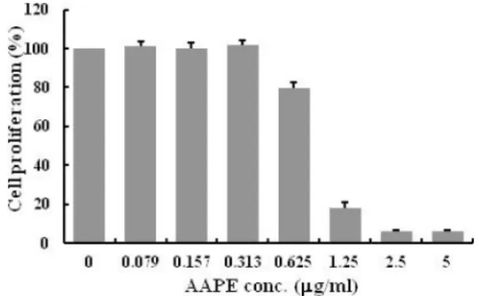
AAPE is a component of ADSC-CM, cell culture medium for ADSC. Since AAPE had the effect of the cell growth, we first examined the effect of AAPE on HDF prolifer- ation. However, AAPE concentrations ranging from 0 to 0.313 µg/ml did not show significant proliferative effect on HDF (Fig. 1). Instead, we observed inhibition of HDF proliferation at AAPE concentrations greater than 0.625 µg/ml (Fig. 1). This suggests that it is dependent on cell types about AAPE effect for cell proliferation.
Collagen synthesis in HDFs
Promotion of collagen synthesis was evaluated in HDF treated with AAPE. As shown in Fig. 2, the amount of type
I collagen in the culture medium of HDF was increased by treatment with AAPE for 42 h, in a dose-dependent manner.
Compared with untreated fibroblasts, 0.32 µg/ml AAPE increased collagen production of HDF by a maximum of 142.7%, whereas 35.2 µg/ml vitamin C increased collagen production by 161.0%.
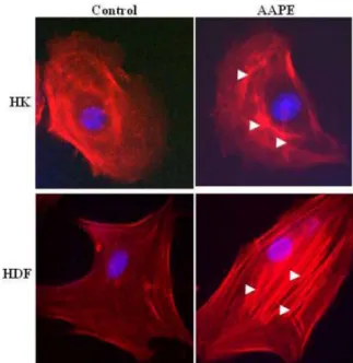
AAPE augments stress fiber formation in HDF and HK
Stress fibers are composed of bundles of approximately 10-30 actin filaments [6] held together by the actin- crosslinking protein such as fascin, espin and filamin [1, 4, 24, 28]. This serve as a cross-linker between the towing and
Fig. 1. Human dermal fibroblast (HDF) proliferation. The amount of HDF proliferation is represented by the cell prolifera- tion (%) in the MTS assay (n=3). The amount HDF proliferation was significantly less in AAPE (more than 0.625 µg/ml) treated group compared to the group treated with AAPE (from 0 to 0.313 µg/ml) by Student’s t-test.Fig. 2. Effects of AAPE and Vitamin C on synthesis rate of pro-collagen type I in normal HDF. The HDF was incubated with medium and samples. The cells were incubated for 42 h at 37oC incubator. Collagen content was measured by amount of pro- collagen type I C-peptide in the supernatant after incubation. Each value represents the mean ± S.D. of three experiments, and values containing asterisks are significantly from the nontreatment group (Cont.) at p<0.01(**) by Dunnett’s t-test.
trailing adhesions, and their organization reflects the direction of the traction force. In motile fibroblasts, ventral stress fibers are oriented parallel to the axis of locomotion [6], which suggests that force generated by contraction of these structures could drive tail retraction. Therefore, these structures provide mechanical contractile force for cell migration. Because stress fiber formation is a cell response characteristic of HK [28] and fibroblast [19] migration, we investigated whether stress fiber formation is induced by AAPE. Stress fiber formation was markedly enhanced by the stimulation of AAPE (Fig. 4) in both cells. We therefore propose that the induction of stress fiber requires the transduction of AAPE signals.
DNA microarray of AAPE
HKs up-regulates collagen type I and III, fibroblast proliferation, and extracellular matrix accumulation to normal dermal fibroblasts. Therefore, the activation of HK is very important about fibroblast activation. In order to address the gene alterations of HK on AAPE we compared the panel of transcripts whose expression was altered in AAPE-treated HKs compared to AAPE-untreated HKs. We screened DNA chip arrays using RNA isolated from HKs.
Our results demonstrate that AAPE in HKs (p < 0.05)
affected expression of 133 identified transcripts regulated minimally by greater than or equal to a 2-fold change. The identified transcripts were associated with 4 functional classes (Fig. 4A). Of the identified regulated genes, 94 were up-regulated (Fig. 4C, Table 1, 2, 3, 4) and 39 were down-regulated (Fig. 4B, Table 1, 2, 3, 4). Of the genes regulated, notable fractions are known to affect cell proli- feration and cell adhesion.
Fig. 3. AAPE induce actin stress fiber formation. HK and HDF cells were left untreated or challenged for 1 h with AAPE (1.22 µg/ml). The cells were then fixed, permeabilized, and stained with rhodamine phalloidin to visualize the actin stress fibers by fluores- cence microscopy. The results are representative of two or three experiments.
Fig. 4. DNA chip analysis. Functional classes of differentially regulated genes in HK incubated with AAPE. Regulated genes were grouped into 4 functional categories and graphed as a per- centage of the total based on their GeneGo designation. 133 genes were differentially regulated based on analysis of the array data (A) Of the regulated genes, 39 were down-regulated (B) and 94 were up-regulated (C). A number of down-regulated genes (12) are associated with cell adhesion.
Table 1. Gene list related to cell proliferation.
Gene Name Gene_info (Synonyms//chromosome//description) Probe ID Regulation
STIL SIL // 1 // SCL/TAL1 interrupting locus 7915926 UP/ Positive
FGF5 HBGF-5 // 4 // fibroblast growth factor 5 8096050 UP/ Positive
TTK MPS1 // 6 // TTK protein kinase 8120838 UP/ Positive
IL31RA GLM-R // 5 // interleukin 31 receptor A 8105411 UP/ Positive
KIF2C KNSL6 // 1 // kinesin family member 2C 7901010 UP/ Positive
CDCA7 JPO1 // 2 // cell division cycle associated 7 8046488 UP/ Positive
MTBP MDM2BP // 8 // Mdm2, transformed 3T3 cell double minute 2, p53 binding protein 8148124 UP/Negative
FANCA FAH // 16 // Fanconi anemia, complementation group A 8003503 UP/Positive
ASPM MCPH5 // 1 // asp (abnormal spindle) homolog, microcephaly associated (Drosophila) 7923086 UP/Positive
CYR61 CCN1 // 1 // cysteine-rich, angiogenic inducer, 61 7902687 UP/ Positive
CDC7 HsCDC7 // 1 // cell division cycle 7 homolog (S. cerevisiae) 7902913 UP/ Positive
KIF15 HKLP2// 3 // kinesin family member 15 8079237 UP/ Positive
TPX2 DIL-2 // 20 // TPX2, microtubule-associated, homolog (Xenopus laevis) 8061579 UP/ Positive SKP2 FBL1 // 5 // S-phase kinase-associated protein 2 (p45) 8104912 UP/ Positive
PROX1 - // 1 // prospero homeobox 1 7909681 UP/ND
CDK2 p33(CDK2) // 12 // cyclin-dependent kinase 2 7956076 UP/Positive
PDCD1LG2 PD-L2 // 9 // programmed cell death 1 ligand 2 8154245 UP/Negative
PTHLH HHM // 12 // parathyroid hormone-like hormone 7962000 UP
VEGFC VRP // 4 // vascular endothelial growth factor C 8103822 UP
UHRF1 RNF106 // 19 // ubiquitin-like with PHD and ring finger domains 1 8024900 UP
EREG ER // 4 // epiregulin 8095728 UP
TIMELESS TIM // 12 // timeless homolog (Drosophila) 7964145 UP
BUB1B BUBR1 // 15 // budding uninhibited by benzimidazoles 1 homolog beta (yeast) 7982663 UP CXCL1 GROa// 4 // chemokine (C-X-C motif) ligand 1 (melanoma growth stimulating activity) 8095697 UP
BLM BS // 15 // Bloom syndrome, RecQ helicase-like 7986068 UP
IFITM1 CD225 // 11 // interferon induced transmembrane protein 1 (9-27) 7937335 UP
FOXM1 FKHL16 // 12 // forkhead box M1 7960340 UP
POLA1 p180 // X // polymerase (DNA directed), alpha 1, catalytic subunit 8166525 UP TACSTD2 TROP2 // 1 // tumor-associated calcium signal transducer 2 7916584 UP
CHEK1 CHK1 // 11 // CHK1 checkpoint homolog (S. pombe) 7945014 UP
ZFP36L2 TIS11D // 2 // zinc finger protein 36, C3H type-like 2 8051814 UP
NCAPG2 MTB // 7 // non-SMC condensin II complex, subunit G2 8144153 UP
BUB1 BUB1A // 2 // budding uninhibited by benzimidazoles 1 homolog (yeast) 8054580 UP
BCL6 ZNF51 // 3 // B-cell CLL/lymphoma 6 8092691 UP/Positive
FGFBP1 HBP17 // 4 // fibroblast growth factor binding protein 1 8099467 UP
HELLS PASG // 10 // helicase, lymphoid-specific 7929438 UP
GINS1 PSF1 // 20 // GINS complex subunit 1 (Psf1 homolog) 8061471 UP
MKI67 KIA // 10 // antigen identified by monoclonal antibody Ki-67 7937020 UP
NF2 SCH // 22 // neurofibromin 2 (merlin) 8072242 UP
DLGAP5 DLG7 // 14 // discs, large (Drosophila) homolog-associated protein 5 7979307 UP NASP PRO1999 // 1 // nuclear autoantigenic sperm protein (histone-binding) 7901123 UP TGFBR2 TGFR-2 // 3 // transforming growth factor, beta receptor II (70/80kDa) 8078350 UP
CENPF hcp-1 // 1 // centromere protein F, 350/400ka (mitosin) 7909708 UP
BRCA2 GLM3 // 13 // breast cancer 2, early onset 7968484 UP
RACGAP1 ID-GAP // 12 // Rac GTPase activating protein 1 7963157 UP
CDC25A CDC25A2 // 3 // cell division cycle 25 homolog A (S. pombe) 8086880 UP
BRCA1 PSCP // 17 // breast cancer 1, early onset 8015769 UP
PLK1 STPK13 // 16 // polo-like kinase 1 (Drosophila) 7994109 UP
EPGN EPG // 4 // epithelial mitogen homolog (mouse) 8095723 UP
CKS2 CKSHS2 // 9 // CDC28 protein kinase regulatory subunit 2 8156290 UP
PCNA MGC8367 // 20 // proliferating cell nuclear antigen 8064844 UP
Table 1. Continued.
Gene Name Gene_info (Synonyms//chromosome//description) Probe ID Regulation
TCF19 SC1 // 6 // transcription factor 19 8179228 UP
UTP20 DRIM // 12 // UTP20, small subunit (SSU) processome component 7957890 UP
NRP1 CD304 // 10 // neuropilin 1 7932985 DOWN
L1CAM SPG1 // X // L1 cell adhesion molecule // protein-coding 8175871 DOWN
VIPR1 VPAC1 // 3 // vasoactive intestinal peptide receptor 1 8079060 DOWN
DDR2 TKT // 1 // discoidin domain receptor tyrosine kinase 2 7906900 DOWN
WARS IFP53 // 14 // tryptophanyl-tRNA synthetase 7981290 DOWN
ANG RNASE4 // 14 // angiogenin, ribonuclease, RNase A family, 5 7973084 DOWN
FGF2 HBGF-2 // 4 // fibroblast growth factor 2 (basic) 8097256 DOWN
ADM AM // 11 // adrenomedullin 7938390 DOWN
GPNMB HGFIN|NMB // 7 // glycoprotein (transmembrane) nmb 8131844 DOWN
LRP1 CD91// 12 // low density lipoprotein-related protein 1 (alpha-2-macroglobulin receptor) 7956301 DOWN
LAMP3 CD208 // 3 // lysosomal-associated membrane protein 3 8092348 DOWN
HBEGF HEGFL // 5 // heparin-binding EGF-like growth factor 8114572 DOWN
IGFBP4 IBP4 // 17 // insulin-like growth factor binding protein 4 8007100 DOWN
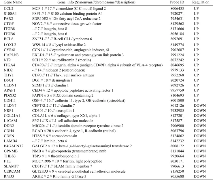
Table 2. Gene list related to cell adhesion.
Gene Name Gene_info (Synonyms//chromosome//description) Probe ID Regulation
CCL2 MCP-1 // 17 // chemokine (C-C motif) ligand 2 8006433 UP
S100A4 FSP1 // 1 // S100 calcium binding protein A4 7920271 UP
FAR2 SDR10E2 // 12// fatty acyl CoA reductase 2 7954631 UP
CTGF NOV2 // 6 // connective tissue growth factor 8129562 UP
ITGB8 - // 7 // integrin, beta 8 8131666 UP
ITGB6 - // 2 // integrin, beta 6 8056184 UP
BCL6 ZNF51 // 3 // B-cell CLL/lymphoma 6 8092691 UP
LOXL2 WS9-14 // 8 // lysyl oxidase-like 2 8149774 UP
CYR61 CCN1 // 1 // cysteine-rich, angiogenic inducer, 61 7902687 UP
HAPLN3 EXLD1 // 15 // hyaluronan and proteoglycan link protein 3 7991224 UP
NF2 SCH // 22 // neurofibromin 2 (merlin) 8072242 UP
ITGA4 CD49D// 2 // integrin, alpha 4 (antigen CD49D, alpha 4 subunit of VLA-4 receptor) 8046695 UP
NID2 - // 14 // nidogen 2 (osteonidogen) 7979133 UP
THY1 CD90 // 11 // Thy-1 cell surface antigen 7952268 UP
DSG1 DG1 // 18 // desmoglein 1 8020724 UP
CLDN1 SEMP1 // 3 // claudin 1 8092726 UP
APAF1 CED4 // 12 // apoptotic peptidase activating factor 1 7957759 UP
PDZD2 PAPIN // 5 // PDZ domain containing 2 8104693 UP
CDH11 OSF-4 // 16 // cadherin 11, type 2, OB-cadherin (osteoblast) 8001800 UP
CLDN7 CEPTRL2 // 17 // claudin 7 8012126 DOWN
NRP1 CD304 // 10 // neuropilin 1 7932985 DOWN
COL21A1 COLA1L // 6 // collagen, type XXI, alpha 1 8127201 DOWN
L1CAM SPG1 // X // L1 cell adhesion molecule 8175871 DOWN
DDR2 MIG20a // 1 // discoidin domain receptor tyrosine kinase 2 7906900 DOWN
CDH4 RCAD // 20 // cadherin 4, type 1, R-cadherin (retinal) 8063796 DOWN
CDSN HTSS // 6 // corneodesmosin 8124862 DOWN
LAMB4 - // 7 // laminin, beta 4 8142232 DOWN
B4GALNT2 GALGT2 // 17 // beta-1,4-N-acetyl-galactosaminyl transferase 2 8008172 DOWN
GPNMB NMB // 7 // glycoprotein (transmembrane) nmb 8131844 DOWN
THBS3 TSP3 // 1 // thrombospondin 3 7920664 DOWN
FTL MGC71996 // 19 // ferritin, light polypeptide 8030171 DOWN
SLAMF7 CD319 // 1 // SLAM family member 7 7906613 DOWN
CERCAM GLT25D3 // 9 // cerebral endothelial cell adhesion molecule 8158250 DOWN
RND3 ARHE // 2 // Rho family GTPase 3 8055688 DOWN
Proteome analysis via antibody array
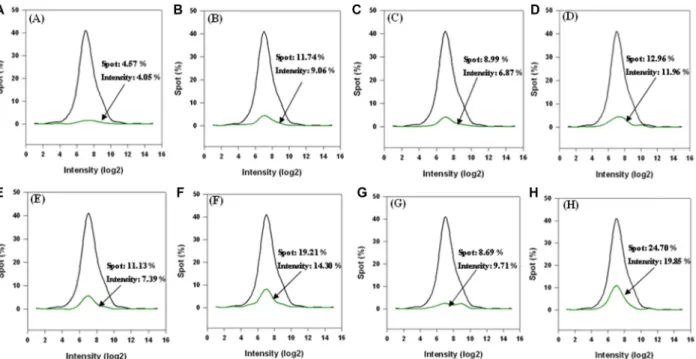
In order to visualize protein expression for proteome analysis, an explorer antibody array chip coated with 656 antibodies was conducted to identify proteins in AAPE (Fig. 5). Protein level measurements were clustered by the ratio of hormone (Fig. 5A), cytokine (Fig. 5B), cell cycle (Fig. 5C), cancer marker (Fig. 5D), apoptosis (Fig. 5E), angiogenesis (Fig. 5F), stem cell (Fig. 5G), and signal transduction (Fig. 5H) level by comparing the signal intensity of soluble protein expression in AAPE to a negative control. Furthermore, a cytokine-focused antibody
array with 77 antibodies was conducted to identify proteins that may be involved in mediating the effect of AAPE on proliferation, migration or regeneration (Fig. 6) by comparison with explorer antibody array data. p value < 0.05 was con- sidered significantly. Analysis of the array results revealed high protein expression level of VEGF, TGF- β2, CD54 and TGF- β3 compared to HGF, shows low protein expression level of FGF-1, FGF-2, IL-4, IL-6, G-CSF, GM-CSF com- pared to HGF (Fig. 6). HGF is composed of 60 kDa alpha- chain and a 34 kDa beta-chain and induced HK in vitro scratch-wound healing in a dose-dependent manner [21].
Table 3. Gene list related to cell migration.
Gene Name Gene_info (Synonyms//chromosome//description) Probe ID Regulation
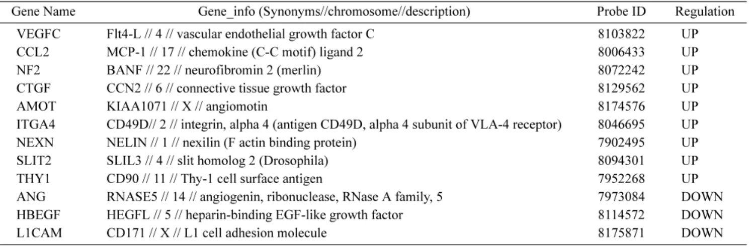
VEGFC Flt4-L // 4 // vascular endothelial growth factor C 8103822 UP
CCL2 MCP-1 // 17 // chemokine (C-C motif) ligand 2 8006433 UP
NF2 BANF // 22 // neurofibromin 2 (merlin) 8072242 UP
CTGF CCN2 // 6 // connective tissue growth factor 8129562 UP
AMOT KIAA1071 // X // angiomotin 8174576 UP
ITGA4 CD49D// 2 // integrin, alpha 4 (antigen CD49D, alpha 4 subunit of VLA-4 receptor) 8046695 UP
NEXN NELIN // 1 // nexilin (F actin binding protein) 7902495 UP
SLIT2 SLIL3 // 4 // slit homolog 2 (Drosophila) 8094301 UP
THY1 CD90 // 11 // Thy-1 cell surface antigen 7952268 UP
ANG RNASE5 // 14 // angiogenin, ribonuclease, RNase A family, 5 7973084 DOWN
HBEGF HEGFL // 5 // heparin-binding EGF-like growth factor 8114572 DOWN
L1CAM CD171 // X // L1 cell adhesion molecule 8175871 DOWN
Table 4. Gene list related to response to wounding.
Gene Name Gene_info (Synonyms//chromosome//description) Probe ID Regulation
PLAT TPA // 8 // plasminogen activator, tissue 8150509 UP
CXCL1 CYB1//4//chemokine (C-X-C motif) ligand 1 (melanoma growth stimulating activity) 8095697 UP
CCL2 MCP-1 // 17 // chemokine (C-C motif) ligand 2 8006433 UP
MAP2K3 MEK3 // 17 // mitogen-activated protein kinase kinase 3 8005707 UP
ELK3 ERP // 12 // ELK3, ETS-domain protein (SRF accessory protein 2) 7957665 UP
HRH1 H1-R // 3 // histamine receptor H1 8077851 UP
EREG ER // 4 // epiregulin 8095728 UP
PLSCR4 TRA1 // 3 // phospholipid scramblase 4 8091306 UP
CTGF NOV2 // 6 // connective tissue growth factor 8129562 UP
SLC7A2 HCAT2 // 8 // solute carrier family 7 (cationic amino acid transporter) 8144786 UP
ITGB6 ITGB6 // 2 // integrin, beta 6 8056184 UP
KRT1 KRT1A // 12 // keratin 1 7963491 UP
BCL6 LAZ3 // 3 // B-cell CLL/lymphoma 6 8092691 UP
TFPI2 REF1 // 7 // tissue factor pathway inhibitor 2 8141016 UP
SYT7 IPCA-7 // 11 // synaptotagmin VII 7948588 DOWN
CD55 DAF // 1 // CD55 molecule, decay accelerating factor for complement 7909332 DOWN
ADM AM // 11 // adrenomedullin 7938390 DOWN
C1RL CLSPa // 12 // complement component 1, r subcomponent-like 7960757 DOWN
HBEGF HEGFL // 5 // heparin-binding EGF-like growth factor 8114572 DOWN
NFE2L1 NRF1 // 17 // nuclear factor (erythroid-derived 2)-like 1 8008087 DOWN PTX3 TSG-14 // 3 // pentraxin-related gene, rapidly induced by IL-1 beta 8083594 DOWN
BLNK SLP65 // 10 // B-cell linker 7935270 DOWN
IL-6 has a proliferative and migration effect on mouse HKs [23, 8]. GM-CSF also stimulates of HK proliferation, granu- lation tissue formation, and vascularization [14]. TGF- β3 promotes wound healing by recruiting fibroblast to the wound site. Other protein contained AAPE. Anyway, these proteins support evidences of the application of AAPE in the skin regeneration.
Disscusion
In this study, the ability of ADSC secretomes to promote skin cell regeneration was investigated. We called condition- medium of ADSC obtained under hypoxia condition an AAPE. Skin wound healing is a complex process having combined efforts of various types and lineages of skin cells, ECMs, and soluble factors. Immune activation, in- flammation, re-epithelialization, ECM reorganization and tissue remodeling are sequential events to repair skin wounds. HKs stimulated during wound repair secrete several growth factors and multiple cytokines that activate fibroblasts and endothelial cells, initiate the infiltration of immune cells, and produce systemic effects [22, 25]. They also produce extracellular matrix proteins, and adhesive molecules. This study investigated whether or not locally applied AAPE can accelerate skin regeneration process in vitro system. The collagen neo-synthesis of HDF, which is
Fig. 5. Antibody array of AAPE. Result of quantile normalization with explorer array data. Total data in explorer array: black line;hormone, cytokine, cell cycle, cancer marker, apoptosis, angiogenesis, stem cell, and signal transduction data in explorer array: green line.
Fig. 6. Heatmap analysis of AAPE followed by antibody array.
AAPE was subjected to antibody array. Portions of the array illus- trating the differential expression of proteins between HGF and sample antibody are shown. Heat map were analyzed by Genowiz 4.0TM as described in Materials and Methods. The median strepta- vidin-Cy3 fluorescence from all microsphere subsets was exported.
The heat map shows antibody reactivity intensity (i.e., values above HGF) as red pixels and (values below HGF) as green pixels.
one major cell type present in skin, was increased by AAPE treatment. AAPE are thought to trigger the prolifer- ation, cell migration, response to wound, and adhesion of HK through DNA chip assay. Moreover, AAPE induced the stress fiber formation of HK that are able to migrate efficiently (Fig. 4). Cell mobility is generally described as a periodic process between alternating phases of protrusion and adhesion. Cell adhesion provides the direction point required for generating pulling forces, and the cell migrates forward direction by the tension driven by contraction of the cell body and retraction of the tail [20]. Because stress fiber supplies contractile force derived from the contractile nature, cell movement is dependent on reorganization of cell cytoskeleton, predominantly actin filaments. Thus, the stress fiber was formed during cell traffic, suggesting that this architecture might be important for efficient cell motility.
The regeneration ability of AAPE is owed to several secretome identified by proteome analysis via an antibody array. Hepatocyte growth factor (HGF) triggers influx of neutrophils, monocytes and mast cells into wounded areas [3] and facilitates the release of pro-angiogenesis factors [5]. Scratch assays were performed in the presence and absence of HGF treatment to assess the response of HaCaT cells to HGF. PAI-1, a member of the serine protease inhibitor (serpin) superfamily, involved in wound healing [9]. In vivo introduction of the IL-6 gene into human HKs induces epidermal proliferation [23]. CTGF expression is increased during wound repair and is involved in inflam- mation, matrix deposition and remodeling in human dermal fibroblast [10]. In skin wounds, granulocyte-macrophage colony stimulating factor (GM-CSF) mediates HK prolifer- ation at the wound edges [14]. Many other cells are involved in wound healing including macrophages, lymphocytes, fibroblasts, endothelial cells, and dendritic cells, which is target for GM-CSF. Therefore, GM-CSF is a pleiotropic cytokine controlling organized processes during wound repair. AAPE also contained TGF- β3, which promotes wound healing by facilitating HK migration of in vivo studies [2]. Collectively, present study provides the clues of the regenerative effect of the AAPE. However, for the proper application of AAPE into skin biology, more in vivo studies about the action mechanisms of AAPE on skin are needed and clinic standard application method of AAPE should be established. In conclusion, the present researches have been reported about HDF and HK biological effect of AAPE in vitro. AAPE activates HK proliferation and migra-
tion. Moreover, AAPE induces the collagen synthesis of HDF. Thus, AAPE is suitable for application in the treatment of skin problems.
Acknowledgement
This study was supported by a grant from Dong Eui University (2011AA197).
R
EFERENCES1. Adams, J. C. 1995. Formation of stable microspikes contain- ing actin and the 55 kDa actin bundling protein actin and the 55 kDa actin bundling protein, fascin, is a consequence of cell adhesion to thrombospondin-1: Implications for the anti- adhesive activities of thrombospondin-1. J. Cell Sci. 108:
1977-1990.
2. Bandyopadhyay, B., J. Fan, S. Guan, Y. Li, M. Chen, D. T.
Woodley, and W. Li. 2006. A “traffic control” role for TGFbeta3: Orchestrating dermal and epidermal cell motility during wound healing. J. Cell Biol. 172: 1093-1105.
3. Bevan, D., E. Gherardi, T. P. Fan, D. Edwards, and R. Warn.
2004. Diverse and potent activities of HGF/SF in skin wound repair. J. Pathol. 203: 831-838.
4. Chen, B., A. Li, D. Wang, M. Wang, L. Zheng, and J. R.
Bartles. 1999. Espin contains an additional actin-binding site in its N terminus and is a major actin-bundling protein of the sertoli cell-spermatid ectoplasmic specialization junctional plaque. Mol. Biol. Cell 10: 4327-4339.
5. Dong, G., T. L. Lee, N. T. Yeh, J. Geoghegan, C. Van Waes, and Z. Chen. 2004. Metastatic squamous cell carcinoma cells that overexpress c-Met exhibit enhanced angiogenesis factor expression, scattering and metastasis in response to hepato- cyte growth factor. Oncogene 23: 6199-6208.
6. Cramer, L. P., M. Siebert, and T. J. Mitchison. 1997. Identifi- cation of novel graded polarity actin filament Bundles in locomoting heart fibroblasts: implications for the generation of motile force. J. Cell Biol. 136: 1287-1305.
7. Efimenko, A., E. E. Starostina, K. A. Rubina, N. I. Kalinina, and E. V. Parfenova. 2010. Viability and angiogenic activity of mesenchymal stromal cells from adipose tissue and bone marrow in hypoxia and inflammation in vitro. Tsitologiia 52:
144-154.
8. Gallucci, R. M., D. K. Sloan, J. M. Heck, A. R. Murray, and S. J. O’Dell. 2004. Interleukin 6 indirectly induces HK migration. J. Invest. Dermatol. 122: 764-772.
9. Huber, K. 2001. Plasminogen activator inhibitor type-1 (part one): Basic mechanisms, regulation, and role for throm- boembolic disease. J. Thromb. Thrombolys 11: 183-193.
10. Igarashi, A., H. Okochi, D. M. Bradham, and G. R. Groten- dorst. 1993. Regulation of connective tissue growth factor gene expression in human skin fibroblasts and during wound repair. Mol. Biol. Cell 4: 637-645.
11. Kim, W. S., B. S. Park, H. K. Kim, J. S. Park, K. J. Kim, J.
S. Choi, S. J. Chung, D. D. Kim, and J. H. Sung. 2008. Evi- dence supporting antioxidant action of adipose-derived stem cells: Protection of human dermal fibroblasts from oxidative stress. J. Dermatol. Sci. 49: 133-142.
12. Kim, W. S., B. S. Park, and J. H. Sung. 2009. Protective role of adipose-derived stem cells and their soluble factors in photoaging. Arch. Dermatol. Res. 301: 329-336.
13. Kim, W. S., B. S. Park, J. H. Sung, J. M. Yang, S. B. Park, S.
J. Kwak, and J. S. Park. 2007. Wound healing effect of adi- pose-derived stem cells: A critical role of secretory factors on human dermal fibroblasts. J. Dermatol. Sci. 48: 15-24.
14. Mann, A., K. Breuhahn, P. Schirmacher, and M. Blessing.
2001. HK-derived granulocyte-macrophage colony stimulat- ing factor accelerates wound healing: Stimulation of HK pro- liferation, granulation tissue formation, and vascularization.
J. Invest. Dermatol. 117: 1382-1390.
15. Mansbridge, J. 2008. Skin tissue engineering. J. Biomater.
Sci. Polym. Ed. 19: 955-968.
16. Nobes, C. D., and A. Hall. 1999. Rho GTPases control polar- ity, protrusion, and adhesion during cell movement. J. Cell Biol. 144: 1235-1244.
17. Nolte, S.V., and W. Xu, H. O. Rennekampff, and H. P. Rode- mann. 2008. Diversity of fibroblasts a review on implica- tions for skin tissue engineering. Cells Tissues Organs 187:
165-176.
18. Ren, H., Y. Cao, Q. Zhao, J. Li, C. Zhou, L. Liao, M. Jia, Q.
Zhao, H. Cai, Z. C. Han, Q. Zhao, R. Yang, G. Chen, and R.
C. Zhao. 2006. Proliferation and differentiation of bone mar- row stromal cells under hypoxic conditions. Biochem. Bio- phys. Res. Commun. 347: 12-21.
19. Rid, R., N. Schiefermeier, I. Grigoriev, J. V. Small, and I.
Kaverina. 2005. The last but not the least: The origin and significance of trailing adhesions in fibroblastic cells. Cell Motil. Cytoskeleton 61: 161-171.
20. Ridley, A. J., M. A. Schwartz, K. Burridge, R. A. Firtel, M.
H. Ginsberg, G. Borisy, J. T. Parsons, and A. R. Horwitz.
2003. Cell migration: Integrating signals from front to back.
Science 302: 1704-1709.
21. Peura, M., J. Bizik, P. Salmenperä, A. Noro, M. Korhonen, T. Pätilä, A. Vento, A. Vaheri, A.; Alitalo, R.; Vuola, J. A.
Harjula, and E. Kankuri. 2009. Bone marrow mesenchymal stem cells undergo nemosis and induce HK wound healing utilizing the HGF/c-Met/PI3K pathway. Wound Repair Regen. 17: 569-577.
22. Shephard, P., G. Martin, S. Smola-Hess, G. Brunner, T. Krieg, and H. Smola. 2004. Myofibroblast differentiation is induced in HK-fibroblast co-cultures and is antagonistically regu- lated by endogenous transforming growth factor beta and interleukin-1. Am. J. Pathol. 164: 2055-2066.
23. Sato, M., D. Sawamura, S. Ina, T. Yaguchi, K. Hanada, and I. Hashimoto. 1999. In vivo introduction of the interleukin 6 gene into human HKs: Induction of epidermal proliferation by the fully spliced form of interleukin 6, but not by the alternatively spliced form. Arch. Dermatol. Res. 291: 400- 404.
24. Wang, K., J. F. Ash, and S. J. Singer. 1975. Filamin, a new high-molecular-weight protein found in smooth muscle and non-muscle cells. Proc. Natl. Acad. Sci. USA 72: 4483-4486.
25. Werner, S. T. Krieg, and H. Smola. 2007. HK-fibroblast interactions in wound healing. J. Invest. Dermatol. 127: 998- 1008.
26. Wright, C. S., M. A. Van Steensel, M. B. Hodgins, and P. E.
Martin. 2009. Connexin mimetic peptidesimprove cell migration rates of human epidermal HKs and dermal fibro- blasts in vitro. Wound Repair Regen. 17: 240-249.
27. Xia, W., T. T. Phan, I. J. Lim, M. T. Longaker, and G. P.
Yang. 2004. Complex epithelial-mesenchymal interactions modulate transforming growth factor-beta expression in kel- oid-derived cells. Wound Repair Regen. 12: 546-556.
28. Zhang, L., M. Deng, R. Parthasarathy, L. Wang, M. Mon- gan, J. D. Molkentin, Y. Zheng, Y. Xia. 2005. MEKK1 trans- duces activin signals in HKs to induce an actin stress fiber formation and migration. Mol. Cell Biol. 25: 60-65.
국문초록
인체 섬유아세포 및 케라티노사이트에 대한 지방줄기세포 분비물의 세포생물학적 기능
이재설1·이종환1,2,3*1동의대학교 바이오물질제어학과
2동의대학교 블루바이오RIC센터
3동의대학교 생명공학과
피부재생에 대한 지방줄기세포 배양상등액(ADSC-CM)의 효능에 대한 연구를 진행하였다. ADSC-CM이 피부재생 에 기여하는 기작은 명확하지 못하지만, ADSC-CM은 다양한 분비물을 포함하고 있고 따라서 피부트러블 처리를 위 한 훌륭한 재료이다. 저 산소 상태에서 생산된 ADSC-CM, 즉 advanced adipose-derived stem cell protein extract
(AAPE)는 피부재생에 보다 좋은 재료이다. 본 연구는 피부 재생에 결정적 역할을 하는 인체 primary 세포인 섬유아
세포(HDF)와 케라티노사이트(HK)를 이용하여 AAPE의 효능을 검증하였다. 0.32 µg/ml AAPE에서 콜라겐 합성이 관 찰 되었으며 AAPE는 stress fiber 형성을 강화하였다. DNA microarray 결과에서는 세포증식, 세포이동, 세포부착, 상 처반응에 관여하는 133개의 유전자 발현이 조절되는 것을 알았다. Antibody array를 통해 CD54, FGF-2, GM-CSF, IL-4, IL-6, VEGF, TGF-β2, TGF-β3, MMP-1, MMP-10, 그리고 MMP-19와 같은 MMP, 성장인자, 사이토카인등 25개의 알려진 단백질이 포함되어 있다는 것을 알았다. 따라서, AAPE는 HK의 세포생물학적 기능을 활성화 할 수 있다고 사료되며 HDF에서는 콜라겐 합성을 유도하였다. 이러한 결과는 AAPE가 피부재생에 임상적 적용이 가능하 리라는 것을 의미한다.