Research Article Open Access
Difference in Muscle Activities According to Stability on Support Surface During Plank Exercise
Yong-Ho Cho, PT, PhD⋅Jin-Ho Choi, PT, PhD
†Dept. of Physical Therapy, Daegu Haany University
Received: July 17, 2017 / Revised: July 19, 2017 / Accepted: July 28, 2017
ⓒ 2017 J Korean Soc Phys Med
| Abstract |
1)PURPOSE: The present study aimed to measure muscle activities in the pectoralis major, the erector spinae, and the quadriceps femoris according to support surface states of arms and legs during plank exercise.
METHODS: The subjects of this study were 21 healthy males in their 20s and their muscle activities at three states were measured as follows: The first state was where the support surface of arms and legs was stable. The second state was where only arms were unstable, and the third state was where only legs were unstable. Electromyography (EMG) was used to measure muscle activities. Pectoralis major, quadriceps femoris, and elector spinae were measured for muscle activities.
RESULTS: The muscle activities in the pectoralis major were statistically high when arms were unstable. The muscle activities in the quadriceps femoris were statistically high when legs were unstable. The muscle activities in the erector spinae were higher when arms and legs were unstable compared to that at the stable support surface. No significant
† Corresponding Author : [email protected]
This is an Open Access article distributed under the terms of the Creative Commons Attribution Non-Commercial License (http://creativecommons.org/licenses/by-nc/3.0) which permits unrestricted non-commercial use, distribution, and reproduction in any medium, provided the original work is properly cited.
difference was revealed statistically when arms and legs were unstable.
CONCLUSION: If the instability of arms and legs is employed during plank exercise, exercise on the upper and lower bodies or the erector spinae is expected to be more effective.
Key Words: EMG, Muscle activity, Plank exercise
Ⅰ. Introduction
The muscles in the trunk play an important role in supporting the center of body. Thus, the stability of trunk muscles is highly important to basic stability for movements of upper extremities and correct alignment of spine (Lehman et al., 2005). A lower back pain, which is one of the most common musculoskeletal disorders in modern people, is also caused by instability around the trunk and imbalance of muscles (Hodges, 2003). A sit-up exercise is frequently performed to increase muscle strength in trunk muscles but it can increase a pressure on the spine thereby causing a problem in the lumbar spine. In particular, it can be more aggravated in patients with lower back pain (McGill, 2010).
In recent years, the plank has been widely done instead
of doing sit-ups to overcome the above problems. The plank
increases activities of abdominal muscles among trunk muscles thereby strengthening trunk muscles without too much pressure on the spine (Snarr and Esco, 2014). The movements and activities of arms and legs affect the motion of the trunk. An increase in abdominal muscles was exhibited through limb movements, and activities in abdomen were higher when alternating movements of limbs were performed. Although differences can be found in the plank via limb's movements, differences can also be found via differences in support surface of arms and legs that support the floor (Hodges et al., 1999; Lee et al., 2013). Although a number of studies have been conducted on the plank as an exercise to increase muscle activities and muscle strength in abdomen, few studies have been conducted on muscles according to a support surface (Kim et al., 2016). Depending on the support surface of the floor, changes in muscle activities in trunk, arms, and legs were found during push-ups (Cho and Choi, 2016; Seo et al., 2013). Studies on the plank have been focused on strengthening abdominal muscles among trunk muscles (Tong et al., 2014).
Thus, the this study aimed to determine muscle activities according to a state of support surface in light of the consideration that muscle activities in trunk and limb muscles would be different according to a support surface during the plank as same as muscle activities are different according to a difference in support surface during push-ups. That is, the plank is an exercise that places lower arms and legs on the floor, so this study expected muscle activities would be different according to the stability state in the floor, which was why this study aimed to measure changes in muscle activities according to the state of arms and legs at the floor during the plank.
Ⅱ. Methods
1. Subjects
The present study was conducted with 21 healthy adult
males in their 20s who had no neurological and muscu- loskeletal disorders. The subjects were informed about the study method and consented to participate in the study.
2. Study procedure
The present study was conducted to determine changes in muscle activities of body muscles according to the state of support surface during the plank. The state of support surface was divided into three: both of arms and legs were all stable (Double-Stable Support: D-SS), only arms were unstable (Arm-UnStable Support: A-US), and only legs were unstable (Leg-UnStable Support: L-US). The Togu ball was used to make an unstable state in the arms and legs as well as two pads to make arms unstable, and one pad to make legs unstable. The patients were performed 3 times exercise for precise movement. After 10 seconds of the plank, data were collected for five seconds, and a rest time was given for five minutes between exercises (Lee et al., 2012). Furthermore, every seven subjects had a different exercise order to reduce a difference due to exercise order. Electromyography (EMG) was used to measure muscle activities. Pectoralis major, quadriceps femoris, and elector spinae were measured for muscle activities. In previous studies, many muscles were measured for muscle activity during plank or push up exercise.
Among them, we chose pectoralis major, quadriceps femoris, erector spinae for measuring trunk and low extremity activity (Byrne et al., 2014; Harris et al., 2017;
Snarr and Esco, 2014).
Their muscles activities were measured using an MP150
(BIOPAC systems, Inc., Goleta, CA, USA), surface electro-
myography equipment, and Ag-Ag/Cl electrodes. The at-
tachment position of the electrodes was in each muscle
belly. The ground electrode was attached to an area not
associated with signals. Signals collected at a sampling
rate of 1,000 Hz were notch filtered at 60 Hz after full-wave
rectification to remove electrical noise. Then, 30 to 500
band-pass filtering was conducted. % Maximal Voluntary
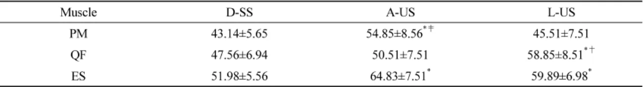
Muscle D-SS A-US L-US
PM 43.14±5.65 54.85±8.56
*‡45.51±7.51
QF 47.56±6.94 50.51±7.51 58.85±8.51
*†ES 51.98±5.56 64.83±7.51
*59.89±6.98
*DSS: Double-Stable Support, A-US: Arm-Unstable Support, L-US: Leg-Unstable Support PM: pectoralis major, QF: Quadriceps Femoris, ES: Erector Spinae
CG: control group, EG: experimental group
* significant difference compared to D-SS
†
significant difference compared to after A-US
‡