336 책임저자:최영현, ꂕ 614-052, 부산광역시 부산진구 양정동 산 45
동의대학교 한의과대학 생화학교실 Tel: 051-850-7413, Fax: 051-853-4036 E-mail: [email protected]
접수일:2006년 8월 18일, 게재승인일:2006년 9월 18일
Correspondence to:Yung Hyun Choi
Department of Biochemistry, Dongeui University College of Oriental Medicine, San 45, Yangjeong-dong, Busanjin-gu, Busan 614-052, Korea Tel: +82-51-850-7413, Fax: +82-51-853-4036
E-mail: [email protected]
세신 열수 추출물에 의한 Caspase 활성과 Bcl-2 발현 저하에 의한 인체 폐암세포의 Apoptosis 유발
1경성대학교 자연과학대학 생물학과, 동의대학교 한의과대학 2생화학교실 및 6해부학교실,
4자연과학대학 생명응용과학과 및 대학원 바이오물질제어학과,
3부산대학교 자연과학대학 생물학과, 5제주대학교 해양과학부
김옥숙1․박 철2,3․정경태4․김기영5․문성기1․이원호3․최병태6․최영현2
Induction of Apoptosis by Water Extract of Asarum sieboldii in Human Lung Carcinoma A549 Cells through Activation of Caspases
and Down-regulation of Bcl-2
Ok-Suk Kim1, Cheol Park2,3, Kyung Tae Chung4, Gi Young Kim5, Sung-Gi Moon1, Won Ho Lee3, Byung Tae Choi6 and Yung Hyun Choi2
1Department of Biology, Kyungsung University, Busan 608-736, Departments of 2Biochemistry and 6Anatomy, Dongeui University College of Oriental Medicine and Department of Biomaterial Control,
Dongeui University Graduate School, 4Department of Life Biotechnology, Dongeui University, Busan 614-052,
3Department of Biology, Busan National University, Busan 609-735,
5Faculty of Applied Marine Science, Cheju National University, Jeju 690-756, Korea
Asarum sieboldii has been used to treat pain and inflammation in traditional Korean medicine as a medicinal and food plant. In the previous study, we reported that water extracts of A. sieboldii (WEAS) has cytotoxic effects on A549 human lung carcinoma cells. In this study, it was analyzed that the effects of WEAS on the apoptotic activity and the expression levels of apoptosis-related genes in A549 cells.
Treatment of WEAS to A549 cells resulted in the induction of apoptotic cell death in a dose-dependent manner as measured by hemocytometer counts, fluorescent microscope and cytometry analysis. The induction of apoptosis of A549 cells by WEAS treatment was associated with an up-regulation of pro-apoptotic Bax expression, and a down-regulation of anti-apoptotic Bcl-2 and inhibitor of apoptosis proteins (IAPs) expression. WEHC treatment induced the proteolytic activation of caspase-3, -8 and -9, and a concomitant degradation of poly (ADP-ribose) polymerase (PARP) and β-catenin protein. Taken together, these results indicated that the inhibitory effect of WEAS on the cell viability in A549 cells was associated with the induction of apoptotic cell death through regulation of several major growth regulatory gene products such as Bcl-2 family expression and caspase protease activity. Though further studies will be needed to identify the active compounds that confer the anti-cancer activity of WEAS, the present findings provide important new insights into the possible molecular mechanisms of the apoptotic activity of WEAS in cancer cells. (Cancer Prev Res 11, 336-345, 2006)
ꠏꠏꠏꠏꠏꠏꠏꠏꠏꠏꠏꠏꠏꠏꠏꠏꠏꠏꠏꠏꠏꠏꠏꠏꠏꠏꠏꠏꠏꠏꠏꠏꠏꠏꠏꠏꠏꠏꠏꠏꠏꠏꠏꠏꠏꠏꠏꠏꠏꠏꠏꠏꠏꠏꠏꠏꠏꠏꠏꠏꠏꠏꠏꠏꠏꠏꠏꠏꠏꠏꠏꠏꠏꠏꠏꠏꠏꠏꠏꠏꠏꠏꠏꠏꠏꠏꠏꠏꠏꠏꠏꠏꠏꠏꠏꠏꠏꠏꠏꠏꠏꠏ Key Words: Asarum sieboldii, Apoptosis, Bcl-2, Caspase, Lung carcinoma
서 론
세포의 죽음은 크게 necrosis와 apoptosis로 구분되며 이 것은 세포의 형태적 및 생화학적인 특성에 의하여 구분 될 수 있다. Necrosis는 생리적, 화학적인 외상에 의한 세 포의 죽음이고 apoptosis는 개체의 정상적인 발달과 분화 에 관여하며, 태아의 형태형성, 난자의 배란, 신경세포의 시냅스 형성 등과 연관된 체내 비정상적인 세포들을 제 거하는 기전이다. 그리고 apoptosis는 DNA 손상, 바이러 스 감염 등에 의한 유전적 조절 하에서 일어나는 정교한 방어기전이라는 점에서 생리적이거나 화학적인 외상에 의한 세포의 죽음인 necrosis와는 구별된다.1∼3) 또한 apop- tosis는 개체보존의 수준에서 손상된 세포들의 제거를 위 한 중요한 수단이기도 하다. Apoptosis의 유발에 종양억 제유전자 p53, Bcl-2 family 및 caspase member 등과 같은 유전자가 관여한다는 사실이 알려지면서 apoptosis와 연 관된 분자적 기전이 최근 많이 밝혀지고 있는데, Bcl-2 family에 속하는 Bcl-2는 apoptosis를 억제하는 반면, Bax의 과발현이 이루어졌을 때는 apoptosis를 유발시킨다고 알 려져 있다.4) 그리고 caspase라고 이름 붙여진 ICE/CED-like protease family 역시 apoptosis 유발에 중요한 역할을 한다 고 알려져 있으며, 이들은 proenzyme 형태로 존재하다가 apoptosis 유도를 활성화시키는 신호에 의해 활성화된 cysteine-related proteases로 되어 직접 또는 간접으로 세포 내에 존재하는 많은 표적 단백질의 분해에 관여하게 된 다.5,6) 그리고 이 인자들에 관한 내용은 아직도 많은 연구 가 진행되고 있는 실정이며, 특히 apoptosis의 유발은 암 세포의 성장, 증식의 억제와 암세포 제거의 한 방법으로 써 널리 연구되고 있다.7)
족도리풀(Asarum sieboldii Miq)은 우리나라 전역에 분포 하는 쥐방울덩굴과에 속하는 다년생 초본식물로서, 그 서식 환경이 조도는 낮고 고도는 높은 북향 내지 북동향 에 치우쳐 있으며 토양의 수분 함량이 많고 부엽층이 두 꺼운 아주 오래된 활엽수림 아래에서 주로 자생하는 것 으로 보고된 바 있다.8) 족도리풀의 뿌리는 가늘고 길며, 맛이 맵다고 하여 세신(細辛)이라고 부르며 예로부터 한 약 재료로서 널리 사용되고 있다.9) 세신에 함유된 주 생 리활성 물질은 methyleugenol 및 xanthoxylol과 같은 정유 성분으로 기침억제 작용 및 항알레르기에 효과가 있으
며,10,11) 살충작용 및 항균활성이 있으며, 뇌세포 보호기
능과 치매 예방 효과 등도 보고된 바 있다.12) 이러한 선 행연구의 내용으로 미루어 세신에 함유되어 있는 다양 한 성분들이 암의 발생이나 암세포의 증식 억제 가능성
이 매우 높을 것으로 예상되지만, 세신 추출물을 대상으 로 항암작용에 관한 연구는 현재까지 이루어진 바가 거 의 없는 실정이다. 본 연구실에서는 세신 추출물의 항암 작용에 관한 연구를 시도하기 위하여 우선 세신 열수 추 출물에 의한 암세포주기 조절 관련 연구를 수행한 바 있 으며, 세신 추출물 처리에 의한 암세포의 증식 억제에 cyclooxygenase-2 선택적 발현 저하가 연관되어 있음을 확 인하였다.13)
본 연구에서는 선행 연구의 결과를 바탕으로 인체 암 세포의 생존율에 미치는 증식에 미치는 족도리풀 추출 물의 영향을 조사하고 세신 추출물 처리에 의한 생존율 저하는 apoptosis 유발과 연관성이 있음을 확인하였으며, A549 인체 폐암세포를 선택하여 세신 추출물에 의한 apoptosis의 유발과 연관성을 가지는 주요 인자들의 발현 변화를 조사하였다.
재료 및 방법 1. 실험 재료 및 세포배양
본 실험에 사용된 세신 열수 추출물(water extract of A.
sieboldii, WEAS)은 Kim 등의 방법에 준하여 추출하였으 며,13) 3차 증류수에 용해하여 멸균과 여과 과정을 거쳐 배지에 적정 농도로 희석하여 처리하였다. 실험에 사용 된 인체 폐암세포(A549 및 NCI-H460)와 대장암세포 (HT29)는 생명공학연구소(KRIBB, Taejeon, Korea)에서 분 양 받았으며, 세포의 배양과 시약처리를 위해 10%의 우 태아혈청(fetal bovine serum, FBS, Gibco BRL, Grand Island, NY, USA)과 1%의 penicillin-streptomycin (Gibco BRL) 등이 포함된 RPMI-1640 배지(Gibco BRL)를 사용하였다. 세포 는 37oC, 5% CO2 조건하의 CO2 incubator에서 배양하였 고, 세포수의 증식에 따른 과밀도 현상에 의한 세포의 성장억제나 각종 오염을 방지하기 위해 0.05% trypsin- ethylenediamine tetraacetic acid (EDTA, Gibco BRL)를 처리 하여 세포를 부유시킨 다음 적정한 수의 세포를 분주하 여 재배양하였다.
2. Hemocytometer를 이용한 세포 생존률의 측정
세포배양용 6-well plate에 암세포를 3×10⁴개/ml 정도 를 분주하고 24시간 동안 안정화시킨 다음 WEAS를 배 지에 희석하여 처리한 후 배양하였다. 72시간 후 배지를 제거하고 0.05% trypsin-EDTA를 처리하여 세포를 부유시 킨 후 phosphate-buffered saline (PBS)를 가하여 세포를 모 은 다음 1,000 rpm으로 5분간 원심분리를 하였다. 상층액 을 제거하고 세포만 남긴 다음 다시 PBS를 1 ml 가하여
충분히 섞은 후 세포 부유액과 0.5% trypan blue (Gibco BRL)를 동량으로 섞어 2분간 처리하였다. Pasteur pippette 의 모세관 현상을 이용하여 세포를 hemocytometer로 옮 긴 후 위상차 현미경(inverted microscope, Carl Zeiss, Ger- many)을 이용하여 200배의 배율로 관찰하여 푸른색으로 염색된 세포를 죽은 세포로 추정하고 염색이 되지 않은 살아있는 세포의 수를 측정하였다. 이에 따른 결과를 Sigma Plot 4.0 프로그램(SPSS Ins.)을 사용하여 분석하였다.
3. Flow cytometry에 의한 sub-G1기 세포 빈도의 분석
정상 및 세신 추출물을 처리한 배지에서 72시간 동안 배양시킨 암세포를 PBS로 씻어 내고 0.05% trypsin-EDTA 를 처리하여 부유시킨 다음 1,000 rpm으로 10분간 원심 분리하여 상층액을 버리고 세포들만 모았다. 여기에 다 시 PBS를 첨가하여 충분히 씻은 다음 1,000 rpm으로 10 분간 원심분리한 후 상층액만 버리고 남은 세포에 0.5 ml의 PBS로 잘 부유시키고, 차가운 ethanol 0.5 ml을 첨가 하여 4oC에서 한 시간 동안 고정시켰다. 5×10⁶개의 고정 된 세포들을 원추형 vial에 넣어서 1,000 rpm으로 수 분간 원심 분리하여 상층액을 제거하고, 1% bovine serum al- bumin (BSA, Sigma)이 함유된 PBS로 2∼3회 washing 과정 을 거친 후 다시 수 분간 원심 분리하였다. 세포 침전물 을 1% BSA를 함유한 PBS 0.8 ml로 부유시키고 핵산에 특이적으로 결합하는 형광물질인 DNA intercalating dye propidium iodide (PI, concentration, 50μg/ml; Sigma)와 0.1 mg/ml의 RNase (Sigma)를 처리하여 암실 (4oC)에서 1시간 동안 염색과정을 거쳤다. PBS로 두 번 washing 과정을 거 친 후, 부유액을 만들고, 35μm pore size의 nylon mesh에
부유액을 pipette으로 통과시켜 단일 세포로 분리시킨 후 DNA flow cytometry (Becton Dickinson, San Jose, CA, USA) 에 적용시켜 형광반응에 따른 sub-G1기에 속하는 세포 의 빈도를 ModiFit LT (Becton Dickinson) program을 사용하 여 분석하였다.
4. DAPI staining에 의한 세포핵의 형태 관찰
WEAS 처리에 의한 암세포의 apoptosis 유발 여부 확인 을 위한 핵의 형태적 변화를 관찰하기 위하여 정상 및 WEAS가 처리된 배지에서 72시간 동안 배양된 세포를 모은 다음 37% formaldehyde 용액과 PBS를 1:9의 비율 로 섞은 fixing solution을 모아진 세포에 500μl 첨가하여 잘 섞어준 후, 실온에서 10분 동안 고정하였다. 1,000 rpm으로 5분간 원심분리한 후 상층액을 제거하고 PBS 200μl를 넣어서 충분히 섞은 후, slide glass 위에 80μl 정 도 떨어뜨려 900 rpm에서 5분간 cytospin하였다. PBS로 2∼3회 washing하고 4’,6-diamidino-2-phenylindole (DAPI, Sigma) 용액을 세포가 고정된 slide glass 위에 적당량을 떨 어뜨린 후 빛을 차단하고 실온에서 염색시켰다. 15분가 량 염색시킨 후, PBS로 DAPI 용액을 충분하게 washing하 고 1차 증류수로 재빠르게 washing한 다음 absolute alcohol 을 이용하여 탈수과정을 거친 slide glass 위에 mounting solution을 처리하고 cover glass를 덮는다. 그 후 형광 현미 경을 이용하여 400배의 배율로 각 WEAS 처리 농도에 따 른 암세포의 핵의 형태 변화를 관찰하였다.
5. RT-PCR에 의한 mRNA 발현의 분석
동일한 조건에서 준비된 암세포를 대상으로 TRIzol B (Invitrogen, Carlsbad, CA, USA)를 이용하여 total RNA를 분
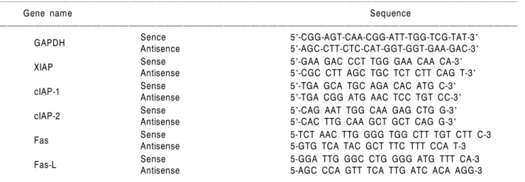
Table 1. Gene-specific primers for RT-PCR
ꠏꠏꠏꠏꠏꠏꠏꠏꠏꠏꠏꠏꠏꠏꠏꠏꠏꠏꠏꠏꠏꠏꠏꠏꠏꠏꠏꠏꠏꠏꠏꠏꠏꠏꠏꠏꠏꠏꠏꠏꠏꠏꠏꠏꠏꠏꠏꠏꠏꠏꠏꠏꠏꠏꠏꠏꠏꠏꠏꠏꠏꠏꠏꠏꠏꠏꠏꠏꠏꠏꠏꠏꠏꠏꠏꠏꠏꠏꠏꠏꠏꠏꠏꠏꠏꠏꠏꠏꠏꠏꠏꠏꠏꠏꠏꠏꠏꠏꠏꠏꠏꠏꠏꠏꠏꠏꠏꠏꠏꠏꠏꠏꠏꠏꠏ
Gene name Sequence
ꠏꠏꠏꠏꠏꠏꠏꠏꠏꠏꠏꠏꠏꠏꠏꠏꠏꠏꠏꠏꠏꠏꠏꠏꠏꠏꠏꠏꠏꠏꠏꠏꠏꠏꠏꠏꠏꠏꠏꠏꠏꠏꠏꠏꠏꠏꠏꠏꠏꠏꠏꠏꠏꠏꠏꠏꠏꠏꠏꠏꠏꠏꠏꠏꠏꠏꠏꠏꠏꠏꠏꠏꠏꠏꠏꠏꠏꠏꠏꠏꠏꠏꠏꠏꠏꠏꠏꠏꠏꠏꠏꠏꠏꠏꠏꠏꠏꠏꠏꠏꠏꠏꠏꠏꠏꠏꠏꠏꠏꠏꠏꠏꠏꠏꠏ
Sence 5'-CGG-AGT-CAA-CGG-ATT-TGG-TCG-TAT-3'
GAPDH
Antisence 5'-AGC-CTT-CTC-CAT-GGT-GGT-GAA-GAC-3'
Sense 5'-GAA GAC CCT TGG GAA CAA CA-3'
XIAP Antisense 5'-CGC CTT AGC TGC TCT CTT CAG T-3'
Sense 5'-TGA GCA TGC AGA CAC ATG C-3'
cIAP-1 Antisense 5'-TGA CGG ATG AAC TCC TGT CC-3'
Sense 5'-CAG AAT TGG CAA GAG CTG G-3'
cIAP-2 Antisense 5'-CAC TTG CAA GCT GCT CAG G-3'
Sense 5-TCT AAC TTG GGG TGG CTT TGT CTT C-3
Fas Antisense 5-GTG TCA TAC GCT TTC TTT CCA T-3
Sense 5-GGA TTG GGC CTG GGG ATG TTT CA-3
Fas-L Antisense 5-AGC CCA GTT TCA TTG ATC ACA AGG-3
ꠏꠏꠏꠏꠏꠏꠏꠏꠏꠏꠏꠏꠏꠏꠏꠏꠏꠏꠏꠏꠏꠏꠏꠏꠏꠏꠏꠏꠏꠏꠏꠏꠏꠏꠏꠏꠏꠏꠏꠏꠏꠏꠏꠏꠏꠏꠏꠏꠏꠏꠏꠏꠏꠏꠏꠏꠏꠏꠏꠏꠏꠏꠏꠏꠏꠏꠏꠏꠏꠏꠏꠏꠏꠏꠏꠏꠏꠏꠏꠏꠏꠏꠏꠏꠏꠏꠏꠏꠏꠏꠏꠏꠏꠏꠏꠏꠏꠏꠏꠏꠏꠏꠏꠏꠏꠏꠏꠏꠏꠏꠏꠏꠏꠏꠏ
리하였다. 분리된 RNA를 정량한 후, ONE-STEP RT-PCR PreMix Kit (Intron, Biotechnology, Korea)를 이용하여 2μg 의 RNA에서 cDNA를 합성하였다. 이 cDNA를 template로 사용하여 관찰 대상 유전자(Table 1)를 polymerase chain reaction (PCR) 방법으로 증폭하였다. 이때 housekeeping 유 전자인 glyceraldehyde-3-phosphate dehydrogenase (GAPDH) 유전자를 internal control로 사용하였다. 각 PCR 산물들을 1% agarose gel을 이용하여 전기영동하고 ethidium bro- mide (EtBr, Sigma)로 염색한 후 ultra violet (UV) 하에서 확인하였다.
6. Western blot analysis에 의한 단백질 발현의 분석
정상 및 세신 추출물이 처리된 배지에서 자란 암세포 들을 lysis buffer로 용해한 후, 고속원심분리로 세포 내 잔 사물을 분리시킨 후 동량의 단백질을 SDS-polyacrylamide gel 전기영동으로 분리하였다. 분리된 단백질을 함유한 acrylamide gel을 nitrocellulose membrane (Schleicher and Schuell, Keene, NH, USA)으로 electroblotting에 의해 전이 시킨 후, 10% skim milk를 함유한 PBS-T (0.1% Tween 20 in PBS)에 4oC에서 1시간 이상 배양하면서 비특이적인 단 백질들에 대한 blocking을 실시하였다. 그리고 특정 단백 질에 대한 항체를 membrane에 적용시켜 항원 항체 반응 을 일으킨 후, PBS-T로 씻어내고 특정 항체에 대한 이차 항체 반응을 실시한 후 ehanced chemiluminoesence (ECL) 용액 (Amersham Life Science Corp., Arlington Heights, IL, USA)을 적용시킨 다음 X-ray film에 감광시켜 특정 단백 질의 양을 분석하였다. 본 실험에 사용된 항체들은 Santa Cruz Biotechnology Inc. (Santa Cruz, CA, USA) 및 Calbiochem (Cambridge, MA, USA)에서 구입하였으며, 이차 항체로 사 용된 horseradish peroxidase-labeled donkey anti-rabbit immu- noglobulin 및 peroxidase-labeled sheep anti-mouse immu- noglobulin은 Amersham Corp.에서 구입하였다.
7. Caspase-3, -8 및 -9의 활성 측정
Caspase의 in vitro 활성 측정을 위한 colorimetric assay kits 는 R&D Systems (Minneapolis, MN, USA)에서 구입하였다.
활성 측정을 위하여 정상 및 세신 추출물이 처리된 배지 에서 72 시간 배양된 세포를 모은 뒤 단백질을 추출하고 정량하여 각각 150μg의 단백질을 fluorogenic peptide 기 질 100μM이 함유된 extraction buffer 50μl에 혼합하였으 며, microtiter plate에 다시 extraction buffer에 희석하여 각 sample 당 총 volume이 100μl가 되게 하였다. 실험에 사용 된 기질은 caspase-3의 경우에는 Asp-Glu-Val-Asp (DEVD)- p-nitroaniline (pNA)이었고 caspase-8의 경우에는 Ile-Glu-
Thr-Asp (IETD)-pNA이었으며, caspase-9은 Leu-Glu-His-Asp (LEHD)-pNA였다. 준비된 plate를 37oC에서 2시간 동안 incubation 시킨 후 ELISA reader를 이용하여 405 nm의 흡 광도를 이용하여 반응의 정도를 측정하였다.
결과 및 고찰
1. WEAS가 암세포의 생존율에 미치는 영향
3가지 종류의 인체 암세포(A549 폐암세포, NCI-H460 폐암세포, HT29 대장암세포)의 세포 생존율에 WEAS가 각 처리 농도별로 어떤 영향을 미치는지에 대하여 조사 하기 위하여 trypan blue 염색에 의한 hemocytometer count- ing을 이용하여 조사하였다. Fig. 1A의 결과에서 알 수 있 듯이 WEAS를 72시간 동안 처리한 결과 HT29 인체 대장 암세포의 경우 거의 변화가 보이지 않았고 NCI-H460 인 체 폐암세포에서는 약간의 생존율 감소가 나타난 반면 A549 인체 폐암세포의 경우는 처리농도 의존적으로 세 포의 생존율이 많이 감소하였음을 알 수 있었다. 특히 WEAS 처리 농도가 4 mg/ml 이상부터는 생존율이 50%
이상 억제되었으며, 최종 농도인 5 mg/ml에서는 세포의 수가 정상적인 배지에서 배양된 세포보다 약 30% 정도 로 줄어들었다. 따라서 WEAS를 처리하였을 경우 A549 인체 폐암세포의 성장과 생존율에 처리 농도 의존적인 억제 효과가 있음을 알 수 있었다. 본 연구에서 사용된 최고농도인 5 mg/ml까지는 정상세포에서 세포독성을 보 이지 않는 범위였으며, 이러한 결과는 선행연구에서 조 사된 MTT assay의 방법에 의한 결과에서도 유사한 경향 성을 보인 바 있다.13)
2. Apoptotic body의 형성에 미치는 WEAS의 영향
WEAS의 처리에 의한 생존율의 감소가 apoptosis의 유 발과 연관성이 있을 것으로 기대되어 WEAS 처리 후 암 세포 핵의 형태변화를 조사하였다. 이를 위하여 실험방 법에서 서술한 바와 같이 정상 및 WEAS이 처리된 배지 에서 자란 3종류의 암세포를 대상으로 핵산에 특이적으 로 결합하는 형광물질인 DAPI 염색을 통하여 핵의 형태 변형 여부를 조사하였다. Fig. 1B에서 나타난 바와 같이 정상 배지에서 배양된 세포의 경우 핵의 전체가 완전한 형태로 염색되는 양상을 보였으나 WEAS의 농도가 증가 할수록 염색체 응축에 의한 apoptosis가 일어난 세포에서 전형적으로 관찰되는 apoptotic body의 형성 정도가 증가 되었다.2,3) 그중에서 특히 A549 인체 폐암세포에서 그 양 상이 가장 뚜렷하였다. 이는 WEAS의 처리에 따른 암세 포의 생존율의 감소 및 형태 변화가 apoptosis 유발과 밀
접한 연관성이 있음을 보여주는 결과로 생각되어진다.
이상의 생존율 감소 및 핵 내 apoptotic body 출현 등의 실험에서 조사된 3가지 암세포 중 특히 A549 인체 폐암 세포에서 가장 현저한 변화를 나타내었으므로 이하의 실험 과정은 A549 인체 폐암세포만을 대상으로 하여 apoptosis 유발관련 기전 연구를 실시하였다.
3. WEAS 처리에 의한 암세포의 apoptosis의 유발
다음은 A549 세포를 대상으로 WEAS 처리에 의한 apoptosis 유발에 관한 추가적인 증거를 제시하기 위하여 apoptosis 유발 시 특이하게 분해가 일어나는 몇 가지 표 적 단백질의 발현에 미치는 WEAS의 영향을 Western blot 분석법으로 조사하였다. 세포의 내부나 외부의 자극에 의해 apoptosis가 일어나면 poly (ADP-ribose) polymerase (PARP) 단백질이 부분적으로 잘리는 분해과정이 나타난
다.14,15) PARP 단백질은 정상적인 세포의 DNA repair나
genomic stability의 유지에 중요한 역할을 하며,16) apoptosis 과정 중 caspase-3의 활성에 의해 단백질 분해가 일어나
게 되면 PARP의 효소적 기능의 상실로 정상적인 repair 기능의 상실이 일어난다는 사실이 밝혀져 있다.6) 정상적 인 세포의 경우 PARP 단백질은 116 kDa의 분자량을 가 지지만 apoptosis가 일어난 경우 85 kDa 크기의 단편이 관 찰되거나 주 band의 발현이 감소된다.
또한 β-catenin 단백질은 세포 내 골격 유지와 부착성 세포의 전사조절 및 세포유착에 관계된 apoptosis 조절과 관련이 있다.17,18) 그리고 β-catenin 단백질 역시 세포 유 착성 apoptosis가 유발되면 caspase-3의 활성과 연관되어 단편화가 일어나는 것으로 알려져 있다.19,20) 정상 세포의 경우 β-catenin은 92 kDa의 분자량을 가지나 세포 유착성 apoptosis (adherent cell apopotosis)가 일어나면 62∼72 kDa 로 단편화가 일어난다. 따라서 WEAS 처리에 의한 apoptosis 유발에도 이러한 현상들이 관련되어 있지는의 여 부를 조사한 결과, Fig. 2A에서 알 수 있듯이 PARP와 β- catenin의 발현양 감소 및 단편화 현상이 WEAS 처리 농 도 의존적으로 나타났다.
따라서 WEAS 처리에 따른 apoptosis 유발의 정도를 정 Fig. 1. Effect of water extract of A.
sieboldii (WEAS) on the cell viability and nuclear morphology of human lung carcinoma (A549 and NCI- H460) and colon cancer (HT29) cells.
(A) The cells cultured in the absence or in the presence of increasing concentrations of WEAS for 72 h.
Viable cell number was determined by hemocytometer counts of trypan blue-excluding cells. Results are ex- pressed as the means±S.E. of three independent experiments. (B) After 72 h incubation with WEAS, cells were fixed and stained with DAPI for 10 min incubation at room tempera- ture. The cells were washed with PBS and nuclear morphology was photographed with a fluorescence microscope using blue filter (Magnifi- cation, ×400).
0 1 2 3 4 5
120
Relative cell viability
WEAS (mg/ml) 0
HT29 A549 NCI-H460 100
60 80
40 20
A
A549
B
NCI-H460
HT29
0 1 3 5
WEAS (mg/ml)
량적으로 비교 평가하기 위하여 동일한 조건으로 배양 된 세포들을 대상으로 PI 염색액을 이용하여 핵을 염색 한 후 DNA flow cytometry 분석을 이용하여 세포주기의 sub-G1기에 해당되는 세포들의 빈도를 조사하였다. Fig.
2B에 나타낸 결과에서도 알 수 있듯이 WEAS의 처리 농 도의 증가에 따라 sub-G1기에 해당하는 세포의 빈도가 증가하여 농도 5 mg/ml에서는 생존율이 50% 이상 감소 하였음을 알 수 있다. 이상의 결과에서 WEAS 처리에 의 한 A549 인체 폐암세포의 생존율 감소가 apoptosis 유발 과 밀접한 관련이 있음을 알 수 있었다.
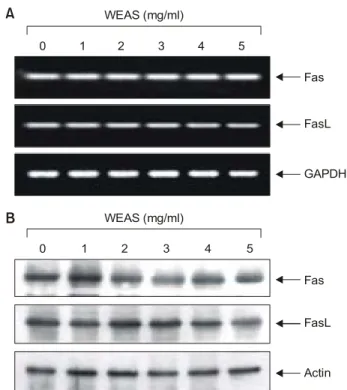
4. Fas 및 FasL의 발현에 미치는 WEAS의 영향
Apoptosis를 유발하는 경로는 크게 mitochondrial path- way 와 death receptor pathway의 두 가지로 구분할 수 있는 데, death receptor는 세포사멸의 기능을 갖는 tumor necro-
sis factor (TNF) 수용체군을 의미한다.21) 현재 가장 잘 알 려진 다섯 종류 중 하나가 APO-1 또는 CD95 라고도 알 려진 Fas에 의한 신호전달계이다. 그리고 TNF 수용체군 에 작용하는 대표적인 리간드(ligand) 중의 하나가 FasL이 다.22) FasL는 type II transmembrane protein으로서 tumor necrosis factor receptor super-family인 Fas와 결합하여 활성 화되면 death domain을 매개로 Fas는 caspase와 같은 apop- tosis 유발에 중요한 단백질들과 반응하여 활성화를 유도 하게 된다.21) 따라서 WEAS의 처리에 따른 apoptosis 유발 에 관여하는 분자생물학적 기전해석을 위한 시도로서 이러한 death receptor가 관여하는지의 여부를 알아보기 위하여 death receptor를 통한 apoptosis 유발의 대표적인 Fas 및 FasL 유전자의 발현을 조사하였다. Fig. 3A 및 B에 나타낸 바와 같이 WEAS가 Fas 및 FasL의 전사수준 및 번 역수준 모두에서 큰 변화가 관찰되지 않았다. 이는 A549 Fig. 2. Induction of cell death by WEAS treatment in A549
human lung carcinoma cells. (A) Cells were incubated with WEAS for 72 h, lysed and cellular proteins were separated by 8% SDS-polyacrylamide gels and transferred onto nitrocellulose membranes. The membranes were probed with anti-PARP and anti-β-catenin antibodies. Proteins were visualized using ECL detection system. Actin was used as a loading control. (B) After 72 h incubation with WEAS, the cells were fixed and then stained with PI. Apoptotic sub-G1 cells were determined by a DNA flow cytometry. Data are expressed as the means±S.E.
of three independent experiments.
0 1 2 3 4 5
60
% of sub-G1 cells
WEAS (mg/ml) 0
50
30 40
20 10 Actin β-catenin
A PARP
B
Fig. 3. Effects of WEAS in the levels of Fas and FasL expression in A549 cells. (A) After 72 h incubation with WEAS, total RANs were isolated and reverse-transcribed. The resulting cDNAs were sujected to PCR with Fas and FasL primers, and reaction products were subjected to electrophoresis in a 1%
agarose gel and visualized by EtBr staining. GAPDH was used as an internal control. (B) Cells were lysed, and then cellular proteins were separated by 10% SDS-polyacrylamide gels and transferred onto nitrocellulose membranes. The membranes were probed with anti-Fas and anti-FasL antibodies. Proteins were visualized using ECL detection system. Actin was used as a loading control.
0 1 2 3 4
WEAS (mg/ml)
5
Fas
FasL
GAPDH
0 1 2 3 4
WEAS (mg/ml)
5
Fas FasL
Actin
A
B
인체폐암세포에 WEAS을 처리하였을 경우 death receptor pathway 중 Fas 및 FasL를 통한 apoptosis는 일어나지 않았 음을 보여주는 결과라고 사료된다.
5. Bcl-2 및 Bax의 발현에 미치는 WEAS의 영향
WEAS의 처리로 인한 apoptosis 유발 기전을 해석하기 위해 다음으로 조사된 apoptosis 관련 유전자는 Bcl-2 family이다. Bcl-2 및 Bcl-xL은 apoptosis 유발을 억제하는 anti-apoptotic 인자이며, Bax 및 Bad는 apoptosis 유발을 촉 진하는 pro-apoptotic 인자이고, Bid는 death receptor의 활 성화와 연관된 apoptosis 촉진 인자이다.23,24) 이 유전자들 은 상대적인 발현의 차이 및 세포내 존재하는 위치에 따 라서 mitochondria로부터 cytochrome c를 유리시켜 종양 억 제 유전자인 p53, caspases 및 DNA 단편화와 연관된 endonuclease 등의 활성을 조절한다.23,25,26) 이들 두 유전자 는 dimer를 이루고 있는데 Bax/Bad의 발현이 증가하고 Bcl-2/Bcl-xL의 발현이 상대적으로 감소하면 apoptosis가 유발될 수 있는 것으로 알려져 있다.23,27) Fig. 4의 Western blot 분석 결과, Bax의 단백질 발현의 정도는 WEAS의 처 리농도 의존적으로 그 양이 증가하였으나, Bad 및 Bid의 경우는 WEAS 처리에 따라 큰 변화가 없었다. 그러나 apoptosis 억제에 관여하는 대표적인 유전자인 Bcl-2의 경 우 WEAS 처리 농도가 증가될수록 단백질 수준에서의 발현이 매우 감소되어 5 mg/ml 처리군에서는 거의 발현 이 되지 않았다. 이는 결국 WEAS 처리에 의한 apoptosis 이 유발에는 최소한 Bcl-2가 중요한 역할을 하고 있음을 의미하는 것이며, Bax의 발현 증가 및 Bcl-2의 발현 감소 로 인한 apoptosis 유발 관련 효소들의 활성화가 이루어지 고 있음을 시사하여 주는 것이다.
6. Caspases의 활성에 미치는 WEAS의 영향
한편 caspase protease라는 효소 역시 apoptosis 유발에 중 요한 조절인자로서 작용하는데, 이들 family에 속하는 단 백질들은 세포에서 핵과 mitochondria의 외막에 불활성 상태로 존재하며,25) Bcl-2/Bax family 발현의 변화에 따라 이들의 활성도가 조절될 수 있다.6) 이들은 proenzyme 형 태로 존재하다가 apoptosis 유도를 활성화시키는 신호에 의해 활성화된 protease로 전환되어 직접 또는 간접적으 로 세포 내에 존재하는 많은 표적 단백질의 분해에 관여 한다. 따라서 caspases의 활성화는 apoptosis의 유발에 대한 또 다른 증거가 될 수 있으며 많은 선행연구 등에서 검 증되어 왔다. 그러나 많은 경우에 apoptosis는 이러한 유 전자들의 발현이나 세포주기 비의존적으로도 일어날 수 도 있다. 지금까지 알려진 caspases 중 대부분의 apoptosis
가 유발된 세포에서 caspase-3, caspase-8 및 caspase-9이 높 은 활성도를 나타내므로 이들 caspases의 발현에 미치는 WEAS의 영향을 Western blot 분석을 통하여 먼저 조사하 였다. Fig. 5A의 결과에서 볼 수 있듯이 불활성인 pro- caspase-3과 pro-caspase-9의 발현이 WEAS의 처리 농도 의 존적으로 감소되었고, caspase-3의 경우 활성형이 검출되 었다. 그리고 WEAS 처리에 의한 A549 세포에서 caspases 의 활성화 여부를 정량적으로 조사하기 위하여 colo- rimetric assay kits를 이용하여 조사한 결과, Fig. 5B에 나타 난 바와 같이 WEAS가 처리된 A549 세포에서 조사된 3 가지 casapse 모두가 처리 농도 의존적으로 활성이 모두 증가되었음을 알 수 있었는데, 특히 caspase-3의 활성이 가장 높게 관찰되었다. 따라서 WEAS 처리에 의한 A549 인체 폐암세포의 apoptosis 유발은 Bcl-2/Bax family의 발현 변화와 연관된 caspases 효소 단백질의 활성에 따른 PARP 및 β-catenin과 같은 단백질의 분해(Fig. 2A)와 연관성이 있음을 알 수 있었다.
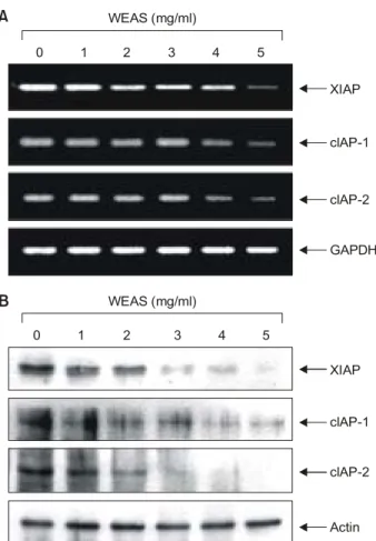
7. IAP family의 발현에 미치는 WEAS의 영향
Apoptosis에 관여하는 여러 인자들 중 최근 밝혀진 또 다른 class인 IAP family에 속하는 여러 유전자 산물은 곤 충세포에서 바이러스 감염에 의한 apoptosis를 억제하기 Fig. 4. Effects of WEAS treatment on the levels of Bcl-2 family proteins in A549 human lung carcinoma cells. Cells were incubated with WEAS for 72 h, lysed and cellular proteins were separated by 12% SDS-polyacrylamide gels and transferred onto nitrocellulose membranes. The membranes were probed with indicated antibodies. Proteins were visualized using ECL detection system. Actin was used as a loading control.
0 1 2 3 4
WEAS (mg/ml)
5
Bax
Bad
Bcl-2
Bcl-XL
Bid
Actin
위한 baculoviral 단백질 군들로서 외부 신호에 의한 세포 내 anti-apoptotic 활성을 지닌다. 그리고 이들 중 어떤 IAPs는 caspases와의 직접적인 결합을 통하여 그들의 apoptotic 활성을 억제할 수 있을 것으로 밝혀져,28∼30) apoptosis 조절에 IAP family들이 결정적인 역할을 하고 있 음이 밝혀지고 있다. 이상의 결과에서 WEAS 처리에 의 한 apoptosis 유도는 caspases의 활성화가 매우 중요한 역 할을 할 것으로 관찰되었기 때문에 WEAS 처리에 따른 이들의 활성화 IAP family에 속하는 주요 인자들 간의 연
관성을 조사하였다. Fig. 6A 및 B의 결과에서 알 수 있듯 이 조사된 IAP family인 XIAP, cIAP-1, cIAP-2는 모두 전사 수준 및 단백질 발현 양쪽 모두에 WEAS의 처리농도 의 존적으로 그 양이 매우 감소하였다. 따라서 WEAS 처리 에 의한 A549 인체 폐암세포의 apoptosis 유발에서 IAP family 인자들의 발현 감소가 caspases의 활성화에 중요한 역할을 하는 것으로 생각할 수 있다.
결 론
본 실험에서는 진해거담작용으로 널리 쓰이고 있는 족도리풀(Asarum sieboldii Miq)의 뿌리(세신)의 열수 추출물 Fig. 5. Effects of WEAS treatment on the expression and
activity of caspases in A549 human lung carcinoma cells. (A) Cells were incubated with WEAS for 72 h, lysed and cellular proteins were separated by 12% SDS-polyacrylamide gels and transferred onto nitrocellulose membranes. The membranes were probed with indicated antibodies. Proteins were visualized using ECL detection system. Actin was used as a loading control. (B) After 72 h incubation with WEAS, aliquots (150 μg protein) were incubated with substrates, DEVD-pNA, IETD-pNA and LEHD-pNA, for in vitro caspase-3, -8 and -9 activity, respectively, at 37oC for 3 h. The released fluorescent products were measured. Results are expressed as the means±S.E. of three independent experiments.
0 1 2 3 4 5
Relative activity
WEAS (mg/ml) 0
Caspase-9 Caspase-3 Caspase-8 5
3 4
2 1
A
Pro-caspase-3 Active-caspase-3 Pro-caspase-8
Pro-caspase-9 Actin
0 1 2 3 4
WEAS (mg/ml)
5
B 6
Fig. 6. Effects of WEAS in the levels of IAP family expression in A549 cells. (A) After 72 h incubation with WEAS, total RNAs were isolated and reverse-transcribed. The resulting cDNAs were sujected to PCR with indicated primers, and reaction products were subjected to electrophoresis in a 1% agarose gel and visualized by EtBr staining. GAPDH was used as an internal control. (B) Cells were lysed, and then cellular proteins were separated by 10% SDS-polyacrylamide gels and transferred onto nitrocellulose membranes. The membranes were probed with indicated antibodies. Proteins were visualized using ECL detection system. Actin was used as a loading control.
0 1 2 3 4
WEAS (mg/ml)
5
XIAP
clAP-1
clAP-2
GAPDH
0 1 2 3 4
WEAS (mg/ml)
5
XIAP
clAP-1
clAP-2
Actin
A
B
(WEAS)을 이용하여 인체 암세포의 apoptosis 유발에 관한 내용을 분석하였다. 조사된 인체 암세포에서 WEAS의 처리 농도 의존적으로 세포의 생존율이 감소되었으며, apoptotic body의 형성이 증가되어 apoptosis의 유발과 밀 접한 관련이 있었음을 알 수 있었다. WEAS에 의한 apop- tosis 유발을 폐암세포 A549를 대상으로 PARP 및 β-cate- nin 단백질의 발현 변화로 재확인하였으며, DNA flow cytometry 분석을 통하여 정량화하였다. RT-PCR과 Wes- tern blot analysis를 통해 WEAS에 의한 인체 폐암세포의 apoptosis의 유발과 관련이 있는 몇 가지 주요 인자들의 발현 정도를 조사하여 본 결과에서 apoptosis 억제에 관여 하는 중요한 인자인 Bcl-2의 단백질 발현이 WEAS 처리 에 따라 매우 강하게 감소되었음을 확인하였다. 그러나 Fas/FasL system에 속하는 인자들의 발현은 WEAS 처리에 의하여 큰 변화가 없었으며, WEAS 처리는 caspases (-3, -8 및 -9)의 활성화를 유도하였으며, IAP family 인자들의 발현 저하를 유도하였음을 알 수 있었다. 이상의 결과로 부터 WEAS가 암세포에서 효과적인 화학요법제로 사용 될 수 있을 가능성을 제시할 수 있으나, 나아가 더 많은 연구를 통해 WEAS 처리에 의한 항암 효과에서의 주 성 분물질에 대한 분석이 필요할 것이다. 또한 그러한 성분 물질들이 어떤 특수한 기전들을 통하여 항암 효과를 나 타내는지에 대해서도 밝혀져야 할 것이다.
감사의 글
본 연구는 농림부 농림기술개발사업(현장적용기술개 발)의 연구비 지원에 의해 이루어졌음을 밝히며 이에 감 사 드립니다.
참 고 문 헌
1) Searle J, Kerr JF, Bishop CJ. Necrosis and apoptosis: distinct modes of cell death with fundamentally different significance.
Pathol Annu 17, 229-259, 1982.
2) Lieberthal W, Koh JS, Levine JS. Necrosis and apoptosis in acute renal failure. Semin Nephrol 18, 505-518, 1998.
3) Zimmermann KC, Bonzon C, Green DR. The machinery of programmed cell death. Pharmacol Ther 92, 57-70, 2001.
4) Chiarugi V, Magneli L, Cinelli M, Basi G. Apoptosis and the cell cycle. Cell Mol Biol Res 40, 603-612, 1994.
5) Evans VG, Mutiple pathways to apoptosis. Cell Biol Int 17, 461-476, 1993.
6) Nagata S. Apopotosis by death factor. Cell 88, 355-365, 1997.
7) Schultz DR, Harrington Jr WJ. Apoptosis: programmed cell death at a molecular leve. Semin Arthritis Rheum 32, 345-369,
2003.
8) Kim YS, Park HD, Kim EC. Taxonomic study of the genus Asarum in Korea. J Plant Taxonomic Soc Kor 8, 19-31, 1978.
9) Yook CS, Kim JG. A new species and two new forma of Asiasarum. Kor J Pharmacol 27, 342-346, 1996.
10) Zhang F, Wang LX, Luo Q, Xiao HB, Liang XM, Cai SQ.
Analysis of volatile constituents of root and rhizome of Asa- rum heterotropoides Fr. var. mandshuricum (Maxim.) Kitag. by gas chromatography-mass spectrometry. Se Pu 20, 467- 470, 2002.
11) Hashimoto K, Yanagisawa T, Okui Y, Ikeya Y, Maruno M, Fujita T. Studies on anti-allergic components in the roots of Asiasarum sieboldi. Planta Med 60, 124-127, 1994.
12) Villegas M, Vargas D, Msonthi JD, Marston A, Hostettmann K. Isolation of the antifungal components Falcarindiol and Sarisn from Heteromorpha trifoliata. Planta Medica 36-37, 1998.
13) Kim OS, Park C, Moon SK, Choi YH. Anti-proliferative effects of water extract of Asarum sieboldii in human cancer cells. Cancer Prevention Res 11, 240-247, 2006.
14) Kaufmann SH, Desnoyers S, Ottaviano Y, Davidson NE, Poirier GG. Specific proteolytic cleavage of poly (ADP-ribose) polymerase: an early marker of chemotherapy-induced apop- tosis. Cancer Res 53, 3976-3985, 1993.
15) Lazebnik YA, Kaufmann SH, Desnoyers S, Poirier GG, Earnshaw WC. Cleavage of poly (ADP-ribose) polymerase by a proteinase with properties like ICE. Nature 371, 346-347, 1994.
16) Tewari M, Quan LT, O'Rourke K, Desnoyers S, Zeng Z, Beidler DR, Poirier GG, Salvesen GS, Dixit VM. Yama/
CPP32, a mammalian homolog of CED-3, is a CrmA-inhi- bitable protease that cleaves the death substrate poly (ADP- ribose) polymerase. Cell 81, 801-809, 1995.
17) Rowlands TM, Symonds JM, Farookhi R, Blaschuk OW.
Cadherins: crucial regulators of structure and function in reproductive tissues. Rev Reprod 5, 53-61, 2000.
18) Wijnhoven BP, Dinjens WN, Pignatelli M. E-cadherin-catenin cell-cell adhesion complex and human cancer. Br J Surg 87, 992-1005, 2000.
19) Fukuda K. Apoptosis-associated cleavage of β-catenin in human colon cancer and rat hepatoma cells. Int J Biochem Cell Biol 31, 519-529, 1999.
20) Steinhusen U, Badock V, Bauer A, Behrens J, Wittman- Liebold B, Dorken B, Bommer K. Apoptosis-induced cleavage of β-catenin by caspase-3 results in proteolytic fragments with reduced transactivation potential. J Biol Chem 275, 16345- 16353, 2000.
21) Sheikh MS, Huang Y. Death receptors as targets of cancer therapeutics. Curr Cancer Drug Targets 4, 97-104, 2004.
22) Klas C, Debatin KM, Jonker RR, Krammer PH. Activation interferes with the APO-1 pathway in mature human T cells.
Int Immunol 5, 625-630, 1993.
23) Antonsson B, Martinou JC. The Bcl-2 protein family. Exp Cell Res 256, 50-57, 2000.
24) Jurgensmeier JM, Xie Z, Deveraux Q, Ellerby L, Bredesen D, Reed JC. Bax directly induces release of cytocrome c from isolated mitochondria. Proc Natl Acad Sci USA 95, 4997-5002, 1998.
25) Reed JC. Bcl-2 family proteins. Oncogene 17, 3225-3236, 1998.
26) Rosse T, Olivier R, Monney L, Rager M, Conus S, Fellay I, Jansen B, Borner C. Bcl-2 prolongs cell survival after Bax- induced release of cytochrome c. Nature 391, 496-499, 1998.
27) Lenaz G, Bovina C, Formiggini G, Castelli GP. Mitochondria,
oxidative stress, and antioxidant defences. Acta Biochim Pol 46, 1-21, 1999.
28) Holcik M, Gibson H, Korneluk RG. XIAP: apoptotic brake and promising therapeutic target. Apoptosis 6, 253-261, 2001.
29) Hussein MR, Haemel AK, Wood GS. Apoptosis and mela- noma: molecular mechanisms. J Pathol 199, 275-288, 2003.
30) Salvesen GS, Duckett CS. IAP proteins: blocking the road to death's door. Nat Rev Mol Cell Biol 3, 401-410, 2002.