pISSN 1738-3544 eISSN 2288-1662
Inactivation of Candida albicans Biofilm by Radachlorin-Mediated Photodynamic Therapy
Pil Seung Kwon
Department of Clinical Laboratory Science, Wonkwang Health Science University, lksan 54538, Korea
라다클로린으로 매개된 광역학치료에 의한 백색 캔디다 바이오필름의 비활성
권필승
원광보건대학교 임상병리과
The purpose of this study was to evaluate the in-vitro efficacy of PDT using red light emitting diode (LED) with Radachlorin for biofilm inhibition of clinical Candida albicans isolates. The suspensions containing C. albicans at 9×10⁸ CFU/mL were prepared on yeast nitrogen base containing 5%
glucose. The biofilm formation was grown for 3 h after seeding suspensions each 100 ul on a 96-well plate and then supernatant was discarded. Each well was treated with 0.39 g/mL from 50
g/mL concentrations of Radachlorin on adherent biofilm. After a 30-minute incubation, light was irradiated for 30, 60, or 90 minutes using the following light source of wavelength 630 nm LED, at energy densities of 14, 29, and 43 J/cm2. Afterwards, all supernatant was removed and dried.
Adherent cells were stained with safranin O and dried. The cell viability was measured using a microplate reader at 490 nm. Also, a fluorescent signal on C. albicans was observed by saturation of a photosensitizer. In conclusion, a significant inhibition of 72.5% was observed to C. albicans on biofilm at the Radachlorin dose of 50 g/mL with 630 nm LED. The Photosensitizer (Radachlorin) was adequate at 30 minuttes for C. albicans. Overall, the results showed that inhibition of biofilm formation was Radachlorine dose-dependent. The results suggest that PDT, using Radachlorin with 630 nm LED, is able to decrease biofilm formation of C. albicans.
Keywords: Biofilm, Candida albicans, Light emmiting diode, Photodynamic therapy, Radachlorin
Corresponding author: Pil Seung Kwon Department of Clinical Laboratory Science, Wonkwang Health Science University, lksan 54538, Korea
Tel: 82-63-840-1212 E-mail: [email protected]
This is an Open Access article distributed under the terms of the Creative Commons Attribution Non-Commercial License (http://creativecommons.org/licenses/by-nc/4.0) which permits unrestricted non-commercial use, distribution, and reproduction in any medium, provided the original work is properly cited.
Copyright © 2015 The Korean Society for Clinical Laboratory Science. All rights reserved.
Received: October 19, 2015 Revised 1st: October 22, 2015 Revised 2nd: October 26, 2015 Revised 3rd: November 3, 2015 Accepted: November 4, 2015
서 론
Candida albicans
의 기회감염 진균증은 면역기능이 악화된 환 자로부터 표재성과 전신성 감염을 일으킨다(Mayer 등, 2013). 캔 디다 종들은 병원입원환자의 혈류감염을 일으키고 유병률이 20∼60% 지속적으로 증가하고 있고 사망률도 높아지고 있다(Morgan 등, 2005). 대부분의 캔디다증 환자에서 발견되는
C. albicans
는 건강한 사람의 점막 상재균으로서, 심각한 질환에 걸렸거나 암, 장기 이식, AIDS 또는 수술 후의 환자처럼 면역력이 약화된 환자의 표재 성감염과 점막상피를 통한 전신성 혈류감염을 유발하는 기회감염 균이다(Odds 등, 2001).
C. albicans
는 효모형, 가균사(pseudohyphae), 균사의 세 가지 형태로 변환할 수 있는 능력이 있으며, 병원성 결정인자로는 숙주 상피세포 부착 능력과 부착소들, 조직의 단백질이나 인지질을 가수분해하는 세포외 분비 효소들, 균사 형성과 형태 변환 능력 등이 알 려져 있다. 주요독성인자로 세포외 물질의 표면에 고착되어 균막 (biofilm)형태로 존재한다. 생체에 생기는 균막은 캔디다증의 발전 과정에 결정적인 역할을 하며 생체뿐만 아리라, 카테터, 삽입보철 물 등의 의학적 처치기구에 형성되는 균막도 캔디다증 감염원으로 중요하다(Ramage 등, 2006).
C. albicans
의 균사는 균막을 형성하여 집락화를 이루는 결정적 단계로서 캔디다가 숙주세포의 상피를 통과하는 것을 돕는 독성요 인으로 작용하며, 결합이 불가능한 효모형이나 균사형의C.
albicans
는 독성이 약한 것으로 보고되어 있다(Thompson 등, 2011).C. albicans
와 관련된 대부분의 질환에서 균막이 관련되어 있는 것으로 보고되고 있고, 균막은Candida
의 은신처로 작용하여 숙주의 면역체계를 회피하고 다른 병원체로부터의 경쟁적 압박에 서도 생존하게 할 수 있게 한다(Blankenship와 Mitchell, 2006).특히
C. albicans
에 의해 형성된 균막은 일반적으로 다양한 항진균 제에 대한 내성을 나타내기 때문에 통상적으로 사용하는 항진균제 를 지속적으로 사용할 수 없게 하는 어려움을 가중시키고 있다 (Shapiro 등, 2011).현재 심부 진균증의 치료에 사용되는 항진균제는 작용 메카니즘 에 따라 세포막 기능의 변화(amphotericin B), Deoxyribonucleic acid (DNA)나 Ribonucleic acid (RNA)합성의 저해(flucytosine), ergosterol 생합성의 저해(azole [fluconazole]), glucan 합성을 저 해(echinocandin [caspofungin])하는 네 가지 부류로 나눌 수 있 다(Oliver 등, 2008). 그러나 항진균제의 반복적 사용으로 말미암 아 내성균이 출현하여 캔디다증에 대한 치료에 어려움을 겪고 있는 실정이며, 새로운 효과적인 항진균치료방법이 대두되고 있다.
광역학치료는 가시광선과 독성이 없는 광감작제가 결합하여 활 성산소(reactive oxygen species, ROS)가 생성되어 세포를 사멸 시키는 치료법이다(Vera 등, 2012). 이 치료법은 광감작제를 세균 에 침투시킨 후 빛을 쬐어 세포독성을 많이 내는 단일항 산소 (singlet oxygen)과 ROS 의 생산을 유도하여 세균의 손상을 일으 키게 하는 치료법으로 생성된 ROS는 세균의 DNA, 단백질, 세포막 등의 손상을 일으켜 세균을 사멸하게 만드는 치료법이다(Imlay, 2003; Hong 등, 2007; Kwon, 2011; Kwon, 2015).
C. albicans
의 광역학치료는 몇몇 연구자들에 의해 광감작제와 결합시키는 빛의 파장별로 효과가 다른 것으로 보고되었다 (Demidova 등, 2005; Lam 등, 2011). 광역학치료에서 항진균효 과를 보고한 광감작제에는 phenothiazinium salts, toluidine blue O (TBO), methylene blue (MB)를 사용하였다(Giroldo 등, 2009; Dai 등, 2011; Prates 등, 2011). 효모형으로부터 사상형으 로 이행되어 발아되는 균막도 치료가 가능하며, 캔디다의 균막형성은 집락화가 필수적이며 독력과 연관되어 있고
C. albicans
의 균막 형성은 항진균제의 내성을 일으킨다(Shapiro 등, 2011). 지금까지C. albicans
의 광역학치료에 관한 연구자들은 균주부유액을 사용 하여 효과를 입증하였거나, 다른 계열의 광감작제를 이용하였다.그래서 본 연구의 목적은 최근 개발된 광감작제인 클로린계열의 Rhadachlorin을 이용하여
C. albicans
의 균막형성을 제거 될 수 있는지를 광역학적 치료의 최적조건을 평가하고자 하였다.재료 및 방법
1. 사용균주
단국대학교병원 환자의 pus에서 나온
Candida albicans
를 API 20C Aux (bioMerieux, Marcy 'Etoile, France)와 Vitek Yeast Biochemical Card (YBC, Hazelwood, Mo, USA)로 균 동정하여 API 20C에서는 97.2%, Vitek II에서는 95% 정확도로 동정되었다.2. 균주 배양
C. albicans
균막은 Yeast extract peptone dextrose (YEPD) agar에 도말하여 37oC 배양기(VS-9108MS, Vision, Seoul, Korea) 에서 하룻밤 산소성 배양하였다. YEPD agar는 Yeast extract 21 g, peptone 7 g, glucose 14 g, agar 14 g, 증류수 700 mL을 혼합하여 직접 제조해서 사용하였다.3. 광감작제 및 광원
본 연구에 사용된 광감작제인 RadachlorinⓇ (RADA-PHARMA Co, Ltd., Moscow, Russia, 라다클로린)은 액체앰플을 Dulbecco's phosphate buffered saline (Hyclone, Logan, UT, USA)에 1,000
g/mL로 희석하여 사용하였다.
광원은 Light emmiting diode (LED)를 자체 제작하여 사용하였 고, 사용된 LED는 5의 원통모양으로 시험관에 조사하기 위해 690 nm LED array를 제작하였다. LED array는 가로 75 mm×세로 110 mm Printed circuit board에 총 192개의 LED가 장착되었다.
LED의 회로 연결은 직렬과 병렬을 병합하였고, 직류 전원을 사용 하였고, 수명 연장과 손상을 방지하기 위하여 안정적으로 저항을 150Ω으로 하였으며, LED array와 조사받는 96 well plate와의 거 리는 100 mm로 하고 에너지밀도는 파워미터 Power Max R (Coherent Inc., Portland, USA)로 측정하여 690 nm LED array는 평균 8 mW/cm2의 30, 60, 90분 동안 조사하여 최종 에너지밀도는 14, 29, 43 J/cm2이었다.
Fig. 1. Biofilm inhibition assay for C. albicans after Radachlorine dose-dependent PDT. Biofilms were grown in 96-well plate for 3 h at 37oC and then discard supernatant. Each wells were treated to 0.39 g/mL from 50 g/mL concentration of Radachlorinon on adherent cells. After 30 minutes incubation, light was irradiated for 60 minutes using the following light source of wavelength 630 nm LED, each energy density 14 J/cm2. After removes all super- natant and dries. Adherent cells were stained with safranin O and dried. The optical density at 490 nm was determinated using Microplate Reader. Values are expressed in mean±SD.
Fig. 2. Biofilm inhibition assay for C. albicans after irradiation dose-dependent PDT. Biofilms were grown in 96-well plate for 3h at 37oC and then discard supernatant. Each wells were treated to 50 g/mL of Radachlorinon on adherent cells. After 30 minutes incubation, light was irradiated for 30 to 90 minutes using the following light source of wavelength 690 nm, each energy density 14, 29 and 43 J/cm2. After removes all supernatant and dries.
adherent cells were stained with safranin O and dried. The optical density at 490 nm was determinated using Microplate Reader.
Values are expressed in mean±SD.
4. 균막 형성
Ramage 등(2001)의 방법을 변형하여 이용하였다. YEPD agar 에 도말한 후 37oC 배양기에 24시간 배양 한 후 생성된 집락을 배양 된 균주를 다른 균의 오염이 없는지 집락을 관찰 후 오염된 것이 없 을 시에 멸균된 면봉을 이용하여 집락을 취하여 5% Glucose가 첨 가된 Yeast Nitrogen Base (YNB; Difco, Detroit, MI, USA)를 12×75 mm Polyethylene cap tube (SPL Life Sciences, Pocheon, Korea)에 현탁시킨 다음 탁도계(Densicheck plus;
biomeriex, Durham, NC, USA)를 이용하여 McFarland No. 3.0 로 맞춘 후 균막형성시험에 사용하였다.
희석한 균주부유액을 96 well plate (Falcon; Franklin Lakes, NJ, USA)에 100 uL씩 분주하였다. 그 후 37oC 산소성배양기에서 3시간 을 배양한 후 상층액을 제거하였다. 균막이 형성된 96 well plate를 microplate reader (Emax; Molecular Devices, Sunnyvale, CA, USA)에 장착하여 540 nm에서 흡광도를 측정하여 흡광도 값이 0.5 이상을 균막형성으로 판정하였다.
5. 광역학치료
균막이 형성된 96 well plate 각 well에 Radachlorin 50 g/mL 부터 0.39 g/mL까지 농도의 광감작제를 50 uL 씩 분주하였다. 그 후 은박지로 96 well plate를 싸서 37oC 산소성배양기에서 3시간 을 배양하였다.
배양기에서 꺼낸 96 well plate의 은박지를 벗기고 자체 제작된 690 nm LED를 30분, 60분, 90분 빛을 쬐어 주었다. 조사가 끝난 96 well plate를 상층액을 모두 제거 후 Heilmann등(1996)과 같
은 바이오필름형성능의 정량측정법과 같이 Safranin O 염색액으 로 30초 동안 염색하였다. 그 후 PBS로 세척하고 microplate reader (Emax; Molecular Devices, Sunnyvale, CA, USA)에 장착 하여 490 nm파장으로 흡광도를 측정하여 균막형성의 억제를 3회 측정하고 표준편차를 구하였다.
6. 광감작제의 균막내 흡수시험
균막형성된 96 well plate well에 Radachlorin 50 g/mL을 50 uL씩 분주 후 30분후에 광감작제의 균막내 흡수를 확인하고자 Dulbecco's phosphate buffered saline (Hyclone, Logan, UT, USA)로 세척 후 형광현미경으로 검경하였다.
결 과
1. 라다클로린의 농도별 균막억제효과
형성시킨 균막에 광감작제인 라다클로린의 농도 50 g/mL부터 0.39 g/mL까지 희석된 농도로 처리 후 LED 빛을 조사하였을 때 균막제거능의 결과는 대조군은 흡광도 값 평균이 1.193이였으며, 라다클로린의 농도 50 g/mL에서는 0.328로 대조군을 100% 기 준으로 할 때 27.4%만이 형성을 보여 72.6%의 균막이 제거되어 가 장 많은 억제능을 보였다.
25, 12.5, 6.25, 3.12, 1.56, 0.78, 0.39 g/mL 까지 농도에서는 각 각 42, 76, 78, 85, 86, 91, 97% 의 균막형성을 보였다(Fig. 1).
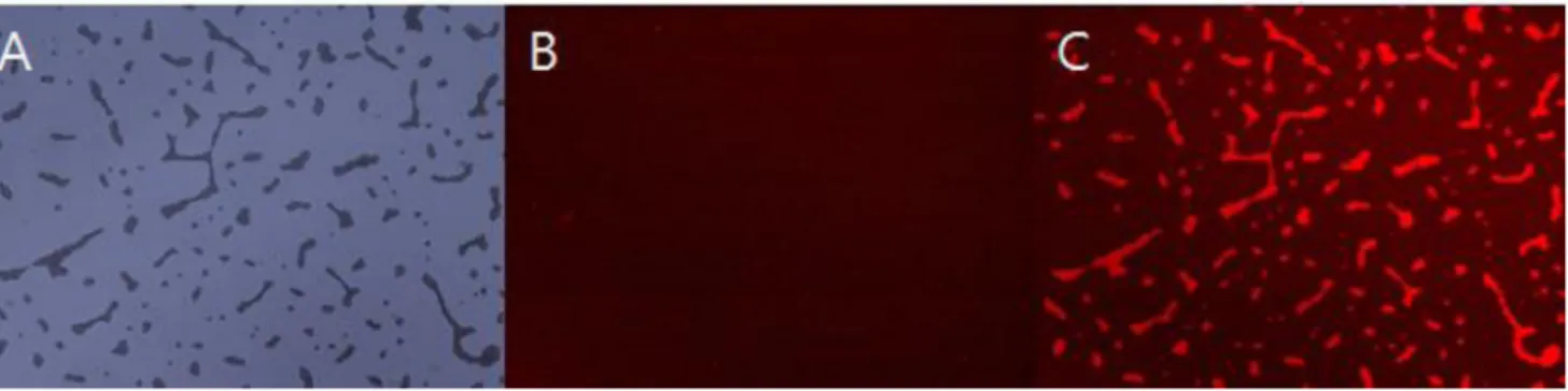
Fig. 3. The fluorescence images were shown to accumulation of Radachlorine in C. albicans. The suspensions containing Candida albicans at 9×10⁸ CFU/mL were prepared to yeast nitrogen base containing 5% glucose. and than treated to 50 g/mL Radachlorinon for 30min incubation. The images were observed to accumulation of Radachlorine in C. albicans using fluorescent microscope with exitation wavelength 510∼560 nm. (A) light image, (B) No treated radachlorin, (C) treated radachlorin. Original magnification (×200).
2. 광원 에너지밀도별 균막억제효과
형성시킨 균막에 광감작제인 라다클로린의 농도 50 g/mL 처 리 후 690nm LED 빛을 30분, 60분, 90분을 조사하여 최종 에너지 밀도 14, 29, 43 J/cm2에서 균막의 억제효과결과는 대조군의 흡광 도 값 평균이 0.68이였으며, 30분 조사(14 J/cm2)일 때, 흡광도 값 평균은 0.33으로 대조군과 비교하여 47% 형성율을 보였고, 60분 조사(29 J/cm2)일 때흡광도 값 평균은 0.28로 대조군과 비교하여 40% 형성율을 보였고, 90분 조사(43 J/cm2)일 때 흡광도 값 평균은 0.22 로 대조군과 비교하여 31% 형성율을 보였다. 에너지밀도에는 균막형성율이 90분간 조사하면 균막억제효과가 가장 뛰어났지만, 30분과 60분은 근소한 차이로 측정되었다(Fig. 2).
그리고 라다클로린 처리없이 LED 빛만을 조사시에는 흡광도 값 이 0.52가 나와 조금은 억제가 되는 것으로 나타났으며, 오히려 광 감작제만을 처리시에는 흡광도 값이 0.56으로 나와 빛 조사군보다 균막제거에 영향이 없는 것으로 결과가 나왔다.
3. 광감작제의 균막내 흡수확인
라다클로린의 균막내 흡수과정의 확인을 위해 형광현미경 관찰 결과는 Fig. 3A는 투과광이미지이며, Fig. 3B는 라다클로린을 처리 하지 않은 상태의 균막이며, Fig 3C는 라다클로린 50 g/mL를 처 리하고 30분 후의 형광이미지이다. 이 결과로 30분의 반응시간이 면 라다클로린의 균막침투를 확인이 증명되었다.
고 찰
진균 감염에 있어서 항진균제의 내성 획득 문제는 에이즈 환자 와 같은 면역저하자의 증가, 악성종양 환자, 스테로이드 치료의 증 가와 관련하여 진균 감염의 치료 실패 및 사망률의 증가와 관련한
매우 중요한 문제로 대두되고 있다.
C
.albicasn
의 바이오필름의 구조는 Hawser와 Dougglas (1994)는 전자 현미경 촬영으로 카테터 (catheter)에 형성된 바이 오필름의 형태를 처음으로 확인하였다.C. albicans
가 접촉면에 부 착하면, 이후 3∼6시간 후부터 발아관을 형성하기 시작하고, 24∼48시간의 배양 시간이 지나면 완전히 성숙하게 된다. 지속적으로
C. albicans
가 증식하고, 미세집락을 형성하며 다양한 외부 물질들 도 함께 침착된다. 이렇게 일단의 지지물이 형성된 후 효모형 세포 는 균사체로 전환되어 삼차원적 구조를 만들며 수많은 미세집락과 수분이 통할 수 있는 공간이 서로 얽혀 보통 수백 m 크기의 바이오 필름을 형성하게 된다. 바이오필름의 성장촉진은 접촉면의 친수 성, 고혈당 환경,C. albicans
의 이형태성(dimorphism)은 바이오 필름 형성에 주요한 요인으로 작용된다.우리의 연구는 클로린 계열 광감작제를 이용한 광역학치료가
C.
albicans
바이오필름형성을 억제하는지를 농도별과 광원의 세기로 비교하였고, 가장 좋은 조건은 라다클로린 50 g/mL의 농도로 광역학 치료시 바이오필름형성을 억제할 수 있음을 제시하였다.
Dovigo 등(2010)은 포토프린 광감작제와 455 nm 청색 LED를 이 용하여 캔디다 4개의 균종을 에너지밀도별로 비교하였는데, 이는 균주부유액을 광역학적 치료 후 집락형성수를 측정하였지만,
C.
albicans
종에 대해 18 J/cm2의 에너지밀도의 빛 조사에 50 g/mL 에서 우수한 효과를 발표하였다. 이는 Fig. 2번과 같이 우리의 연구 에 사용된 라다클로린의 결과와 같은 농도에서 효과가 보였음을 알 수 있다.광역학치료에서 세포사멸은 Reactive Oxygen Species (ROS) 의 생성이며, 이것은 광감작제가 특정의 파장의 빛을 흡수하면서 발생되는 것으로 세포의 DNA, 단백질, 세포막 등을 파괴하는 원리 이다. 본 연구에서는 광감작제가 세포내에 흡수가 되는 것은 확인 하였으나, 이와 더불어 빛 조사 시 발생되는 ROS의 생성유무의 관
찰이 앞으로 증명해야 할 것이며, 최근 2014년 Rosseti 등은
C.
albicans
바이오필름형성억제를 ROS 생성과 세포막 침투를 입증 한 바가 있으며 우리의 라다클로린에 매개된 광역학치료도 동일할 것으로 추정된다. 그리고 Rosseti 등은 광감작제로 0.1 mg/mL의 Toluidine blue를 사용하였는데, 치료 후 50% 생성율을 보여 본 연 구보다는 미진한 억제효과를 보여 광감작제인 라다클로린이 적은 농도에서도 더 좋은 효과를 보였다. 또한, ROS의 생성능은 빛을 조 사한 후 1시간, 2시간, 3시간을 비교하였는데, 시간이 길수록 생성 능이 많아진 것을 확인할 수 있었다. 그래서 본 연구에서도 광역학 치료 시 빛 조사 후 바로 염색하여 흡광도를 측정하였는데, 시간을 3시간 후 흡광도 측정을 하였으면, 더 좋은 억제효과가 있으리라 짐 작할 수 있다.Junqueira 등(2012)은 human immunodeficiency virus (HIV) 양성환자의 구강에서 분리된 9가지 효모균을 대상으로 ZnPc 광감 작제와 660 nm gallium-aluminum-arsenide (GaAlAs) laser를 이용하여 광역학 치료를 한 결과에서
C. albicans
균주와 비 Candida 종들을 비교하였으나 큰 차이점을 나타내지 않았다. 앞으 로 연구에는 많은 Candida 균종을 치료대상으로 고려하여야 하고, 이 결과를 미루어 볼 때C. albicans
균주와 비Candida 종들은 라다 클로린으로 매개된 광역학 치료에서도 억제 효과는 비슷할 것으로 사료된다.C. albicans
의 바이오필름은 접촉면의 표면에 강하게 부착되어 있어 제거가 어렵고, 생체 내에서는 지속적 접촉에 의한 만성 염증 의 원인이 된다. 바이오필름의 형성이 안정화되고, 성숙화 되는 시 기에는 바이오필름형성 초기와 비교하여 에르고스테롤 농도가 급 격히 감소되는 것으로 알려져 있고(Douglas, 2002) 이에 바이오필 름의 구조가 성숙되고 안정화되면서, 항진균제의 주요 표적인 에르 고스테롤의 합성 역시 저하되고, 이는 항진균제의 효과 감소로 이 어지게 되는 것이다. 이렇게 바이오필름을 형성한C. albicans
의 경우 항진균제 내성 발현과 관련하여 복잡하고, 다양한 기전이 함 께 작용하고 있기에, 칸디다 항진균제에 대한 내성감염증에 대한 적절한 치료를 위해서는C. albicans
의 바이오필름과 관련된 내성 기전에 대한 연구가 더 필요하고, 항진균제 내성에서 자유로울 수 있는 치료방법이 고려되어야 할 것이다. 우리의 연구결과와 같이 라다클로린에 매개되는 광역학치료법은C. albicans
의 바이오필 름의 예방과 치료방법으로 고려할 수 될 수 있음을 입증하였다.요 약
이 연구의 목적은 임상
Candida albicans
의 바이오필름억제을 위한 Rhadachlorin과 적색발광다이오드를 이용한 광역학치료의체외적 효과를 평가하고자 하였다.
Candida albicans
부유액을 9×10⁸ CFU/mL을 5% 당이 함유된 YNB배지에 준비하였다.Candida albicans
의 바이오필름은 96웰 플레이트에 100 uL씩 분 주후 3시간 배양하고 상층액을 제거하였다. 각 웰에 50 g/mL 부 터 0.39 g/mL 농도로 Rhadachlorin을 부착된 세포위에 처리하 였다. 30분간 배양 후 빛을 30, 60, 90분을 630 nm 광원의 LED를 이용하여 조사하였고, 이때 각각의 에너지밀도는 14, 29, 43 J/cm2 이였다. 모든 상층액을 제거후에 건조시켰다. 부착된 세포를 safranin O로 염색하였다. 490 nm 파장으로 마이크로플레이트 리 더로 흡광도를 측정하였다. 또한, 광감작제포화에 의해Candida albicans
의 형광시그널을 관찰하였다. 결론적으로 확연한 억제는 Rhadachlorin의 농도 50 g/mL과 630 nm LED치료에서Candida albicans
의 바이오필름의 72.5%가 감소되었다. 또한 광 감작제 Rhadachlorin은Candida albicans
의 30분에서 충분한 축 적을 보였다. 전체적으로 이 결과들은 바이오필름형성의 억제는 Rhadachlorin의 농도에 의존되었다. 이 결과들은 Rhadachlorin 과 630 nm LED의 광역학치료는Candida albicans
의 바이오필름 억제가 가능한 것으로 제의된다.Acknowledgements:
이 논문은 2015년도 원광보건대학교 교내 연구비 지원에 의해서 수행됨.Funding:
2015년도 원광보건대학교 교내연구비Conflict of interest:
NoneReferences
1. Blankenship JR, Mitchell AP. How to build a biofilm: a fungal perspective.
Curr. Opin. Microbiol
. 2006, 9:588-594.2. Dai T, Bil de Arce VJ, Tegos GP, Hamblin MR. Blue dye and red light, a dynamic combination for prophylaxis and treatment of cutaneous Candida albicans infections in mice.
Antimicrob Agents Chemother
. 2011, 55:5710-5717.3. Demidova TN, Hamblin MR. Effect of cell-photosensitizer binding and cell density on microbial photoinactivation.
Antimicrob Agents Chemother
. 2005, 49:2329-2335.4. Douglas LJ. Medical importance of biofilms in Candida infections.
Rev Iberoam Micol
. 2002, 19:139-1435. Dovigo LN, Pavarina AC, Ribeiro DG, Adriano CS, Bagnato VS.
Photodynamic inactivation of four Candida species induced by photogem.
Braz J Microbiol
. 2010, 41:42-49.6. Giroldo LM, Felipe MP, de Oliveira MA, Munin E, Alves LP, Costa MS. Photodynamic antimicrobial chemotherapy (PACT) with methylene blue increases membrane permeability in Candida albicans.
Lasers Med Sci
. 2009, 24:109-112.7. Hawser SP, Douglas LJ. Biofilm formation by Candida species on the surface of catheter materials in vitro.
Infect Immun
. 1994, 62:915-921.8. Heilmann C, Gerke C, Perdreau-Remington F, Götz F. Charac- terization of Tn917 insertion mutants of Staphylococcus epi- dermidis affected in biofilm formation.
Infect Immun
. 1996, 64:277-82.9. Hong SN, Kwon PS, Kim DS. Photodynamic inactivation of Moraxella catarrhalis.
Korean J Clin Lab Sci
. 2007, 39:19-24.10. Imlay JA. Pathways of oxidative damage.
Annu Rev Microbiol
. 2003, 57:395-418.11. Junqueira JC, Jorge AO, Barbosa JO, Rossoni RD, Vilela SF, Costa AC,
et al
. Photodynamic inactivation of biofilms formed by Candida spp., Trichosporon mucoides, and Kodamaea ohmeri by cationic nanoemulsion of zinc 2,9,16,23-tetrakis(phenyl- thio)-29H, 31H-phthalocyanine (ZnPc).Lasers Med Sci
. 2012, 27:1205-1212.12. Kwon PS. The effects of photodynamic therapy for vancomycin resistant enterococci.
Korean J Clin Lab Sci
. 2011, 43:124-132.13. Kwon PS. Antimicrobial effects of photodynamic therapy using blue light emitting diode with photofrin and radachlorine against Propionibacterium acnes.
Korean J Clin Lab Sci
. 2015, 47:6-10.14. Lam M, Jou PC, Lattif AA, Lee Y, Malbasa CL, Mukherjee PK,
et al
. Photodynamic therapy with Pc 4 induces apoptosis of Candida albicans.Photochem Photobiol
. 2011, 87:904-909.15. Mayer FL, Wilson D, Hube B. Candida albicans pathogenicity mechanisms.
Virulence
. 2013, 4:119-128.16. Morgan J, Meltzer MI, Plikaytis BD, Sofair AN, Huie-White S, Wilcox S,
et al
. Excess mortality, hospital stay, and cost due to candidemia: a case–control study using data from pop- ulation-based candidemia surveillance.Infect Control Hosp Epidemiol
. 2005, 26:540-547.17. Odds FC, Gow NA, Brown AJ. Fungal virulence studies come of age.
Genome Biol
. 2001, 2:reviews1009.1-reviews1009.4.18. Oliver BG, Silver PM, Marie C, Hoot SJ, Leyde SE, White TC.
Tetracycline alters drug susceptibility in Candida albicans and other pathogenic fungi.
Microbiology
. 2008, 154:960-970.19. Prates RA, Kato IT, Ribeiro MS, Tegos GP, Hamblin MR.
Influence of multidrug efflux systems on methylene blue-medi- ated photodynamic inactivation of Candida albicans.
J Antimi- crob Chemother
. 2011, 66:1525-1532.20. Ramage G, Martínez JP, Lípez-Ribot JL. Candida biofilms on implanted biomaterials: a clinically significant problem.
FEMS Yeast Res
. 2006, 6:979-986.21. Ramage G, Vandewalle K, Wickes BL, Lopez-Ribot JL.
Characteristics of biofilm formation by Candida albicans.
Rev Iberoam Micol
. 2001;18:163-70.22. Rosseti IS, Chagas LR, Costa MS. Photodynamic antimicrobial chemotherapy (PACT) inhibits biofilm formation by Candida al- bicans, increasing both ROS production and membrane permeability.
Laser Med Sci
. 2014, 29:1059-106423. Shapiro RS, Robbins N, Cowen LE. Regulatory circuitry govern- ing fungal development, drug resistance, and disease.
Microbiol Mol Biol Rev
. 2011, 75:213-267.24. Thompson DS, Carlisle PL, Kadosh D. 2011. Coevolution of morphology and virulence in Candida species.
Eukaryot
.Cell
. 2011, 10:1173-1182.25. Vera DM, Haynes MH, Ball AR, Dai T, Astrakas C, Kelso MJ,