INTRODUCTION
Transitional cell carcinoma of the upper urinary tract (TCC- UUT) is a relatively uncommon disease, accounting for 4.5- 9% of all renal tumors and 5-6% of all urothelial tumors (1).
The incidence of TCC-UUT appears to have increased recent- ly, probably due to increased environmental exposure and aging population (2). The upper urinary tract has anatomi- cal characteristics such as a thin muscle layer, proximity to the kidney and rich lymphatic drainage. Tumor invasion may significantly influence distant metastasis and progression in patients with TCC-UUT (3). However, the exact molecular mechanisms of tumor invasion, recurrence, progression, and prognosis of this disease remain largely unclear.
Caveolins are the major structural proteins of caveolae, the vesicular invaginations of the plasma membrane. The caveolin family includes caveolin-1, -2, and -3. Caveolin-1 and -2 are co-expressed in many cell types including adipocytes, endothe- lial cells, smooth muscle cells, and fibroblasts, whereas the expression of caveolin-3 is muscle-specific (4-6). Caveolin-1 has been implicated in intracellular transport, membrane trafficking, and signal transduction (5), and has also been shown to play an important role in various human patho- logical conditions, including cancer, diabetes, bladder dys- function, muscular dystrophy, and those related to the car- diovascular system such as atherosclerosis, cardiac hypertro-
phy, cardiomyopathy, pulmonary hypertension, and neointi- mal hyperplasia (6, 7).
The role of caveolin-1 in cancers remains controversial, because caveolin-1 has been reported to be dysregulated in various cancers, but the pattern of dysregulation appears to vary with cancer types. The caveolin-1 expression is decreased in several cancers such as ovarian carcinoma, pulmonary ade- nocarcinoma, various soft tissue sarcomas, and breast cancer and thought to function as a tumor suppressor gene (8-11).
On the other hand, the caveolin-1 expression is upregulated in the other cancers such as renal cell carcinoma, prostate can- cer, bladder cancer, colonic adenocarcinoma, and esophageal squamous cell carcinoma and associated with higher stage, cancer progression, and poor prognosis (12-17).
There have been no previous studies about caveolin-1 expres- sion in TCC-UUT. In the present study, therefore, we inves- tigated the relationship between caveolin-1 expression and prognosis in patients with TCC-UUT.
MATERIALS AND METHODS Patients and specimens
Formalin-fixed, paraffin-embedded, archival surgical speci- mens that had been obtained from 98 patients (76 men and
296
Dae Sung Cho, Hyunee Yim*, Kang Su Cho�, Sung Joon Hong�, Nam Hoon Cho�, Sun Il Kim, Hyun Soo Ahn, and Se Joong Kim
Departments of Urology and Pathology*, Ajou University School of Medicine, Suwon; Departments of Urology�and Pathology�, Yonsei University College of Medicine, Seoul, Korea
Address for correspondence Se Joong Kim, M.D.
Department of Urology, Ajou University School of Medicine, San-5 Wonchon-dong, Yeongtong-gu, Suwon 443-721, Korea
Tel : +82.31-219-5272, Fax : +82.31-219-5276 E-mail : [email protected]
DOI: 10.3346/jkms.2008.23.2.296
Impact of Caveolin-1 Expression on the Prognosis of Transitional Cell Carcinoma of the Upper Urinary Tract
This study aimed to investigate the relationship of caveolin-1 expression with prog- nosis in patients with transitional cell carcinoma of the upper urinary tract (TCC- UUT). Formalin-fixed, paraffin-embedded tissue sections of TCC-UUT from 98 patients, who had undergone radical nephroureterectomy, were stained immuno- histochemically using antibodies against caveolin-1. The expression pattern of cave- olin-1 was compared with the clinicopathological variables. The caveolin-1 expres- sion was significantly correlated with T stage (p<0.001) and grade (p=0.036). The survival rate of patients with caveolin-1 positive tumors was significantly lower than that of patients with caveolin-1 negative tumors (p<0.0001). The univariate analy- ses identified T stage, grade, and caveolin-1 expression as significant prognostic factors for cancer-specific survival, whereas the multivariate analyses indicated that T stage and caveolin-1 expression were independent prognostic factors. These results show that the increased expression of caveolin-1 is associated with tumor progression and poor prognosis in TCC-UUT, suggesting that caveolin-1 may play an important role in the progression of TCC-UUT.
Key Words : Caveolin 1; Prognosis; Carcinoma, Transitional Cell; Upper Urinary Tract
Received : 2 May 2007 Accepted : 14 August 2007
22 women; mean age of 61.7 yr, range 33-85 yr), who had been diagnosed with TCC-UUT, were assessed. All patients had undergone radical nephroureterectomy either at Ajou University Hospital or Yonsei Medical Center between Novem- ber 1994 and December 2004. Tumors were staged using the 2002 TNM staging system (18) and graded according to the World Health Organization (WHO)/International Society of Urological Pathology (ISUP) grading criteria (19).
Immunohistochemistry
Immunohistochemical staining for the expression of cave- olin-1 was carried out as previously described (12). Briefly, paraffin-embedded 4- m tissue sections were deparaffinized, treated with 3% hydrogen peroxide, followed by incubation with the blocking reagent, and then incubated with mouse monoclonal antibody against caveolin-1 (clone 2297, dilut- ed 1:200; BD Biosciences, San Diego, CA, U.S.A.) for 1 hr at 37℃. Staining was carried out by the standard streptavidin- biotin-peroxidase complex method. Positive caveolin-1 stain- ing of smooth muscle cells or endothelium, known to be
abundant in caveolin-1, served as an internal positive con- trol. Negative controls were obtained by omitting the pri- mary antibody.
Evaluation of immunohistochemical staining
The immunostaining was independently evaluated by two pathologists who were unaware of the clinical data. The cave- olin-1 expression was based on the presence of cytoplasmic and/or membranous staining, which was semi-quantitative- ly estimated according to the methods described by Sinicrope et al. (20), with minor modifications. On the basis of the per- centages of immunopositive cells, the data were subdivided into five categories: 0, ≤10%; 1, 11-25%; 2, 26-50%; 3, 51-75%; and 4, >75% positive cells. The immunointensity was also subclassified into four categories: 0, negative; 1, weak;
2, moderate (same intensity of smooth muscle cells); and 3, strong (Fig. 1). The immunoreactive score (caveolin-1 expres- sion) for each case was generated by multiplying the values for the two parameters. A score of 0 was considered as nega- tive, while all other scores were considered as positive.
Fig. 1. Immunohistochemical staining for caveolin-1. (A) Cancer cells were not stained (caveolin-1 intensity 0). (B) Smooth muscle cells were stained as an internal control. Cancer cells were weakly stained, compared to an internal control (caveolin-1 intensity 1). (C) Cancer cells were stained in same intensity as internal control (caveolin-1 intensity 2). (D) Cytoplasms of cancer cells were strongly stained for caveolin-1 (caveolin-1 intensity 3). Original magnification, ×400.
A B
C D
Statistical analysis
The correlation between caveolin-1 expression and various clinicopathological variables was analyzed by the chi-square test. The cancer-specific survival calculations were illustrat- ed with Kaplan-Meier curves, and univariate and multivari- ate analyses were performed using the log-rank test and the Cox proportional hazards regression model, respectively. The values of p<0.05 were considered to be statistically signifi- cant in all of the analyses. All statistical analyses were per- formed using SPSS�, version 12.0.
RESULTS
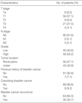
The clinicopathological characteristics of the 98 patients with TCC-UUT are summarized in Table 1. There were no
distant metastases in any patients at the time of radical neph- roureterectomy. Seventy patients were disease-free at a medi- an follow-up of 52.5 months (range, 12-162 months). The other 28 patients developed metastases at a median of 28 months (range, 4-86 months) after radical nephroureterec- tomy. Twenty-seven patients died of disease during the fol- low-up period.
The patterns of caveolin-1 expression in the 98 patients are summarized in Table 2. Of the 98 sections, positive immunos- taining for caveolin-1 was observed in 10 sections (10.2%).
The caveolin-1 expression was significantly correlated with T stage (p<0.001) and grade (p=0.036), but not with N stage (p=0.149), tumor location (p=0.847), previous history of blad- der cancer (p=0.711), coexisting bladder cancer at diagnosis (p=0.211), or bladder cancer recurrence during the follow-
Characteristics No. of patients (%)
T stage
Ta 8 (8.2)
T1 56 (57.1)
T2 9 (9.2)
T3 21 (21.4)
T4 4 (4.1)
N stage
N0 90 (91.8)
N1 5 (5.1)
N2 3 (3.1)
Grade
Low 40 (40.8)
High 58 (59.2)
Tumor location
Renal pelvis 56 (57.1)
Ureter 42 (42.9)
Previous history of bladder cancer
No 91 (92.9)
Yes 7 (7.1)
Coexisting bladder cancer
No 89 (90.8)
Yes 9 (9.2)
Bladder cancer recurrence
No 63 (64.3)
Yes 35 (35.7)
Table 1. Clinicopathological data of the 98 patients included in
the study Variables No. of
patients (%)
p value No. of caveolin-1 positive tumors (%) T stage
Superficial (Ta+T1) 64 (65.3) 1 (1.6) <0.001 Invasive (T2-T4) 34 (34.7) 9 (26.5)
N stage
N0 90 (91.8) 8 (8.9) 0.149
N1+N2 8 (8.2) 2 (25.0)
Grade
Low 40 (40.8) 1 (2.5) 0.036
High 58 (59.2) 9 (15.5)
Tumor location
Renal pelvis 56 (57.1) 6 (10.7) 0.847
Ureter 42 (42.9) 4 (9.5)
Previous history of bladder cancer
No 91 (92.9) 9 (9.9) 0.711
Yes 7 (7.1) 1 (14.3)
Coexisting bladder cancer
No 89 (90.8) 8 (9.0) 0.211
Yes 9 (9.2) 2 (22.2)
Bladder cancer recurrence
No 63 (64.3) 7 (11.1) 0.691
Yes 35 (35.7) 3 (8.6)
Table 3. Relationship between caveolin-1 expression and clini- copathological variables
Intensity Proportion Total
0 1 2 3 4
0 86 0 0 0 0 86
1 2 4 1 0 0 7
2 0 1 0 1 0 2
3 0 1 2 0 0 3
Total 88 6 3 1 0 98
Table 2. Pattern of caveolin-1 expression in the 98 patients
Variables Univariate
p value
Multivariate p value Hazards ratio (95% CI) T stage <0.0001 3.068 (1.078-8.736) 0.036
N stage 0.0682 1.398 (0.402-4.861) 0.598
Grade 0.0172 1.315 (0.446-3.871) 0.620
Tumor location 0.8689 1.415 (0.574-3.491) 0.451 Previous history of 0.3393 0.793 (0.175-3.600) 0.764
bladder cancer
Coexisting bladder cancer 0.7997 0.812 (0.204-3.235) 0.767 Bladder cancer recurrence 0.3694 1.594 (0.701-3.625) 0.266 Caveolin-1 expression <0.0001 5.080 (1.799-14.342) 0.002 Table 4. Univariate and multivariate survival analyses
CI, confidence interval.
up (p=0.691) (Table 3).
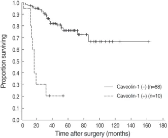
A Kaplan-Meier survival curve showed that the survival rate of patients with caveolin-1 positive tumors was signifi- cantly lower than that of patients with caveolin-1 negative tumors (p<0.0001) (Fig. 2).
The univariate analyses identified T stage (p<0.0001), grade (p=0.0172), and caveolin-1 expression (p<0.0001) as signif- icant prognostic factors for cancer-specific survival, whereas the multivariate analyses indicated that T stage (p=0.036) and caveolin-1 expression (p=0.002) were independent prog- nostic factors (Table 4).
DISCUSSION
The role of caveolin-1 in cancers still remains controver- sial. Several studies have suggested that caveolin-1 could func- tion as a tumor suppressor gene. The genes encoding cave- olin-1 and -2 are localized at the D7S522 locus located on human chromosome 7q31.1, a region frequently deleted in various types of human cancers (21). The allelic loss at chro- mosome 7q31.3 occurred in all grades and stages of invasive ovarian carcinomas, but not in borderline ovarian tumors, suggesting that the inactivation of a tumor suppressor gene in this region is an early event in ovarian tumorigenesis (22).
Caveolin-1 (-/-) null mice showed acceleration of the devel- opment of dysplastic mammary lesions (23), and caveolin-1 transfected human breast cancer cell line MCF-7 cells showed less proliferation than the vector control transfectants (11).
In addition, the caveolin-1 expression has been found to be down-regulated in ovarian carcinoma, pulmonary adenocar- cinoma, and sarcomas (8-10). Taken together, these findings suggest that caveolin-1 has tumor suppressor characteristics.
In contrast, however, several studies have shown elevated
caveolin-1 expression levels in some cancers and cancer cell lines. The increased expression of caveolin-1 has been found to be associated with drug-resistant cancer cell lines, such as paclitaxel or etoposide-resistant human lung cancer cells and androgen-insensitive mouse prostate cancer cells (24-26). Fur- thermore, the increased caveolin-1 expression has been report- ed in renal cell carcinoma, prostate cancer, bladder cancer, colonic carcinoma, and esophageal squamous cell carcinoma.
In many of these tumors, caveolin-1 overexpression was asso- ciated with higher tumor stage, tumor invasion, tumor size, higher grade, and poor prognosis (12-17). These findings indicate that caveolin-1 may function as a tumor promoter during carcinogenesis rather than as a tumor suppressor.
The differential expression of caveolin-1, depending on the histologic subtypes of cancers or different malignant stages, has also been reported. In the majority of small cell lung can- cers, caveolin-1 expression was lost through promoter methy- lation, the major mode of inactivation of many tumor sup- pressor genes in human cancers, whereas caveolin-1 expres- sion in the majority of non-small cell lung cancers was retain- ed and appeared to be required for tumor growth through the activation of FAK and Ra1A signaling (27). In lung ade- nocarcinomas, the caveolin-1 served as a tumor suppressor with the loss of caveolin-1 regulation, resulting in tumor extension and dedifferentiation, whereas caveolin-1 overex- pression in lung squamous cell carcinoma was correlated with tumor extension, suggesting that these reciprocal functions of caveolin-1 are due to different activation states of the dif- ferent domains of caveolin-1 and altered interactions with binding partners (28). In oral squamous cell carcinomas, the rate of caveolin-1 immunoreactivity increased progressively from normal oral mucosa, oral pre-cancer lesions to primary carcinomas, whereas it decreased from primary to metastatic carcinomas. These findings indicated the possibility of bipha- sic behavior of caveolin-1 in oral carcinogenesis and metas- tasis (29). Therefore, caveolin-1 might function either as a tumor promoter or as a tumor suppressor, depending on the tumor cell types or malignant stages.
For the expression of caveolin-1 in urothelial carcinomas, studies have been done in bladder carcinomas, but not in TCC-UUT. Rajjayabun et al. (14) found that the caveolin-1 expression was correlated with tumor stage and grade of blad- der TCC, but not with tumor multiplicity, recurrence, pro- gression, or survival of patients. Positive immunostaining for caveolin-1 was detected in 10.1% of tumor specimens. Fong et al. (15) showed that positive caveolin-1 immunostaining was detected in 37% of urothelial carcinoma, but not in nonneoplastic urothelium, and that the elevated expression of caveolin-1 and -2 was correlated with tumor grade and squamous differentiation.
In the present study, we investigated the expression of cave- olin-1 in TCC-UUT and found that the positive immunos- taining for caveolin-1 was observed in 10.2% of sections, sim- ilar to the findings of Rajjayabun et al. (14) in bladder TCC.
Proportion surviving
1.0 0.9 0.8 0.7 0.6 0.5 0.4 0.3 0.2 0.1 0.0
0 20 40 60 80 100 120 140 160 180
Time after surgery (months)
Caveolin-1 (-) (n=88)
Fig. 2. Kaplan-Meier cancer-specific survival curves according to the caveolin-1 expression. The survival rate of patients with cave- olin-1 positive tumors was significantly lower than that of patients with caveolin-1 negative tumors (p<0.0001).
Caveolin-1 (+) (n=10)
Furthermore, the caveolin-1 expression was correlated signif- icantly with T stage and grade. The survival rate of patients with caveolin-1 positive tumors was significantly lower than that of patients with caveolin-1 negative tumors. Also, the caveolin-1 expression was an independent prognostic factor for cancer-specific survival. These findings strongly indicate that caveolin-1 is involved in tumor progression, and that the increased expression of caveolin-1 is a late event in urothe- lial carcinomas.
In conclusion, the increased expression of caveolin-1 is asso- ciated with tumor progression and poor prognosis in TCC- UUT, suggesting that caveolin-1 may play an important role in the progression of TCC-UUT.
ACKNOWLEDGMENT
We especially thank Dr. Seung Soo Shin for statistical advice.
REFERENCES
1. Iborra I, Solsona E, Casanova J, Ricos JV, Rubio J, Climent MA.
Conservative elective treatment of upper urinary tract tumors: a multivariate analysis of prognostic factors for recurrence and pro- gression. J Urol 2003; 169: 82-5.
2. Munoz JJ, Ellison LM. Upper tract urothelial neoplasms: incidence and survival during the last 2 decades. J Urol 2000; 164: 1523-5.
3. Miyata Y, Kanda S, Nomata K, Eguchi J, Kanetake H. Expression of cyclooxygenase-2 and EP4 receptor in transitional cell carcino- ma of the upper urinary tract. J Urol 2005; 173: 56-60.
4. Okamoto T, Schlegel A, Scherer PE, Lisanti MP. Caveolins, a family of scaffolding proteins for organizing ‘‘preassembled signaling com- plexes’’ at the plasma membrane. J Biol Chem 1998; 273: 5419-22.
5. Liu P, Rudick M, Anderson RG. Multiple functions of caveolin-1. J Biol Chem 2002; 277: 41295-8.
6. Williams TM, Lisanti MP. The caveolin genes: from cell biology to medicine. Ann Med 2004; 36: 584-95.
7. Schwencke C, Braun-Dullaeus RC, Wunderlich C, Strasser RH. Cave- olae and caveolin in transmembrane signaling: implications for human disease. Cardiovasc Res 2006; 70: 42-9.
8. Wiechen K, Diatchenko L, Agoulnik A, Scharff KM, Schober H, Arlt K, Zhumabayeva B, Siebert PD, Dietel M, Schafer R, Sers C.
Caveolin-1 is down-regulated in human ovarian carcinoma and acts as a candidate tumor suppressor gene. Am J Pathol 2001; 159: 1635- 43.
9. Wikman H, Kettunen E, Seppanen JK, Karjalainen A, Hollmen J, Anttila S, Knuutila S. Identification of differentially expressed genes in pulmonary adenocarcinoma by using cDNA array. Oncogene 2002; 21: 5804-13.
10. Wiechen K, Sers C, Agoulnik A, Arlt K, Dietel M, Schlag PM, Schnei- der U. Down-regulation of caveolin-1, a candidate tumor suppres- sor gene, in sarcomas. Am J Pathol 2001; 158: 833-9.
11. Hino M, Doihara H, Kobayashi K, Aoe M, Shimizu N. Caveolin-1
as tumor suppressor gene in breast cancer. Surg Today 2003; 33:
486-90.
12. Joo HJ, Oh DK, Kim YS, Lee KB, Kim SJ. Increased expression of caveolin-1 and microvessel density correlates with metastasis and poor prognosis in clear cell renal cell carcinoma. BJU Int 2004;
93: 291-6.
13. Yang G, Truong LD, Wheeler TM, Thompson TC. Caveolin-1 expres- sion in clinically confined human prostate cancer: a novel prognos- tic marker. Cancer Res 1999; 59: 5719-23.
14. Rajjayabun PH, Garg S, Durkan GC, Charlton R, Robinson MC, Mellon JK. Caveolin-1 expression is associated with high-grade bladder cancer. Urology 2001; 58: 811-4.
15. Fong A, Garcia E, Gwynn L, Lisanti MP, Fazzari MJ, Li M. Expres- sion of caveolin-1 and caveolin-2 in urothelial carcinoma of the uri- nary bladder correlates with tumor grade and squamous differenti- ation. Am J Clin Pathol 2003; 120: 93-100.
16. Fine SW, Lisanti MP, Galbiati F, Li M. Elevated expression of cave- olin-1 in adenocarcinoma of the colon. Am J Clin Pathol 2001; 115:
719-24.
17. Kato K, Hida Y, Miyamoto M, Hashida H, Shinohara T, Itoh T, Okushiba S, Kondo S, Katoh H. Overexpression of caveolin-1 in esophageal squamous cell carcinoma correlates with lymph node metastasis and pathologic stage. Cancer 2002; 94: 929-33.
18. Greene FL, Page DL, Fleming ID, Fritz AG, Balch CM, Haller DG, Morrow M. AJCC cancer staging manual. 6th ed. New York: Springer- Verlag, 2002; 329-31.
19. Epstein JI, Amin MB, Reuter VR, Mostofi FK. The World Health Organization/International Society of Urological Pathology con- sensus classification of urothelial (transitional cell) neoplasms of the urinary bladder. Bladder Consensus Conference Committee.
Am J Surg Pathol 1998; 22: 1435-48.
20. Sinicrope FA, Ruan SB, Cleary KR, Stephens LC, Lee JJ, Levin B.
bcl-2 and p53 oncoprotein expression during colorectal tumorigen- esis. Cancer Res 1995; 55: 237-41.
21. Engelman JA, Zhang XL, Lisanti MP. Genes encoding human cave- olin-1 and -2 are co-localized to the D7S522 locus (7q31.1), a known fragile site (FRA7G) that is frequently deleted in human cancers.
FEBS Lett 1998; 436: 403-10.
22. Edelson MI, Scherer SW, Tsui LC, Welch WR, Bell DA, Berkowitz RS, Mok SC. Identification of a 1300 kilobase deletion unit on chro- mosome 7q31.3 in invasive epithelial ovarian carcinomas. Onco- gene 1997; 14: 2979-84.
23. Williams TM, Cheung MW, Park DS, Razani B, Cohen AW, Muller WJ, Di Vizio D, Chopra NG, Pestell RG, Lisanti MP. Loss of cave- olin-1 gene expression accelerates the development of dysplastic mammary lesions in tumor-prone transgenic mice. Mol Biol Cell 2003; 14: 1027-42.
24. Yang CP, Galbiati F, Volonte D, Horwitz SB, Lisanti MP. Upregu- lation of caveolin-1 and caveolae organelles in Taxol-resistant A549 cells. FEBS Lett 1998; 439: 368-72.
25. Belanger MM, Gaudreau M, Roussel E, Couet J. Role of caveolin-1 in etoposide resistance development in A549 lung cancer cells. Can- cer Biol Ther 2004; 3: 954-9.
26. Nasu Y, Timme TL, Yang G, Bangma CH, Li L, Ren C, Park SH,
. .
. .
DeLeon M, Wang J, Thompson TC. Suppression of caveolin expres- sion induces androgen sensitivity in metastatic androgen-insensitive mouse prostate cancer cells. Nat Med 1998; 4: 1062-4.
27. Sunaga N, Miyajima K, Suzuki M, Sato M, White MA, Ramirez RD, Shay JW, Gazdar AF, Minna JD. Different roles for caveolin-1 in the development of non-small cell lung cancer versus small cell lung cancer. Cancer Res 2004; 64: 4277-85.
28. Kato T, Miyamoto M, Kato K, Cho Y, Itoh T, Morikawa T, Okushi-
ba S, Kondo S, Ohbuchi T, Katoh H. Difference of caveolin-1 expres- sion pattern in human lung neoplastic tissue. Atypical adenomatous hyperplasia, adenocarcinoma and squamous cell carcinoma. Can- cer Lett 2004; 214: 121-8.
29. Hung KF, Lin SC, Liu CJ, Chang CS, Chang KW, Kao SY. The biphasic differential expression of the cellular membrane protein, caveolin-1, in oral carcinogenesis. J Oral Pathol Med 2003; 32:
461-7.